Actoplus Met
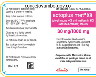
Actoplus Met dosages: 500 mg
Actoplus Met packs: 30 pills, 60 pills, 90 pills, 120 pills, 180 pills, 270 pills, 360 pills
Actoplus met 500 mg generic on-line
They report that there was no difference in survival in these with neurocardiogenic syncope and people with out evaluation blood glucose screening actoplus met 500 mg generic fast delivery. The authors included seizures diabetes in dogs management purchase actoplus met 500 mg without prescription, strokes, and transient ischaemic assaults of their population studied, making interpretation of these information much more difficult. The severity will fluctuate and could be influenced by posture being worse on prolonged standing, with train or post-prandially. They include a non-specific sense of disequilibrium, muzzy heads, poor focus, a fear of collapse, weak legs, and there could additionally be active manoeuvres to abort the symptoms, corresponding to sitting down and resting. They vary with age with extended standing being extra necessary in older populations with as much as 54. Syncope in older age is associated with the act of standing, but may be seen in the sitting position and post-prandially, and is extra commonly seen when cardiovascular active drugs, such as anti-hypertensives are used. Younger sufferers report the consequences of ache, venesection, viscous pain, concomitant illnesses, emotion, menstruation, drugs, fatigue, and fasting (1). Multiple triggers can be reported either on separate events or contributing to one event. The psychological impact of the physical triggers is often uncared for in taking the history from the patient. Their understanding of the potential significance of the physical signs must be acknowledged. A rising sensation within the stomach typically accompanied by a sense of nausea without vomiting can lead to confusion with an epileptic aura, although the presyncopal symptoms are less intense as an experience. It should be famous that complex partial seizures notably of fronto-temporal origin with secondary seizure-induced bradycardia or asystole is a well-recognized syndrome, which is normally a entice for the clinician (21). In the classification of syncope, situational syncope is commonly separated from classical syncope. Examples embody carotid sinus syndrome, swallow, defaecation, micturition, post-exercise, post-prandial, cough, sneeze, laughing, brass instrument playing, weight lifting, medical instrumentation, and prankster valsalva manoeuvres. A detailed assessment of the triggering organ in search of native pathology must be undertaken. If associated to direct intervention, a history of previous neurocardiogenic syncope or presyncope ought to be elucidated (22). The causes for syncope have been shown to be reflex syncope (up to 40%), orthostatic hypotension (6�24%), cardiac syncope (10�20%), and psychogenic causes (1�5%). A number of research have been undertaken so as to introduce order into a posh subject without as yet definite solutions being found. There is an age impact with more cardiac and orthostatic causes seen in older people, however reflex syncope remains the most typical trigger throughout the ages, although it has a much less benign course within the elderly compared with the younger (18). Syncope: the medical syndrome the very word, syncope suggests a sudden event, but the scientific spectrum, actually, is kind of extensive. A detailed historical past must be taken from the affected person who can typically describe the prodrome and post-event options and any eyewitness who can even describe the three parts and may observe external triggers, similar to stress not recognized by the affected person (19). Relatives typically cite a need not to embarrass the patient by videoing their attacks, but the overall benefits of an correct prognosis have to be emphasised. Neurocardiogenic syncope or reflex syncope is most closely related to physiological triggers. Orthostatic syncope because of its name has an orthostatic trigger, while psychogenic syncope might have any of the preceding triggers, however there tends to be extra variability within the historical past, and the attacks could be of longer period and may have some options of psychogenic seizures. The commonest cause is reflex neurocardiogenic syncope and an in depth understanding of the medical options is invaluable for accurate prognosis. Reflex neurocardiogenic syncope the duration of the three parts (prodrome, event, postevent) can range between different attacks in the identical particular person and between different sufferers. Prodromal signs can be present for so much of hours in a light type in sufferers with a significant tendency to collapse, similar to is seen in those with autonomic failure Orthostatic syncope the definition contains syncope related only with orthostatic modifications. Syncope in this medical state of affairs can be due to a failure to keep adequate cerebral perfusion due to blood quantity deficiencies, decreased cardiac output, or impaired venous return with or without reflex neurocardiogenic syncope. Slowly growing autonomic failure as is seen in primary autonomic failure can also be associated with remarkably well-tolerated low blood pressures. The mind is highly dependent on cardiac output to meet its metabolic wants and it takes a disproportionate quantity of the cardiac output. At rest regardless of constituting about 2% of physique mass at 1400 g, it takes 15�20% of cardiac output (27). Cessation of blood flow by neck compression leads to lack of consciousness inside 6 or 7 s (28). In addition to the central function for cardiac output, the baroreflex, which maintains blood strain and cerebral autoregulation, which endeavours to maintain consistent cerebral blood move despite variations in systemic blood stress are the other main controllers of cerebral perfusion. This could be structural or arrhythmogenic and is described as being triggered by rising cardiac workload significantly during train or with cardiac ischaemia. In a large preparticipation screening programme of 7568 athletes, solely 57 had experienced pre-exertion syncope in the preceding 5 years and six had exertion associated syncope. The baroreflex system the baroreflex arc consists of afferent loops taking sensory information from receptors in carotid sinuses via the glossopharangeal nerves and from the aortic arch by way of the vagus to the nuclei of the tractus solitarius in the medulla. Seizures and psychogenic assaults are the primary differential diagnoses that have to be thought-about. The significance of establishing the mechanism of the syncopal attack has acquired consideration (14) and the authors draw consideration to the variability in prognosis and administration of syncope with the implications for reduced quality of life and extra prices. Well-developed pathways with devoted items and skilled clinicians are wanted to manage these sufferers. Given the variability within the prevalence of syncope, being dependent on the population being studied, there are difficulties in determining accurately the incidence of psychogenic attacks. This is harder given the importance of psychological elements in contributing to syncopal events, and the popularity that syncope and psychogenic attacks can co-exist in the same affected person. This makes accurate assessment of incidence and prevalence of psychogenic assaults tough. This displays the populations being studied and the importance of detailed historical past taking and assessment in this population. When resistance to circulate rises, the results are initially seen with a reduction in diastolic flow. Intracranial pressures are influenced by venous pressures, tissue pressures, and the tendency for vessel partitions to collapse. Raised central venous pressures because of obstructive disease or throughout valsalva manoeuvres. Tissue pressures may be significant as a outcome of the cranium is a inflexible restrict on space for mind expansion as is seen with tumours or bleeding. At a cerebral vascular stage, the humoral results are thought to be attenuated by the blood�brain barrier, though differential results of humoral mechanisms on massive versus small vessels could play a component (36). While this has been shown in mammals, it has not been particularly shown in humans. Sensory fibres are associated with modulating cerebral blood move and can be related to increased blood flow as is seen in cortical spreading despair (39), seizures (40), and reactive hyperaemia (41). Autoregulation of blood circulate is best in cerebral, renal, and mesenteric vessels (45), and the mechanisms involved embody myogenic, metabolic, neural, and activation of potassium channels (46�48).
Diseases
- Intercellular cholesterol esterification disease
- Ccge syndrome
- Hydroxycarboxylic aciduria
- Ventricular fibrillation, idiopathic
- Nonmedullary thyroid carcinoma, with cell oxyphilia
- Thyrocerebrorenal syndrome
- Porphyria, Ala-D
- AIDS
- Hereditary hearing loss
- Intoeing
500 mg actoplus met generic with mastercard
However diabetic diet without medication order 500 mg actoplus met amex, sleeves of ventricular myocardium prolong beyond the aortic valve attachments for variable distances (analogous to atrial myocardial extensions in the pulmonary veins) diabetes type 1 paleo diet 500 mg actoplus met generic with amex. Whereas the right coronary cusp and the anterior portions of the left coronary cusp regularly exhibit these myocardial sleeves, the posterior portions of the left coronary cusp and the noncoronary cusp, particularly in relation to the fibrous continuity with the anterior leaflet of the mitral valve (the aortomitral continuity), are solely fibrous and normally devoid of myocardium. Nevertheless, owing to the semilunar configuration of the valvular leaflets, the hinge line of every leaflet crosses the ventriculoarterial junction at two factors. Consequently, there are all the time small segments of myocardium of the infundibulum on the nadirs of the three sinuses. Extensions of ventricular myocardium into the adventitia happen in approximately 20% of individuals and have been traced to a maximal distance of 6 mm beyond the junction. The pulmonic valve, essentially the most superiorly situated of the cardiac valves, lies at the stage similar to the third left costal cartilage at its junction with the sternum. The transverse plane of the aortic valve slopes inferiorly, away from the airplane of the pulmonic valve, such that the orifice of the aortic valve faces rightward at an angle of at least 45 levels from the median plane. The left pulmonic cusp, being essentially the most superficial, lies immediately beneath the pericardium and has no different cardiac constructions related to it. The supravalvular portion of the aorta lies near and in some cases adjacent to the junction and surrounding parts of the proper and posterior pulmonic cusps. As famous, ventricular myocardial sleeves lengthen above the semilunar valves for a variable distance (a few millimeters and as a lot as greater than 2 centimeters). Whereas the myocardial sleeves usually prolong circumferentially around the pulmonic valve between and above all three cusps just above the annulus, more distally the extension is patchy and generally asymmetrical. Myocardial extensions also occur in the intercuspal clefts along with throughout the cusps. Left coronary heart catheterization, right coronary heart catheterization, or both may be warranted. As the origin strikes laterally along the mitral annulus, the R wave in lead I and in the inferior leads decreases in amplitude. Contrariwise, an R wave in lead V2 taller than in leads V1 or V3 suggests an origin in the so-called crux of the heart, close to the posterior descending artery. Furthermore, R wave amplitude in lead V2 or "r" wave amplitude in leads V1 and V2 greater than zero. Additionally, the nearer the origin to the pulmonic valve, the extra rightward and inferior the axis. A simple qR complicated or a monophasic R wave suggests an origin close to the aortomitral continuity. This website is commonly acknowledged by a notch on the downstroke in V1 with an intermediate (relative to the proper and left cusps) precordial transition. A qrS pattern in leads V1 to V3 is very helpful for predicting an origin within the junction between the best and left coronary cusps. In a few of those instances, an insulated myocardial fiber throughout the ventricular outflow septum might exist. This may be defined by a location within the basal-lateral myocardium for the previous and a more anterobasal location for the latter arrhythmias. Induction is exquisitely sensitive to the immediate autonomic standing of the affected person. The space within 1 cm just below the pulmonic valve is outlined as the superior (or distal) facet, and the world greater than 1 cm away is defined as the inferior (or proximal) aspect. The prediction of the precise origin of outflow tract tachycardias could be difficult because of the close anatomical relationship of the completely different anatomical compartments of the outflow tract space. Finally, if all earlier anatomical accesses are unsuccessful, epicardial mapping through a percutaneous pericardial access ought to be thought of. It may be performed by point-by-point mapping with a roving mapping catheter, with the use of a quantity of catheters, or with multielectrode arrays. Once an space of relatively early native activation is discovered, small movements of the catheter tip in that area are undertaken till the location is recognized with the earliest attainable local activation relative to the tachycardia complicated. Fractionated complicated electrograms and mid-diastolic potentials are rarely, if ever, seen and may increase suspicion of underlying heart illness. In some cases, the near-field electrogram is fused with the far-field electrogram during tachycardia. This suggests an origin of arrhythmia exactly on the cusp or passive activation from a true distant web site of origin to both the supravalvular and infravalvular myocardium. Bipolar pacing from the closely spaced distal electrodes of the mapping catheter is more generally used. Use of present strengths close to threshold should enhance accuracy by limiting the dimensions of the virtual electrode within the tissue and preventing seize of myocardium distant from the pacing site. Pacing thresholds of at least 5 to 10 mA usually point out inadequate electrode-tissue contact or inexcitable areas. As a consequence, what mapping defines because the earliest web site of activation relative to neighboring myocardium could be deceptive. The findings on the mapping catheter throughout these catheter-induced complexes are invariably wonderful. When mapping above the aortic or pulmonic valve, near- and far-field potentials are typically recorded. Because of the overlapping nature of the outflow tract and supravalvular area, when two potentials are seen, solely the near-field potential ought to be used for activation timing. In addition to noting the precise timing of activation, the timing of the near-field electrogram relative to the far-field electrogram should be evaluated. On the other hand, automated objective interpretation can provide some benefit to human interpretation. Nonetheless, the effective ablation sites had a high degree of correlation with tempo mapping at efficient ablation sites. Pace mapping can be tough to perform above the semilunar valves (in the aortic cusps or pulmonary artery) and might require high pacing current strengths. Points are added to the map only if stability criteria in area and local activation time are met. The end-diastolic location stability criterion is less than 2 mm and the native activation time stability criterion is less than 2 milliseconds. Activation maps show the native activation time by a color-coded overlay on the reconstructed 3-D geometry. Although the normal single-catheter mapping of the area of interest remains to be required, the flexibility to use the catheter localization system to steer precisely again to previously obtained sites of earliest activation tremendously facilitates the ablation course of. Display of activation instances facilitates comparability of information from nearby sites, overcoming the imprecision of assigned activation instances at single points, and permits fast identification of a putative web site of origin. The activation map may additionally be used to catalogue sites at which pacing maneuvers are carried out during evaluation of the tachycardia. The biggest advantage of noncontact endocardial mapping is its capacity to recreate the endocardial activation sequence from simultaneously acquired a number of information points over a few (theoretically one) tachycardia beats, with out requiring sequential point-to-point acquisitions. The balloon is then deployed; it could be full of distinction dye, permitting it to be visualized fluoroscopically. The highest chamber voltage is on the website of origin of the electrical impulse (see Video 8). The color scale for each isopotential map is about in order that white indicates probably the most adverse potential and blue indicates the least adverse potential.
Generic 500 mg actoplus met fast delivery
The flat return cycle also suggests the presence of fastened websites of entrance and exit from the circuit and glued conduction time from the stimulation website by way of the reentrant circuit over a broad range of coupling intervals diabetes in dogs and symptoms 500 mg actoplus met order with mastercard. If a single extrastimulus produced resetting with a flat response diabetes type 2 uncontrolled buy 500 mg actoplus met visa, the response to double extrastimuli would also be flat. However, as a result of the utilization of double extrastimuli permits engagement of the reentrant circuit at relatively lengthy coupling intervals with larger prematurity, resetting will start at longer coupling intervals and can proceed over a higher vary of coupling intervals than observed with a single extrastimulus. Therefore, double extrastimuli can produce a flat and then growing response curve. The most probable mechanism underlying the decremental conduction is encroachment of the advancing wavefront from the untimely extrastimuli on an increasingly extra refractory tissue inside the reentrant circuit, most probably inside the zone of slow conduction. Mixed Response Curves in Reentrant Rhythms In a blended response curve, the initial coupling intervals reveal a flat portion of the curve of variable length (but lower than 30 milliseconds), adopted by a zone during which the return cycle will increase. With later coupled extrastimuli, the orthodromic wavefront exits from the reentrant circuit, thus capturing a certain portion of myocardium earlier than colliding with the paced antidromic wavefront. If presystolic exercise within the reentrant circuit is recorded earlier than supply of the extrastimulus that resets the tachycardia, one should consider this to characterize local fusion. Resetting with native fusion and a completely paced complicated morphology supplies evidence that the reentrant circuit is electrocardiographically small. Reentrant circuits reset with fusion have a better incidence of flat resetting curves, longer resetting zones, and considerably shorter return cycles measured from the stimulus to the onset of the tachycardia complicated. Resetting with fusion is a potential indication that the pacing web site is situated proximal to the zone of sluggish conduction. Site specificity for resetting is decreased with using multiple extrastimuli. Reentrant rhythms by no means show a lowering resetting curve to single or double extrastimuli. Resetting with Fusion the power to reset tachycardia after it has begun activating the myocardium. In this case, irrespective of how many subsequent extrastimuli are delivered, the return cycle would be the same and equal to that observed through the flat portion of the resetting curve. The conduction time of this impulse to the exit website of the circuit is termed the last entrained interval, and it characterizes the properties of the reset circuit during entrainment. In this case, the return cycle relies upon critically on the variety of extrastimuli delivered that reset the circuit before the return cycle is measured, as a outcome of following the first extrastimulus producing resetting (the nth extrastimulus), subsequent extrastimuli are relatively more untimely and may result in a different return cycle. These measurements shall be qualitatively similar but have totally different absolute values. The capacity to entrain a tachycardia additionally establishes that the reentrant circuit incorporates an excitable hole. Entrainment is the continual resetting of a reentrant circuit by a train of capturing stimuli. This sequence continues till cessation of pacing or block someplace within the reentrant circuit develops. Depending on the degree to which the excitable hole is preexcited (and abbreviated) by the first resetting stimulus, subsequent stimuli fall on totally or partially excitable tissue. Therefore, only resetting phenomena describe the traits of the reentrant circuit. Flat, blended (flat and increasing), and growing curves can be seen during entrainment of macroreentrant circuits. Increasing curves are virtually all the time noticed throughout entrainment of small or microreentrant circuits. The closer the pacing web site is to the circuit, however, the much less untimely a single stimulus may be and reach the circuit and, with pacing trains, the less the variety of stimuli required earlier than a stimulated wavefront reaches the reentrant circuit with out being extinguished by collision with a wave rising from the circuit. A decrease in conduction velocity ends in a reduction within the wavelength and a lessening of the amount of tissue needed to sustain reentry. The pathway between the source and sink contains intracellular resistance (provided by the cytoplasm) and intercellular resistance (provided by the gap junctions). Therefore, the quantity and distribution of hole junctions, in addition to the conductance of the gap junction proteins (connexins), are essential factors for conduction of the action potential. The safety issue for conduction predicts the success of motion potential propagation and is outlined because the ratio of the present generated by the depolarizing ion channels of a cell (source) to the present consumed in the course of the excitation cycle of a single cell in the tissue (sink). By this definition, conduction fails when the security issue drops to lower than 1 and becomes more and more steady as it rises to greater than 1. In essence, local source-sink relationships determine the formation of conduction heterogeneities and supply situations for the development of slow conduction, unidirectional block, and reentry. The extra negative the membrane potential is, the extra Na+ channels are available for activation, the greater the inflow of Na+ into the cell during phase zero, and the larger the conduction velocity. In contrast, membrane depolarization to ranges of -60 to -70 mV can inactivate half the Na+ channels, and depolarization to -50 mV or much less can inactivate all the Na+ channels. Genetic mutations that result in loss of Na+ channel function, as happens in the Brugada syndrome, could cause decreased membrane excitability. This inhomogeneity may be associated to (1) electrical properties of the person cardiac myocyte that generates the action potential (inhomogeneity in electrical excitability or refractoriness, or both), (2) passive properties governing the circulate of present amongst cardiac cells (cell-to-cell coupling and tissue geometry), or (3) combinations of those conditions. Additionally, a few of these modifications are wanted only to set the initial condition for the deviation of the impulse, the so-called unidirectional conduction block. Once the 58 Reduced membrane excitability can also be present in cardiac cells with persistently low ranges of resting potential brought on by disease. With progressive reduction of excitability, less Na+ source current is generated, and conduction velocity and the safety issue lower monotonically. Action potentials with decreased upstroke velocity resulting from partial inactivation of Na+ channels are called depressed quick responses. These motion potential changes are more doubtless to be heterogeneous, with unequal levels of Na+ inactivation that create areas with minimally decreased velocity, more severely depressed zones, and areas of full block. The chance for reentry in such fibers is even higher throughout untimely activation or during rhythms at a speedy rate, as a result of slow conduction or the potential for block is increased even additional. Lateral (sideto-side) gap junctions in nondisc lateral membranes of cardiomyocytes are a lot less ample and happen more often in atrial than ventricular tissues. Additionally, less intercellular hole junctional coupling happens and, hence, greater resistance and slower conduction transversely than longitudinally. Remodeling includes a decrease in some gap junction channels resulting from the interruption of communication between cells by fibrosis and downregulation of connexin-43 (Cx43) formation or trafficking to the intercalated disc. Additionally, hole junctions can become extra outstanding alongside lateral membranes of myocytes (so-called structural remodeling). Similar to its conduct throughout decreased membrane excitability, conduction velocity decreases monotonically with reduction in intercellular coupling. As a end result, individual cells depolarize with a high margin of security, but conduction proceeds with long intercellular delays. At such low levels of coupling, conduction may be very gradual however, paradoxically, very robust. Cx43 expression should decrease by 90% to affect conduction, however even then conduction velocity is decreased only by 20%. In distinction to an uncoupled cell strand, by which the excessive resistance junctions alternate with the low cytoplasmic resistance of the cells, a high diploma of discontinuity may be produced by large tissue segments (consisting of a phase with aspect branches) alternating with small tissue segments having a small tissue mass (connecting segments without branches). This occurs due to sink-source mismatch, during which the current offered by the excitation wavefront (source) is inadequate to charge the capability and thus excite the much bigger volume of tissue ahead (sink).
Effective actoplus met 500 mg
The scientific evaluation and outcomes of first-line investigations ought to allow you to make a differential analysis and working analysis diabetes symptoms memory loss actoplus met 500 mg generic on-line. Pulmonary oedema may be localized and when extreme (alveolar) may produce an air-bronchogram diabetes test log books 500 mg actoplus met amex. The radiological signs of pulmonary oedema are modified by the presence of lung disease. If fever and a productive cough are absent, and the white cell rely is <15 � 109/L or C-reactive protein <10 mg/L, give diuretic alone and assess the response. Breathlessness with a raised jugular venous pressure � this mix may be seen in acute main pulmonary embolism, heart failure with biventricular contain ment, continual hypoxic lung disease complicated by cor pulmonale, and cardiac tamponade. Low or high temperature, raised white count and C-reactive protein, raised lactate. Type 2 respiratory failure is caused by alveolar hypoventilation, with or with out V/Q mismatch and may thus be caused by ailments each intrinsic and extrinsic to the respiratory system. Remember there could usually be a combination of disease processes, for instance pneumonia on a background of pulmonary fibrosis or heart failure. There may be an acute-on-chronic presentation, for example decompensated continual respiratory failure in a affected person with an exacerbation of persistent obstructive pulmonary disease. Management will largely be decided by the working prognosis as properly as the response to the initial therapy. Are there features to suggest infection, for instance fever, cough, purulent sputum or increase in sputum quantity Stephen Chapman, Grace Robinson, John Stradling, Sophie West, and John Wrightson (2014) Oxford Handbook of Respiratory Medicine (3 ed. Evaluation requires clinical assessment, imaging (by chest X-ray and thoracic ultrasonography) and exami nation of pleural fluid. Priorities Clinical evaluation and imaging the scientific evaluation is summarized in Table 12. Pleural effusion ends in basal shadowing obscuring the hemidiaphragm with a concave upper border. The sensitivity of the technique for detecting malignant pleural effusion is round 60%. The effusion is an exudate if it meets one of the following criteria: � Pleural fluid protein/serum protein ratio >0. Cause Heart failure Comment Increased interstitial fluid, which crosses the visceral pleura and enters the pleural area. Investigate additional if atypical features are present (unilateral effusion, fever, chest pain). Usually bilateral effusions, decreased oncotic stress inflicting transudate effusion. May be transudate or exudate, commonly together with ascites, pericardial effusion and cardiac failure. Cause Pleural an infection (parapneumonic effusion and empyema) Malignancy Tuberculosis Chylothorax Oesophangeal rupture Pulmonary embolism Comment Most frequent trigger in young sufferers; empyema is outlined as pus within the pleural cavity. Delayed hypersensitivity response to mycobacteria launched into the pleural area. Often milky effusions, analysis with presence of chylomicrons or pleural fluid triglyceride level >1. Almost all the time exudative; bloody in <50%; it should be suspected when dyspnoea is disproportionate to measurement of effusion, or when patient is hypoxic. Pleural fluid evaluation (1): In all sufferers Test Visual inspection Comment Blood-stained effusion (pleural fluid haematocrit 1�20% of peripheral haematocrit) is prone to be because of malignancy, pulmonary embolism or trauma. An exudate is recognized by a quantity of of the next: � Pleural fluid protein to serum protein ratio >0. The yield of cytology is influenced by the histological type of malignancy: >70% positive in adenocarcinoma, 25�50% in lymphoma, 10% in mesothelioma. Comment eighty three Elevated pleural fluid amylase is seen within the acute pancreatitis and oesophageal rupture. Check triglyceride degree if chylothorax is suspected (opaque white effusion); chylothorax (triglyceride >1. Drainage of a symptomatic effusion � Therapeutic aspiration is a adequate remedy for many symptomatic effusions and could be repeated for effusions that re-accumulate. Pleural aspiration ought to be stopped if the patient develops chest tightness, chest ache or extreme coughing � Effusions which are continual, recurrent and inflicting symptoms, can be handled with pleurodesis or by intermittent drainage with an indwelling catheter. Specific administration for parapneumonic effusions and empyema � Seek recommendation from a chest physician. Talc pleurodesis may be performed both by the insertion of a small chest tube (10�14 F) as a slurry or by medical thoracoscopy as a poudrage. Both strategies have significantly higher results compared to closed pleural biopsies. Thoracoscopy additionally provides diagnostic and therapeutic approaches to patients with pleural effusion. It is mostly because of higher respiratory tract an infection, with a benign course. Clinical indicators indicating a severe cause that warrants inpatient admission are given in Table 13. Duration of cough Associated symptoms: fever, evening sweats, weight loss, haemoptysis, breathlessness, chest pain History of tuberculosis or contact with tuberculosis History of other respiratory illness Foreign travel Symptoms of gastro-oesophageal reflux illness (heart burn, acid reflux disease, pain on swallowing) Post-nasal drip syndrome (feeling of excess mucus accumulating behind the throat, feeling of mucus dripping from the nose into back of the throat) Triggers to cough. Respiratory rate >30 breaths/min Oxygen saturation <92%, (if no historical past of persistent hypoxia) Use of accessory muscles of respiration Tachycardia >130/min Systolic blood strain <90 mmHg or diastolic blood stress <60 mmHg Haemoptysis Fever Chest ache Suspicion of inhaled international body Table thirteen. Full blood depend � raised eosinophil degree could direct you to consider eosinophilic airways disease as the cause for cough. Approximately 30% of chest radiographs requested for investigating the trigger of cough are irregular. Spirometry: reversibility check (for obstructive airways disease), airway provocation test with mannitol or methacholine for cough variant asthma. Fibreoptic laryngoscopy: in sufferers with suspected upper airways pathology as the purpose for cough, laryngoscopy may present modifications related to laryngeal inflammation and oedema. Haemoptysis Haemoptysis is coughing up blood, both combined with sputum or on its own, from the respiratory tract (Table thirteen. Bronchial arteries normally arise directly from the aorta and carry blood at systemic blood pressure. Therefore, bleeding that originates from bronchial circulation causes large haemoptysis; if left untreated it has a mortality rate of up to 80%. A pragmatic definition of massive haemoptysis is 200 mL of blood loss in 24 hours, or haemoptysis significant sufficient to impair fuel change and cause haemodynamic compromise, whatever the length of haemoptysis. Management of large haemoptysis the priority is to stabilize the patient, identify the bleeding web site and cease the bleeding (Table thirteen.
PSK (Coriolus Mushroom). Actoplus Met.
- What other names is Coriolus Mushroom known by?
- Cancer when used with chemotherapy regimens (when PSK products isolated from coriolus mushroom are used).
- Boosting immune function, herpes, chronic fatigue syndrome, hepatitis, lung disorders, body building, ringworm, skin infections (impetigo), urinary and digestive tract infections, poor appetite, and other uses.
- What is Coriolus Mushroom?
- Are there safety concerns?
- Dosing considerations for Coriolus Mushroom.
- How does Coriolus Mushroom work?
Source: http://www.rxlist.com/script/main/art.asp?articlekey=96638
500 mg actoplus met amex
Under such circumstances diabetes mellitus type 2 and obesity actoplus met 500 mg best, constant fusion throughout entrainment is almost impossible (unless a second connection exists between the atria and ventricles; i metabolic disease jobs buy generic actoplus met 500 mg line. The major advantage of this methodology is its independence of tachycardia continuation after cessation of pacing. This target may be outlined by considered one of two approaches: a purely anatomical method and an electroanatomical strategy. Rarely, profitable sluggish pathway ablation might require an utility of energy on the left facet of the posterior septum, alongside the mitral annulus. These potentials have been used by some to outline the positioning of the slow pathway within the triangle of Koch, and they can be used successfully as a guide to target ablation. Whether they symbolize nodal tissue activation, anisotropic conduction through muscle bundles in varied websites in the triangle of Koch, or a combination of both is unclear. The electrogram morphology of the gradual potentials has been variously described as sharp and rapid (representing the atrial connection to the slow pathway; see. Despite these observations, the likelihood of recording putative gradual potentials at the website of efficient sluggish ablation is greater than 90%. Note the sharp (blue arrow, left lower panel) and broad (red arrow, right decrease panel) potentials recorded between the atrial and ventricular electrograms on the ablation sites. Those potentials had been advised to mirror activation of the slow pathway (slow pathway potentials). Moreover, some ablation catheters have asymmetrical bidirectional deflection curves, an possibility that may prove to be of value for catheter reach and stability in some cases. This method also helps evaluate the extension of the zone recording a His potential. Moving the mapping catheter inferiorly, the sluggish pathway potential moves toward the atrial electrogram, and when the optimal web site for gradual pathway ablation is reached, it merges with the atrial electrogram. Catheter-induced junctional ectopy, when present, indicates that the catheter tip is at a good ablation site. Rarely, a superior vena caval approach (through the interior jugular or subclavian vein) is required due to inferior vena caval obstruction or barriers, and one report demonstrated the feasibility of this strategy. Gentle clockwise torque is maintained to hold the catheter in contact with the low atrial septum. This might require cessation of isoproterenol infusion if hyperdynamic contractility is present. However, within the case of slow pathway ablation, the decrement in impedance associated with profitable energy purposes is usually small (approximately 2. Occurrence of this rhythm is strongly correlated with and sensitive to successful ablation websites; it happens more frequently (94% versus 64%) and for an extended period (7. Overdrive atrial pacing at a rate quicker than the junctional rhythm rate was started and confirmed intact atrioventricular conduction. This remark can indicate an excellent ablation website, with no damage to the quick pathway. Whether the fast pathway should be targeted by ablation as an alternative of the gradual pathway in these patients is controversial. In such sufferers, it may be clever to confine additional ablation efforts to the pathway originally targeted for ablation. Most are caused by untimely atrial or ventricular complexes, which subside spontaneously and require no therapy other than reassurance. The anterior strategy selectively ablates or modifies fast pathway retrograde conduction; nevertheless, it could also cause injury to fast or sluggish pathway anterograde conduction. The catheter is then withdrawn whereas a agency clockwise torque is maintained till the His potential turns into small or barely visible or disappears while recording a relatively large atrial electrogram (with an A/V electrogram amplitude ratio larger than 1; see. This normally requires delivery of several cryoapplications at closely adjoining sites. Furthermore, once the catheter tip temperature is lowered to less than 0�C, progressive ice formation at the catheter tip causes adherence to the adjoining tissue (cryoadherence), which maintains secure catheter contact on the web site of ablation and minimizes the risk of catheter dislodgment during changing cardiac rhythm. At this temperature, the cryolesion is reversible (for as a lot as 60 seconds), and the catheter is stuck to the atrial endocardium inside an ice ball that features the tip of the catheter (cryoadherence). Once an ice ball is formed, varied pacing protocols are carried out to test the modification or disappearance of gradual pathway conduction. Thus, other parameters should be used to validate the potential effectiveness of the ablation website. After a few seconds, to enable the catheter to thaw and turn out to be dislodged from the tissue, the catheter is moved to a different web site, and cryomapping is repeated. In Fischer G, editor: Handbuch der vergleichenden und experimentellen entwicklungslehre der wirbeltiere, Jena, Germany, 1906, Semper Bonis Artibus, pp 136�137. Lockwood D, Nakagawa H, Jackman W: Electrophysiological characteristics of atrioventricular nodal reentrant tachycardia: implications for the reentrant circuit. Valderrabano M: Atypical atrioventricular nodal reentry with eccentric atrial activation: is the proper target on the left Morihisa K, Yamabe H, Uemura T, et al: Analysis of atrioventricular nodal reentrant tachycardia with variable ventriculoatrial block: traits of the upper widespread pathway, Pacing Clin Electrophysiol 32:484�493, 2009. Otomo K, Okamura H, Noda T, et al: Unique electrophysiologic traits of atrioventricular nodal reentrant tachycardia with completely different ventriculoatrial block patterns: results of sluggish pathway ablation and insights into the placement of the reentrant circuit, Heart Rhythm three:544�554, 2006. Otomo K, Okamura H, Noda T, et al: "Left-variant" atypical atrioventricular nodal reentrant tachycardia: electrophysiological traits and effect of slow pathway ablation within coronary sinus, J Cardiovasc Electrophysiol 17:1177�1183, 2006. Otomo K, Nagata Y, Uno K, et al: Atypical atrioventricular nodal reentrant tachycardia with eccentric coronary sinus activation: electrophysiological characteristics and essential results of left-sided ablation contained in the coronary sinus, Heart Rhythm 4:421�432, 2007. Gonzalez-Torrecilla E, Almendral J, Arenal A, et al: Combined evaluation of bedside medical variables and the electrocardiogram for the differential prognosis of paroxysmal atrioventricular reciprocating tachycardias in patients with out pre-excitation, J Am Coll Cardiol fifty three:2353�2358, 2009. They have been known as James fibers and are of unsure physiological significance. The term syndrome is used when the anatomical variant is answerable for tachycardia. The yearly incidence of newly identified cases of preexcitation in the general inhabitants was substantially decrease, zero. The lifetime danger of mortality associated to this in asymptomatic individuals can by no means be 415 precisely known however has been estimated at zero. Only a minority of young adult sufferers (10%) developed a first arrhythmic occasion, which was potentially lifethreatening in roughly 5%, however no one died. The incidence of arrhythmias is expounded to the age at the time preexcitation was discovered. Associated congenital heart illness, when present, is more prone to be right-sided than left-sided in location. However, this association might merely mirror the random coexistence of two relatively widespread conditions. Patients usually present in late adolescence or the third decade with syncope or palpitations. The described phenotype of this syndrome is just like the autosomal recessive glycogen storage disease, Pompe illness. This syndrome thus belongs to the group of genetic metabolic cardiomyopathies, rather than to the congenital main arrhythmia syndromes.
500 mg actoplus met discount amex
A combined endocardial and epicardial approach to ablate ligament of Marshall ectopy has a better success price (60% to 70%) blood glucose quality control record purchase 500 mg actoplus met with visa. However managing diabetes brochure generic actoplus met 500 mg otc, no research demonstrated that these findings translate into reduction in general mortality. In many of these research, a single repeat ablation process was required in 10% to 25% of sufferers. More than half of all sufferers undergo at least one repeat ablation process, and when all are full the success fee is roughly 70% to 88%. Considering the intermittent nature of the arrhythmia and the inconsistency of signs, the unique reliance on affected person reporting of symptomatic recurrences results in an underestimation of recurrence of the arrhythmia. Additionally, a major subset of recurrences may be iatrogenic, resulting from macroreentry secondary to gaps in ablation lines or tissue recovery, or each. Repeat ablation procedures must be deferred for at least three months following the preliminary procedure, except in patients with poorly tolerated atrial arrhythmias refractory to medical remedy. The space of restoration within an ablated area of the antrum can potentially create a area of sluggish conduction and the substrate for reentry. Detailed mapping with a high density of points is critical to elucidate the mechanism of the arrhythmias (see Chap. However, when the macroreentrant circuit may be mapped, ablation lesions ought to be tailor-made to interrupt the path of the reentrant circuit. Transition from one tachycardia to another tachycardia incorporating a unique loop of the identical circuit or utilizing a special circuit can be encountered during mapping or after profitable ablation of the preliminary tachycardia. Symptomatic patients manifest with dyspnea and congestive coronary heart failure and experience symptomatic improvement after diuretic therapy. It is important to acknowledge that many of these arrhythmias are self-limited and resolve spontaneously in up to two-thirds of sufferers within the first 3 to 6 months of follow-up. Particular attention ought to be paid at these websites to guarantee continuous transmural lesions. Factors associated with ulcerations were the imply nadir and the cumulative decrease of luminal esophageal temperature, as properly as the variety of lesions with an observed esophageal temperature lower than 30�C. The most frequent causes of mortality were tamponade (25%), stroke (16%), and atrioesophageal fistula (16%). Baseline demographic and medical variables and hospital procedural quantity had comparatively little influence on the general danger of issues. However, an association between hospital procedural quantity and in-hospital demise was observed. This may be significantly true for procedures carried out whereas the patient is beneath conscious sedation, when esophageal peristalsis is prone to happen. Furthermore, the image of the esophagus is obtained with a barium sulfate paste, and the actual dimensions of the esophagus rely upon the amount of contrast media injected. During ablation, the esophagus is empty, and the true dimension and probably the precise location can be misinterpreted. A nasogastric tube is inserted into the esophagus, and the mapping catheter is coated with lubricant and handed down the nasogastric tube under fluoroscopy steering. Acquisition of the catheter tip location is made during pull-back of the catheter out of the nasogastric tube; these data points are saved as a separate map within the electroanatomical mapping system. The EsophaStar catheter can be left in the esophagus and used as a fluoroscopic guide to esophageal location during the ablation procedure. It is necessary to recognize, nonetheless, that the catheter used for tagging could be positioned eccentrically within the esophageal lumen, thus providing misleading info. Another technique to restrict the danger of esophageal harm is real-time imaging of the anatomical course of the esophagus through the ablation procedure by placement of a radiopaque esophageal monitoring probe or use of a viscous radiopaque contrast paste. The best measure to stop atrioesophageal fis- administered barium offers a simple, inexpensive, and secure method to keep observe of the esophagus precisely during an ablation process. In most sufferers, barium paste coats the wall of the esophagus, and residual barium often allows visualization of the esophagus for 1 to 2 hours after the initial barium swallow. However, to keep away from the danger of aspiration, patients should obtain little or no sedation earlier than swallowing the barium. The ablation procedure can additionally be carried out with the patient under general anesthesia with orotracheal intubation and esophagography in the course of the process. General anesthesia ensures sufficient esophageal immobilization as a outcome of the swallow reflex is abolished. Placement of an orogastric tube to permit esophageal localization is carried out before anticoagulation to keep away from any risk of trauma and bleeding. However, the chance of esophageal perforation by the endoscope must be recognized, and the security of this technique must be determined before implementation into scientific apply. Furthermore, apparent displacement of the esophagus with an endoscope can potentially represent mere distortion somewhat than anatomical displacement, during which setting the esophagus could be rendered extra weak to injury. This technique requires general anesthesia to permit tolerability of the esophageal probe. Adjustment of the place of the temperature probe throughout ablation to maintain the probe in shut neighborhood to the ablation catheter tip is crucial to obtain dependable info on the actual present esophageal temperature. Also, as a outcome of the esophagus is broad, a lateral place of the temperature probe might not align with the ablation electrode. The risk of heating of the esophageal wall with out recording a change in central luminal esophageal temperature can be dangerous by offering a false sense of safety. Although research demonstrated the absence of esophageal lesions in patients with a maximal esophageal luminal temperature lower than forty one. Barium paste was given to the patient simply before initiation of sedation for real-time visualization of the esophagus (arrowheads) duringtheablationprocedure. It might be reasonable to enable no much less than 2-minute time intervals before returning to ablate a previously ablated site to permit for heat dissipation and complete cooling of potential esophageal heating. However, this remedy is often offered to patients in whom esophageal lesions are detected by endoscopy or capsule endoscopy following the process. Early prognosis of atrioesophageal fistula is important to give one of the best likelihood for survival and restoration. An audible pop associated with an abrupt rise in impedance is heard in plenty of sufferers who develop tamponade. Percutaneous pericardiocentesis successfully restores hemodynamic perform within the majority of cases. Other sufferers probably have the next fee of arrhythmia recurrence because of pericardial irritation, but in most sufferers this seems to be transient. Although silent cerebral thromboembolism has been reported, its incidence is unknown. This interval is crucial for thromboembolic danger on account of the inflammation and irritation inherently associated with ablation. Although air may be launched via the infusion line, air embolism can also happen with suction when catheters are removed.
Cheap actoplus met 500 mg mastercard
Similar velocities in similar limb segments of the two sides are one other function strongly in favour of a hereditary neuropathy diabetes in dogs and diarrhea cheap actoplus met 500 mg with mastercard. Guillain-Barr� syndrome is properly acknowledged to happen in children although less regularly than in adults diabetic diet outline 500 mg actoplus met cheap fast delivery. The characteristic neurophysiological findings are often various with some kids only having abnormalities of the F waves whereas others demonstrate the entire vary of abnormalities described corresponding to vital prolongation of the distal motor latency, outstanding slowing of the main nerve with marked dispersion on proximal stimulation. The nerve conduction research present focal slowing likely due to the infarction of the nerve. An growing a part of our work has been to monitor the results of neurotoxic medicine, specifically thalidomide, which has made a re-appearance as an efficient remedy particularly in problems of the pores and skin as nicely as gastrointestinal issues (95�100). While one is beneficial to look at many nerves, the examination of the sensory nerves in the legs is adequate to alert the clinicians to the neuropathic change. If the nerves within the leg turn out to be affected, the next study can incorporate the arms as well. Metabolic circumstances such as leucodystrophies are actually a uncommon indication for peripheral nerve studies, having usually been diagnosed by metabolic means. In the previous before such screening became commonplace, the demonstration of a big demyelinating neuropathy in a child showing developmental regression was an essential pointer to these diagnoses. The solely exception to this rule is nerve injury because of trauma, significantly of the higher limb. Worryingly the scenario was that the progression of the disorder from one showing solely sensory abnormalities to full lack of motor responses was very rapid indeed typically occurring in less than six months. These can occur for quite so much of causes, however most commonly as the outcomes of surgical intervention (107). The nerve is especially weak in operations around the pelvis, particularly if involving the lithotomy position (108�110). Also less straightforward to explain are those that have occurred in the context of operations were no intervention both for vascular access or positioning has been made within the pelvic region. Follow-up knowledge is difficult to acquire on these children due to the character of our referral sample, but from the few which were seen again they seem to recover much better than adults. Thoracic outlet syndrome must at all times be sought in situations with numbness within the medial side of the arm and hand, but could be very rare. The presence of cervical ribs, a recognized threat issue for thoracic outlet syndrome, might make the infant extra vulnerable to obstetric brachial plexus injury (111). For a long time that it was the rule to examine around three months of age, but this was pushed by the surgical methods, which would encourage surgery if satisfactory biceps operate had not been achieved by that date (112�114). Around 10 years ago there have been perhaps just one or two, whereas on the final rely there are round 14 (124). This is essential to search for and can, if found, point out the chance of Endplate Acetyl Choline Esterase Deficiency or Slow Channel Syndrome. It is important to recognize these two situations because sufferers deteriorate when given pyridostigmine. Peripheral nerve circumstances of unknown aetiology embody the important illness neuromyopathy. This, despite the high incidence thought to occur in adults (118,119), is rare in children and in particular so in the very youngest (120). Demonstration of involvement of the nerve or muscle by the muscle stimulation strategies is feasible in youngsters, however tough to perform (121). The fact that decreases within the muscle fibre conduction velocity in this condition are associated with prolongation of the compound muscle action potential (122,123) offers a straightforward way to display for this if we had normative information on the duration in regular youngsters. Disorders of the neuromuscular junction the neuromuscular junction in children is affected by either the congenital myasthenic syndromes or the autoimmune type with antibodies towards acetyl choline receptors or, less generally, those. The scenario is kind of different from that in adults the place once the common situations within the differential diagnosis are excluded the finding of an abnormality on the check equates to a analysis of myasthenia. In kids the scientific presentations of myasthenia are protean with various shows corresponding to, feeding problem, stridor, arthrogryposis, apnoea, to record only a few examples. It is therefore very troublesome to exclude different conditions that may influence the differential analysis earlier than doing the check. A bulbar palsy is the commonest cause for an abnormal single fibre in children under one year of age. In the areas of the world where this condition is endemic, with 30�40 cases seen each year, neurophysiology in actuality has no position as clinicians pick it up nearly immediately when the kid is seen. Early analysis has become of appreciable importance due to the use of botulinum immunoglobulin in the therapy of those kids (132�134). The problem therefore on this country, the place the situation is very rare, is that it may take longer than that to make the prognosis (135). It very much depends on how a lot of the neuromuscular junction pool has been affected for these classic findings to be demonstrated. This will lead to a totally totally different direction in their subsequent investigation. While round 30 was commonplace 7 or eight years in the past as many as a hundred can be seen in a single month. However, with its more general acceptance as not solely a humane examination, but also one, which may very quickly give data more simply obtained than by another means, the range of scientific presentations for which it ought to be considered within the investigation technique has expanded. Peripheral motor and sensory nerve conduction research in normal infants and kids. Isolated absence of F waves and proximal axonal dysfunction in Guillain�Barr� syndrome with antiganglioside antibodies. The relative diagnostic sensitivity of different F-wave parameters in various polyneuropathies. Test-retest reliability of contact heat-evoked potentials from cervical dermatomes. Use of repetitive nerve stimulation in the evaluation of neuromuscular junction problems. Electrophysiological and immunological examine in myasthenia gravis: Diagnostic sensitivity and correlation. A comparative examine of single fiber electromyography and repetitive nerve stimulation in consecutive patients with myasthenia gravis. Neurophysiological strategies for the analysis of disorders of the neuromuscular junction in youngsters. Workshop on the usage of stimulation single fibre electromyography for the analysis of myasthenic syndromes in youngsters held in the Institute of Child Health and Great Ormond Street Hospital for Children in London on April twenty fourth, 2009. The non-linear relationship between nerve conduction velocity and pores and skin temperature. Carpal tunnel syndrome in kids with mucopolysaccharidosis and related problems. Electrodiagnostic studies in lipidoses, mucopolysaccharidoses, and leukodystrophies. An algorithm for the protection of costal diaphragm electromyography derived from ultrasound.
Actoplus met 500 mg trusted
Cardiac mapping is a broad term that covers several modes of mapping such as physique floor diabetes insipidus vs psychogenic polydipsia cheap actoplus met 500 mg, endocardial blood sugar 54 buy 500 mg actoplus met mastercard, and epicardial mapping. Mapping throughout tachycardia goals at elucidation of the mechanism or mechanisms of the tachycardia, description of the propagation of activation from its initiation to its completion inside a region of curiosity, and identification of the positioning of origin or a critical website of conduction to serve as a goal for catheter ablation. Activation Mapping Fundamental Concepts Essential to the effective administration of any cardiac arrhythmia is a thorough understanding of the mechanisms of its initiation and maintenance. A document of those electrograms documenting a quantity of websites simultaneously is studied to decide the mechanisms of an arrhythmic occasion. Additionally, electrogram morphology may be of serious importance during mapping. Establishing electrogram criteria, which enable accurate willpower of the moment of myocardial activation at the recording electrode, is important for development of an area map of the activation sequence. Unipolar recordings are used to supplement the knowledge obtained from bipolar recordings. The differences in unipolar and bipolar recordings can be used to help in mapping by concurrently recording bipolar and unipolar indicators from the mapping catheter. The main element of the unipolar electrogram allows willpower of the native activation time, though there are exceptions. The level of maximum amplitude, the zero crossing, the point of most slope (maximum first derivative), and the minimal second derivative of the electrogram have been proposed as indicators of underlying myocardial activation. Using this fiducial level, errors in figuring out the local activation time as compared with intracellular recordings have typically been less than 1 millisecond. The morphology of the unfiltered unipolar recording signifies the direction of wavefront propagation. In that state of affairs, the preliminary unfavorable slope of the recording is often slow, suggesting that the electrogram is a far-field sign, generated by tissue a long way from the recording electrode. One necessary worth of unipolar recordings is that they provide a extra exact measure of local activation. In addition, unfiltered unipolar recordings present information about the path of impulse propagation. Using the unipolar configuration also eliminates a possible anodal contribution to depolarization and permits pacing and recording on the identical location. This usually facilitates the utilization of different mapping modalities, namely pace mapping. This is a big drawback when entrainment mapping is to be performed throughout activation mapping, as a result of recording of the return tachycardia complicated on the pacing electrode instantly after cessation of pacing is required to interpret entrainment mapping outcomes. In a homogeneous sheet of tissue, the preliminary peak of a filtered (30 to 300 Hz) bipolar sign, absolutely the maximum electrogram amplitude, coincides with depolarization beneath the recording electrode, seems to correlate most consistently with local activation time, and corresponds to the maximal adverse dV/dt of the unipolar recording. The signal labeled "Bipolar 30-500 Hz" is identical signal as the proximal His bundle signal (Hisprox)aboveit,displayedatlowergain. Elimination of far-field noise is usually completed by filtering the intracardiac electrograms, sometimes at 30 to 500 Hz. The morphology and amplitude of bipolar electrograms are influenced by the orientation of the bipolar recording axis to the course of propagation of the activation wavefront. In addition, highfrequency components are extra precisely seen, which facilitates identification of local depolarization, especially in irregular areas of infarction or scar. To pace and document simultaneously in bipolar trend at endocardial sites as close together as attainable, electrodes 1 and 3 of the mapping catheter are used for bipolar pacing, and electrodes 2 and four are used for recording. The precision of finding the source of a selected electrical sign is dependent upon the gap between the recording electrodes, as a end result of the sign of curiosity can be beneath the distal or proximal electrode (or both) of the recording pair. In addition, determinations of an electrical reference level, of the mechanism of the tachycardia (focal versus macroreentrant), and, subsequently, of the objective of mapping are important prerequisites. Determination of the mechanism of the tachycardia (focal versus macroreentrant) is important to define the aim of activation mapping. For focal tachycardias, activation mapping entails localizing the positioning of origin of the tachycardia focus. For mapping macroreentrant tachycardias, the objective of mapping is identification of the critical isthmus of the reentrant circuit, as indicated by discovering the location with a continuous exercise spanning diastole or with an isolated mid-diastolic potential. Another epicardial mapping technique utilizes a subxiphoid percutaneous approach for accessing the epicardial floor. The similar basic ideas of activation mapping are used for both endocardial mapping and epicardial mapping. The precision of finding the source of a particular 5 electrical signal depends on the space between the recording electrodes on the mapping catheter. For ablation procedures, recordings between adjacent electrode pairs are generally used. For bipolar recordings, the signal of interest can be beneath the distal or proximal electrode (or both) of the recording pair. As noted, that is germane in that ablation power can be delivered only from the distal (tip) electrode. During preliminary arrhythmia evaluation, recording from this restricted number of websites allows a tough estimation of the site of interest. Mapping concurrently from as many sites as potential significantly enhances the precision, element, and pace of figuring out regions of curiosity. Local activation time is then determined from the filtered (30 to 300 Hz) bipolar signal recorded from the distal electrode pair on the mapping catheter; this time is determined and compared with the timing reference (fiducial point). Recording from a quantity of bipolar pairs from a multipolar electrode catheter is useful in that if the proximal pair has a extra engaging electrogram than the distal, the catheter may be withdrawn barely to obtain the identical place with the distal electrode. Once the site with the earliest bipolar signal is recognized, the unipolar signal from the distal ablation electrode should be used to supplement bipolar mapping. Although this can be a discrete site of impulse formation in focal rhythms, during macroreentry it represents the exit site from the diastolic pathway. These barriers may be scar areas or naturally occurring anatomical or useful (present solely during tachycardia, but not in sinus rhythm) obstacles. The earliest presystolic electrogram closest to mid-diastole is the most commonly used definition for the positioning of origin of the reentrant circuit. However, recording steady diastolic exercise or bridging of diastole at adjacent websites, or both, or mapping a discrete diastolic pathway is extra specific. Therefore, the goal of activation mapping during macroreentry is discovering the site or websites with continuous activity spanning diastole or with an isolated mid-diastolic potential. The abnormal area of scarring, the place the isthmus is located, is frequently giant and accommodates false isthmuses (bystanders) that confound mapping. Additionally, multiple potential reentry circuits can be present, giving rise to multiple different tachycardias in a single patient. Furthermore, in abnormal areas corresponding to infarct scars, the tissue beneath the recording electrode may be small relative to the encompassing myocardium outside the scar; thus, a large far-field sign can obscure the small native potential.
Actoplus met 500 mg safe
Morady F diabetic diet cookbook 500 mg actoplus met cheap with visa, Oral H diabetes type 2 effects generic actoplus met 500 mg on-line, Chugh A: Diagnosis and ablation of atypical atrial tachycardia and flutter complicating atrial fibrillation ablation, Heart Rhythm 6(Suppl):S29�S32, 2009. Deo R, Berger R: the scientific utility of entrainment pacing, J Cardiovasc Electrophysiol 20:466�470, 2009. Weerasooriya R, Jais P, Wright M, et al: Catheter ablation of atrial tachycardia following atrial fibrillation ablation, J Cardiovasc Electrophysiol 20:833�838, 2009. Anselme F: Macroreentrant atrial tachycardia: pathophysiological concepts, Heart Rhythm 5(Suppl):S18�S21, 2008. Each of these classifications has implications relating to mechanisms, as nicely as response to therapy. Identification of these triggers has medical significance as a outcome of remedy approaches directed at elimination of the triggers. V4 V5 V6 292 Based on several features, the thoracic veins are highly arrhythmogenic. Insulated muscle fibers can promote reentrant excitation, automaticity, and triggered exercise. Moe and associates famous that, "The grossly irregular wavefront becomes fractionated because it divides about islets or strands of refractory tissue, and every of the daughter wavelets could now be considered as impartial offspring. Such a wavelet might accelerate or decelerate as it encounters tissue in a roughly superior state of recovery. These wavelets rarely reenter themselves but can reexcite parts of the myocardium lately activated by another wavefront, a process referred to as random reentry. As a result, there are a number of wavefronts of activation that can collide with one another and extinguish themselves or create new wavelets and wavefronts, thereby perpetuating the arrhythmia. The persistence of multiple-circuit reentry is decided by the flexibility of a tissue to keep sufficient concurrently reentering wavefronts in order that electrical activity is unlikely to extinguish simultaneously in all parts of the atria. The variety of wavelets on the heart at any moment depends on the atrial mass, refractory interval, conduction velocity, and anatomical obstacles in several portions of the atria. Studies in isolated human atrial preparations questioned the randomness of atrial activity and suggested the presence of a single supply of stable reentrant activity ("mother rotor") that serves as a periodic background focus, with break-up of emanating waves in atrial tissue of variable electrical properties and anatomical obstacles into a quantity of wavelets spreading in various instructions. Impulses initiated by ectopic focal activity propagate into the atria to encounter heterogeneously recovered tissue. When cardiac impulses are continuously generated at a fast price from any supply or any mechanism, they activate the tissue of that cardiac chamber in a 1:1 manner, up to a crucial fee. However, when this crucial fee is exceeded, in order that not all of the tissue of that cardiac chamber can reply in a 1:1 fashion. Fibrillatory conduction may be attributable to spatially various refractory intervals or by the structural properties of atrial tissue, with source-sink mismatches providing spatial gradients within the response. Autonomic influences (parasympathetic or sympathetic) can cause some of these rapid discharges. Some sufferers have site-specific dispersion of atrial refractoriness and intraatrial conduction delays resulting from nonuniform atrial anisotropy. Atrial fibrosis results from varied cardiac insults that share widespread fibroproliferative signaling pathways. Fibrotic myocardium displays sluggish and inhomogeneous conduction, probably secondary to decreased intercellular coupling, discontinuous branching architecture, and zigzagging circuits. When mixed with inhomogeneous dispersion of refractoriness throughout the atria, conduction block supplies a perfect substrate for reentry. The larger the slowing of conduction velocity is in scarred myocardium, the shorter the anatomical circuit will want to be to sustain a reentrant wavelet. In fact, reentrant circuits need be only a few millimeters in size in discontinuously conducting tissue. These changes are in all probability magnified by the presence of sure disease processes, similar to hypertension, coronary artery disease, and coronary heart failure. In the markedly fibrotic and discontinuous atrial tissue, characterized by discontinuous anisotropy, a marked degree of gap junctional uncoupling, and fiber branching, the security issue for propagation is higher than in normal tissue. As a consequence, blocking of the Na+ current to the same degree as is critical for the termination of useful reentry may not terminate reentry brought on by slow and fractionated conduction in fibrotic scars of reworked atria. Conduction in discontinuous tissue is mostly structurally decided and results in excitable gaps behind the wavefronts. If a niche is of crucial size, the effectiveness of drugs that delay atrial refractoriness might be limited. Furthermore, scar tissue is more probably to exhibit multiple entry and exit points and a number of websites at which unidirectional block happens. However, the relative contribution of triggers versus substrate can differ with the medical context, and the precise nature of the interplay between triggers and substrate remains to be elucidated. Depending on the type, extent, and length of such exterior stressors, a cascade of time-dependent adaptive, as nicely as maladaptive, atrial responses develops so as to keep homeostasis (socalled atrial remodeling), including modifications on the ionic channel degree, cellular level, or extracellular matrix degree, or a combination of these, thus resulting in structural, functional, and electrical penalties. A hallmark of atrial structural reworking is atrial dilation, typically accompanied by a progressive improve in interstitial fibrosis. Importantly, totally different pathological conditions may be associated with a different set of reworking responses within the atria. Acute atrial stretch reduces the atrial refractory interval and action potential period and depresses atrial conduction velocity, probably through a reduction of cellular excitability by the opening of stretch-activated channels or modifications in cable properties (membrane resistance, capacitance, core resistance), or both. Regional stretch for lower than half-hour activates the quick early gene program, thus initiating hypertrophy and altering motion potential period in affected areas. Altered stretch of atrial myocytes additionally ends in opening of stretch-activated channels, rising G protein�coupled pathways. Furthermore, irritation appears to enhance the inhomogeneity of atrial conduction instantly, potentially by way of disruption of expression of connexin proteins, resulting in impaired intercellular coupling. The irritation, in turn, can induce healing and restore that likely improve transforming and promote perpetuation of the arrhythmia. Shortening of the atrial action potential may be caused by a net lower of inward ionic currents (Na+ or Ca2+), a internet enhance of outward currents (K+), or a mixture of each. Atrial ischemia is another possible contributor to electrical transforming and shortening of the atrial refractory interval via activation of the Na+-H+ exchanger. Gap junctional remodeling is manifest as a rise in the expression and distribution of connexin forty three and heterogeneity in the distribution of connexin 40, each of which are intercellular hole junction proteins. Contractile remodeling can potentially cause thrombus formation and atrial dilation. In addition to transforming of the atria, the sinus node can undergo remodeling, resulting in sinus node dysfunction and bradyarrhythmias brought on by decreased sinus node automaticity or extended sinoatrial conduction. The intrinsic system is composed of a large network of autonomic cardiac ganglia buried throughout the epicardial fat throughout the pericardial house. The intrinsic system receives enter from the extrinsic system however acts independently to modulate quite a few cardiac functions, together with automaticity, contractility, and conduction. Conduction turns into slower and fewer organized with growing distance from the rotors, likely because of atrial structural reworking, resulting in fibrillatory conduction. There appears to be a mesh-like arrangement of muscle fascicles made up of circularly oriented bundles (spiraling across the long axis of the vein) that interconnect with bundles that run in a longitudinal orientation (along the lengthy axis of the vein).


