
Altace
Altace dosages: 10 mg, 5 mg, 2.5 mg
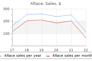
Altace packs: 30 pills, 60 pills, 90 pills, 120 pills, 180 pills, 270 pills, 360 pills
Safe altace 10 mg
Within this cone is the orbital fats blood pressure medication starting with a order altace 2.5 mg, which also contains a quantity of veins and lymphatics blood pressure normal values generic altace 2.5 mg visa. The dural-covered optic nerve sheath courses via this fats centrally and joins the posterior globe to the mind. Traumatic Hyphema Traumatic hyphema is bleeding into the anterior chamber secondary to disruption of blood vessels within the iris or ciliary physique. Although hemorrhage from these vessels can occur in extravasation into both the anterior or posterior chamber, the time period hyphema is reserved for bleeding into the anterior chamber whereas bleeding into the posterior chamber is referred to as vitreous hemorrhage. On medical examination, a blood-fluid degree can be seen, which represents the extravasation of blood into the anterior chamber. The globe lies anteriorly inside this area and is a largely spherical structure with a diameter of approximately 24 mm. The wall of the globe is shaped by three layers: an outermost fibrous layer composed of the sclera and cornea, a center layer made up of the choroid and ciliary body, Corneal Laceration Corneal lacerations are often seen within the context of penetrating trauma. An 128 Section i craniocerebral orbital-Maxillo-Facial eMergencieS 5-30 A, Traumatic hyphema. Also note the triangularshaped metallic foreign body and intraocular air throughout the vitrea. The right lens can be low in attenuation compared to the left, in preserving with acute lens edema (red arrow). Note the adjacent intraocular metallic international body close to the medial facet of the lens (white arrow). The enhance in transverse diameter of the globe causes Chapter 5 stretching of the zonular attachments that maintain the lens in place, which might finally lead to partial or complete dehiscence. Face and Neck Emergencies 129 Dislocation In partial dislocation the zonular fibers tear on just one side while the contralateral fibers remain intact. The intact fibers act as a hinge, resulting in posterior angulation and protrusion of the affected aspect of the lens into the vitreous humor. It is important for the radiologist to be cognizant that not all lens dislocations are the result of trauma. Spontaneous lens dislocations can happen within the context of a quantity of systemic issues, mostly Marfan syndrome, Ehlers-Danlos syndrome, and homocystinuria. An important clue that means a nontraumatic trigger is bilateral lens dislocation. When blunt or penetrating trauma disrupts this capsule, water is allowed to enter the lens, leading to acute lens edema or a "traumatic cataract. Just as bilateral lens dislocations recommend a systemic cause, bilateral lens edema suggests a nontraumatic cause, similar to diabetes. The mechanism in diabetic sufferers begins with an elevated glucose level inside the globe, which creates an osmotic gradient across the lens. However, as a result of the zonular attachments of the lens stay intact, the posterior displacement of the lens ends in growth of the anterior chamber, in keeping with open-globe damage. Just as with lens dislocations, not all contour abnormalities are the consequence of globe rupture. Other necessary pitfalls to avoid are iatrogenic modifications in the globe associated to prior ophthalmologic procedures. Perfluoropropane fuel may be injected into the vitreous as technique of a gasoline tamponade remedy for retinal detachment, resulting in low attenuation areas inside the globe which have an identical appearance to traumatic intraocular air. Other iatrogenic mimics are ocular home equipment corresponding to low-attenuation silicone sponges and high-attenuation scleral buckles, each of which can indent the globe and be mistaken for open-globe damage in the presence of penetrating intraocular international our bodies. Fluid can accumulate within the potential spaces between these layers, leading to retinal or choroidal detachment. Retinal Detachment the retina can become detached from the choroid in the setting of inflammation, neoplasm, or trauma. The majority of the retina is loosely connected to the choroid, although there are very agency attachments seen along its anterior margin, referred to as the ora serrata, and posteriorly on the optic disc. The main mechanism of injury entails perforation of the sclera, usually simply posterior to the insertions of the extraocular muscle tissue the place the sclera is thinnest, with resultant extrusion of the vitreous. The irregular contour of the globe has been descriptively termed the flat tire or mushroom signal, particularly when posterior flattening and volume loss are present (see Choroidal Detachment the underlying explanation for choroidal detachment is ocular hypotony, which could be inflammatory, iatrogenic, or traumatic in cause. There is also a layering assortment of decreased T2 signal inside the posterior globe, according to acute hemorrhage (red arrow). There is complete transection of the left optic nerve (white arrow) and avulsion of the medial rectus muscle, which is enlarged and herniating by way of the lamina papyracea into the adjoining ethmoid sinus (red arrow). This distinction is essential as a end result of serous detachments are normally extra benign and infrequently resolve spontaneously, whereas hemorrhagic detachments are associated with a poorer prognosis. The removal of metallic foreign bodies can be essential as a end result of many metals contain copper or iron, each of which may trigger retinal toxicity and subsequent blindness. Potential pitfalls in the evaluation of intraorbital metallic foreign our bodies are surgical gadgets, corresponding to scleral bands, which might result in a false-positive end result. Glass Computed tomography can be the most sensitive imaging modality for the detection of intraocular glass. Given the bony confines of the orbit, a retrobulbar hematoma can lead to an increase in intraorbital pressure. If the rise in orbital volume is small, compensatory proptosis and prolapse of intraorbital fats can ensue. However, if the intraorbital strain rises quickly, compression of the optic nerve or central retinal artery can happen, leading to ischemia and blindness. Findings must be communicated instantly to the referring clinician as a end result of emergent decompression could be imaginative and prescient saving. First-line surgical intervention is lateral canthotomy, which could be performed on the bedside. Unlike the high-attenuating substances of metal and glass, picket fragments are very low in attenuation as a outcome of their air-filled porous microstructure and thus could be mistaken for intraorbital air. In addition, the attenuation of wooden can increase over time, which is thought secondary to fluid displacing air inside the pores. It is necessary to consider for delicate fractures involving the orbital apex, particularly the optic canal and anterior clinoid process. The most typical extraocular muscles involved are the medial rectus and inferior rectus, doubtless associated to their proximity to the relatively thin lamina papyracea and orbital floor, respectively. Fractures of these structures can lead to entrapment or shearing of the adjoining extraocular muscle. Alternatively, avulsion can occur within the setting of direct penetrating trauma corresponding to stab wounds. Johnson Nontraumatic orbital emergencies generally current with acute visible loss, ache, proptosis, and/or ophthalmoplegia. Alternatively, sufferers may present with a mix of native and systemic signs and signs. Infection, inflammatory problems, neoplasms, and vascular lesions may each present emergently, and imaging could contribute to analysis and delineation of extent of the method or help to inform patient administration. Imaging has little position in the assessment of "floaters" or retinal or choroidal detachments in the nontraumatic setting; nonetheless, associated pathologic conditions could additionally be recognized on routine orbital imaging. For example, though optic neuritis is a scientific diagnosis, the affiliation with multiple sclerosis found on imaging can make clear the diagnosis and assist in remedy choice making.
Order altace 5 mg with amex
Over time heart attack quotes altace 2.5 mg buy cheap, developmental delays and behavioral issues might emerge indicating the necessity for additional psychological and educational intervention hypertension medicines cheap altace 5 mg line. Families ought to be encouraged to communicate freely and repeatedly about adoption with the kid, starting within the toddler years and continuing through adolescence. Various tales from folklore as properly as cases 3006 Child in the Social Milieu known to the child and household may be freely mentioned. Thus, the kid is familiarized with the idea of adoption and looks at it in a positive mild and is healthier ready to settle for it when told about his personal adoption. Adopted kids may face resentment and nonacceptance from the prolonged relations. The pediatrician needs to deal with the dad and mom as well as the kid with empathy and endurance. Disruption charges are greater among kids adopted from foster care homes, which can be related to older age on the time of adoption, a historical past of a number of placements or a historical past of publicity to trauma and abuse previous to adoption. Great care needs to be paid to preparation of adoptive parents, and making certain their availability of a full range of postadoption services; including physical well being, mental well being and developmental services for the adopted children. Adoption is legally defined as the method via which the adopted youngster is permanently separated from his organic mother and father and turns into the legitimate child of his adoptive mother and father with all the rights, privileges and obligations which are attached to the relationship. Adoption of youngsters shall be guided by a set procedure and in a time bound method. Pediatricians can assess precise and potential problems or risks that these kids could have. Families ought to be encouraged to speak freely and repeatedly about adoption with the child. It is critical that the kid be advised about his own adoption by the adoptive dad and mom quite than listening to about it from others. The Right to Participation Children have the best to categorical their opinions and views. They have the best to acquire info, and the proper to freedom of thought and expression. In some instances, countries are obliged to be constant in defining benchmark ages-such because the age for admission into employment and completion of obligatory training; but in different situations the convention is unequivocal in setting an upper limit-such as prohibiting life-imprisonment or capital punishment for those underneath 18 years of age. The convention, in a way, is a method of empowering youngsters and creating an surroundings during which all children are in a position to reside securely and notice their full potential of life. Governments are obliged to bring their legislation, coverage and apply into accordance with the requirements within the Convention; to remodel the requirements into actuality for all children; and to abstain from any motion that may preclude the enjoyment of those rights or violate them. The international neighborhood displays and helps progress on the implementation of the Convention. The Convention supplies a common set of requirements to be adhered to by all international locations. Children are neither the property of their dad and mom nor are they helpless objects of charity. The Convention offers a imaginative and prescient of the child as an individual and a member of a household and a neighborhood, with rights and obligations appropriate to his or her age and stage of improvement. No longer the passive recipient of advantages, the child has become the topic or holder of rights. The Convention is in force in nearly the entire group of nations, thus offering a common ethical and authorized framework to develop an agenda for youngsters. At the same time, it constitutes a common reference in opposition to which progress could also be assessed. They generate obligations and responsibilities that we all should honor and respect. The Convention on the Rights of the Child is essentially the most widely and rapidly ratified human rights treaty in historical past. Somalia is presently unable to proceed to ratification as it has no acknowledged authorities. By signing the Convention, the United States has signaled its intention to ratify-but has but to accomplish that. The Right to Survival this includes proper to life, the attainable standard of health, nutrition and an sufficient standard of living. These rights seek to ensure that kids have nutritious meals, potable consuming water, a safe home and access to health facilities. Every youngster has the proper to live together with his or her parents and the kid shall not be separated from mother and father in opposition to his or her will besides when such separation is important in the best pursuits of the child. The baby also has the right to keep contact with each parents if separated from one or each. The Right to Protection Every youngster has a right to freedom from all types of exploitation including sexual exploitation, abuse, prostitution and involvement in pornography and drug abuse, inhuman or degrading remedy and neglect by mother and father or others liable for care of youngsters. The Right to Development these rights embrace the proper to schooling, help for development and care throughout early childhood, and the best to have enough social security. Children can get pleasure from their own tradition, follow their faith and talk in their own language. India has made some important commitments in the path of ensuring the basic 3008 Table 1 Constitutional provisions protect kids in India Articles of Indian Constitution Article 15 Contents Affirms the best of the State to make special provision for girls and youngsters. Provides that no baby under the age of 14 years shall be employed to work in any hazardous employment. Requires children to be given opportunities and amenities to develop in a healthy method and in circumstances of freedom and dignity, and that childhood and youth be protected towards exploitation and ethical and material abandonment. Provides at no cost and obligatory training for all youngsters until they full the age of 14 years. Every youngster has a right to full time elementary schooling of satisfactory and equitable quality in a formal college which satisfies sure important norms and requirements. The coverage additional goals to awaken the conscience of the community, to shield kids from violation of their rights, and strengthening the family, society and the nation. This coverage describes the steps to be taken by State and group to be certain that every youngster enjoys his or her rights as described within the Convention on Rights of the Children. The Commission focuses on the following tasks: · To construct public awareness and create a moral force within the nation to stand by children and defend their rights. There has been progress in general indicators: infant mortality charges are down, child survival is up, literacy rates have improved and school drop-out charges have fallen. But the issue of kid rights in India continues to be caught between legal and policy commitments to kids on the one hand, and the fallout of the process of globalization on the opposite. Various 5 12 months plans of India have progressively added more schemes for welfare and safety of child. The allied methods are police, health-care system, justice (juvenile) system, training system, transport system, labor division, media, division of telecommunications, company sector, elected representatives, and all of us. It will design a curriculum that will be integrated into the various training programs of academic establishments of the allied techniques. The scheme plans to setup a child safety data management system to formulate and implement efficient intervention methods and monitor their outcomes. Juvenile Justice Act, 2000 the Government of India enacted the JuvenileJusticeAct in 1986.
Altace 5 mg buy mastercard
Sclerosis of thyroid cartilage can indicate invasion by a tumor or reactive modifications heart attack jack smack u blue purchase altace 10 mg on-line, making the diagnosis of cartilage invasion by a tumor troublesome hypertension stage 3 5 mg altace safe. Vocal twine paralysis in hypopharyngeal cancers can subsequently result from direct invasion of the recurrent laryngeal nerve and fixation of the cricoarytenoid joint. Indirect involvement of the recurrent laryngeal nerve by metastatic lymphadenopathy in the tracheoesophageal groove can even happen however is relatively uncommon. The cricoid cartilage articulates with the thyroid, which encloses the supraglottis and glottis. The epiglottic cartilage attaches to the thyroid cartilage anteriorly at the level of the anterior commissure of the glottis. Natural fenestrations are present within the infrahyoid portion of the epiglottic cartilage (see Question 39). They span the supraglottic and glottic larynx, and the vocal strategy of the arytenoid cartilage provides attachment to the vocal ligament. One of the important thing concerns in deciding on the extent of glossectomy is the connection of the tumor to the neurovascular pedicles of the tongue. In doing so for the patient, the surgeon may also should sacrifice the left neurovascular bundle, which is able to result in an anterior subtotal glossectomy. Reconstruction of the resultant defect within the flooring of the mouth would require a microvascular free radial forearm flap. The raphe is separated from the ramus of the mandible by adipose tissue, and the inferior alveolar nerve passes underneath it within the retromolar trigone. The fibers of the buccinator and the superior pharyngeal constrictor muscles interdigitate alongside this raphe in order that involvement by a tumor facilitates tumor unfold anteriorly to the buccal house and posteriorly to the pharynx. The tumor has prolonged on to the best pterygomandibular raphe after which anteriorly to the proper buccinator muscle, which appears thickened (white arrow) in contrast with the other normal buccinator muscle (black arrow). The patient has a great mandibular height, and the tumor will due to this fact be amenable to partial glossectomy with marginal mandibulectomy and free flap reconstruction. In addition, note the intensive gentle tissue modifications in the retromolar trigone persevering with anteriorly along the lateral side of the mandible and posteriorly along the superior constrictor muscle. Regressive reworking of adjoining bone is a radiographic function of a slowgrowing neoplasm. Patient elements embody smoking and alcohol use, particularly continued use throughout remedy, poor dental hygiene, dental extraction after radiation therapy, poor dietary status, and extreme mucositis and trismus. Tumor-related elements embrace a regionally advanced infiltrative ulcerated lesion in shut proximity or a lesion invading bone. Treatment-related elements embody surgery, which requires manipulation of the mandibular bone. Lymphomas of the top and neck also have an affinity for nerves, and an correct histological diagnosis is very crucial in avoiding mistreatment. Over time, he developed facial numbness and then diplopia, for which he was referred to a neurologist for a presumed cavernous sinus major tumor. The imaging features are according to an expansile lesion arising from and centered on the concerned cartilage, which remains largely intact. The cricoid cartilage could be involved by different more frequent illness processes, such as squamous cell carcinoma of the larynx or papillary carcinoma of the thyroid, but the radiographic options in these entities would be consistent with gentle tissue infiltration and cartilage destruction. Papillary carcinoma of the thyroid also can lead to calcification, but this is typically present within the welldifferentiated long-standing part of the thyroid tumor, not within the invasive intralaryngeal element. Adenoid cystic carcinoma is the commonest minor salivary gland tumor of the subglottic larynx and cervical trachea. Chondroradionecrosis after radiation therapy can additionally be seen with harmful cartilage modifications and is commonly troublesome to differentiate from recurrent cancer. However, the imaging studies would also produce other stigmata of radiation remedy, including edema of the airway and soft tissue stranding to point out previous therapy. Note the asymmetrically enlarged and enhancing right inferior alveolar nerve inside the alveolar canal of the mandible. Infiltrative, irregular borders are the only constant feature that permits radiographic diagnosis of a malignant tumor. This lesion is simply too massive and too round to represent a normal intraparotid lymph node and shows no fatty hilum. This is an extremely useful sequence for assessing the extent of the lesion in relation to the parotid parenchyma. However, rupture of the tumor and spillage within the operative subject can end result in seeding of the entire surgical mattress. Primary tumors of this space are unusual, and this area is mostly involved by extension of a deep lobe parotid tumor. Schwannomas seem as well-defined, ovoid masses which could be heterogeneous on imaging. Lingual thyroid represents ectopic thyroid tissue on the opening of the thyroglossal duct within the base of the tongue (foramen cecum). They are typically asymptomatic and are therefore most frequently large at presentation. Chunky calcification does occur with longstanding, well-differentiated papillary thyroid carcinoma, and transformation to poorly differentiated or anaplastic most cancers may find yourself in laryngeal invasion. However, the calcified component of the tumor in these situations is generally positioned exterior the larynx throughout the thyroid gland itself. Although new onset cartilage sclerosis might represent a long-term sequela of radiation remedy, cartilage sclerosis can be brought on by invasion by a tumor or reactive adjustments to an adjoining tumor; due to this fact it should immediate suspicion for recurrent disease. Patients with locally superior, bulky tumors which are in shut proximity to or invading the laryngeal cartilaginous framework are usually at increased threat of creating chondronecrosis. Trauma to the mucosal surface of the larynx, especially from injudicious or random biopsies to rule out a suspected tumor, can considerably elevate the danger of chondronecrosis. Look for focal and asymmetric uptake that coregisters to the positioning of the primary tumor and/or a focal mass on anatomical imaging: diffuse uptake often signifies postradiation irritation and never a recurrent tumor. Increased mild-to-moderate diffuse laryngeal or oropharyngeal uptake might persist for extended durations after chemoradiation remedy. Communication between the treating clinician and the radiologist is of tremendous value, as is a comparability between pretreatment and posttreatment baseline imaging studies. Partial defects of the pharynx are typically reconstructed with a pedicled patch, similar to a pectoralis major myocutaneous flap, or a free flap, corresponding to a radial forearm or rectus abdominis myocutaneous flap. The presence of large amounts of fats within the surgical bed on this patient point to rectus flap reconstruction. There are additional benefits in that this procedure is performed on an outpatient basis, a cytopathologist is present to guarantee sufficient sampling, and no residual scar is current after the process. Superficial lesions such as thyroid nodules, superficial parotid lobe lesions, and small lateral cervical lymph nodes are readily accessible with the usage of real-time ultrasound steerage. The outer guide needle is positioned at the edge of the lesion of curiosity, and a smaller needle is passed through it to sample the lesion for cytological or histological examination with a core needle, if indicated.
10 mg altace purchase otc
The syndrome encompasses a wide spectrum of abnormalities blood pressure medication joint pain altace 5 mg otc, including cardiac heart attack 51 altace 2.5 mg generic without a prescription, palate, immunologic and orthopedic anomalies. At least one anomaly of the occiput or cervical spine is observed in all patients. C1 variations embrace dysmorphic form, open posterior arch, and occipitalization, and axis variations include a dysmorphic dens. Increased segmental motion is noticed on dynamic imaging in higher than 50% of patients, typically at more than one level. Central nervous system anomalies similar to Chiari sort I malformation have additionally been reported. All sufferers ought to have screening radiographs, and heaps of require follow-up for the cervical spine. Neurological signs as a result of involvement of upper cervical backbone could occur due to wire compression, vertebral artery compression, blockage of the aqueduct of Sylvius and cranial nerve compression. Children with these problems want an assessment of cervical backbone stability particularly if being given common anesthesia. Tuberculosis of the spine may be managed effectively with antitubercular chemotherapy and applicable bracing in a majority of sufferers. The scientific presentation and consequence of remedy of congenital muscular torticollis in infants-a study of 1,086 cases. Early detection actually helps to outline remedy and control rapid progression while doubtlessly preventing complications in the lengthy run. Other etiologies of structural scoliosis primarily based on associated circumstances are listed in Table 1. Balanced curves could not get noticed until adulthood after they can present with backache. Childhood scoliosis is usually painless; a grievance of ache should immediate a seek for a neural etiology. On general examination, look for cafй-au-lait spots, facial dysmorphism, ligamentous laxity and different features which would counsel a nonidiopathic etiology for scoliosis. Midline cutaneous abnormalities of the again such as hemangiomas, hair patch, sacral dimple or skin tag are sought. Associated findings can embody breasts and shoulder degree asymmetry, a sideways truncal shift and an apparent discrepancy within the leg-length. The mature spine has four balanced curves within the sagittal aircraft: (1) a cervical lordosis, (2) a thoracic kyphosis (measuring about 20°50°), (3) a lumbar lordosis (measuring about 31°79°) and (4) a kyphotic curve on the sacral level. The lordosis begins to develop within the cervical backbone when the kid begins head holding. It is necessary to preserve the middle of gravity for steadiness and to maintain an upright posture. Spinal deformity in simple term might be stated as an abnormal curvature of the backbone. The deformity is named scoliosis (occurring within the coronal plane), or hyperkyphosis and hyperlordosis (in the saggital plane). A coronal airplane curvature of higher than 10° (using the Cobb method) is termed as scoliosis, while curves lower than this are generally termed spinal asymmetry. Congenital Wedge vertebrae Hemivertebrae Unilateral bar Block vertebra Neuromuscular Upper motor neuron - Cerebral palsy - Spinocerebellar degeneration (Friedreich ataxia, Charcot-MarieTooth disease) - Syringomyelia - Spinal wire tumor - Spinal cord trauma Lower motor neuron - Poliomyelitis - Spinal muscular atrophy Duchenne muscular dystrophy Arthrogryposis Other muscular dystrophies Syndromes Compensatory (Nonstructural) Scoliosis In compensatory scoliosis, the deformity is obviously compensatory to an abnormality outside the backbone, corresponding to within the shortening of the decrease limb, or abduction or adduction contracture of the hip. When the limb size asymmetry is corrected (during sitting or by placing blocks underneath the brief limb), the curve tends to disappear. Children with limb-length inequality can even have a coexisting structural scoliosis. The analysis is confirmed if the curve persists on a standing radiograph obtained after correcting the limb-length discrepancy. Structural Scoliosis Structural scoliosis refers to a noncorrectable deformity of the affected spinal segment. The preliminary curve is probably correctable, however as soon as it exceeds a sure point of mechanical stability, the backbone buckles and rotates into a set deformity. To counterbalance the primary curve and maintain spinal balance, secondary (compensatory) curves almost all the time develop. Secondary curves Compensatory Source: Adapted from the Terminology Committee, Scoliosis Research Society: A glossary of scoliosis terms. Position of occiput might help differentiate balanced from unbalanced (or decompensated) curves. In the sagittal airplane, the auricle and the shoulder ought to be centered over the greater trochanter. A detailed neurological examination is necessary, to detect an underlying spinal twine lesion. Idiopathic scoliosis is further categorized according to the age of the child when the deformity was initially seen. The three subtypes differ in terms of development, prognosis and treatment methods. The degree of curve correctability could be ascertained by X-rays in place of lateral bending. Skeletal age is helpful for planning therapy and ought to be assessed, because the deformity normally worsens in the course of the period of speedy skeletal growth. Etiology Although the precise etiology of idiopathic scoliosis stays an enigma, many theories have been proposed over time. Genetic factors, hormonal and metabolic dysfunction, neurologic dysfunction and biomechanical elements (asynchrony in spinal development and anterior spinal overgrowth) have been implicated. In general, the etiology is considered multifactorial with a genetic predisposition. Systemic Disorders Natural History the pure historical past of idiopathic scoliosis is determined by the age at presentation or prognosis. Thoracic curves greater than 50° and thoracolumbar or lumbar curves larger than 30° are likely to progress at approximately 1°/year throughout life. Curves larger than 70° end in chronic lung disease, and people above 100° might result in cor pulmonale and cardiorespiratory failure. Infantile Idiopathic Scoliosis Prognosis and Treatment Treatment is directed at preventing worsening of the scoliosis and progressive fixed deformity. In common, the youthful the child (more growth remaining years) and higher the curve, worse is the prognosis. Most childish curves (90%) resolve on their own; the remaining are progressive and can become severe. Large curves earlier than the age of 5 years might have an result on respiratory perform and trigger ventilation-perfusion mismatch. Curves which are potentially progressive should endure a scientific and radiographic analysis at about 46-month intervals.
Altace 5 mg buy lowest price
These patients could do as well (if not better) than sufferers present process median sternotomy blood pressure chart british heart foundation altace 10 mg order free shipping. This could come up from the lumen and a tiny ulcerlike projection or from retrograde flow from an intercostal artery prehypertension with low heart rate 10 mg altace order with visa. These blushes can result in confusion within the categorization of which entity of acute aortic syndrome is current. Some of the literature has proven that a blush from an ulcerlike projection is at increased danger for progressing to a true aortic dissection. In another variation a dissection involving the ascending aorta may rupture just above the valve airplane into a shared sheath with the pulmonary artery. The blood in the wall of the pulmonary artery will compress the pulmonary artery and will occlude it. Sometimes the blood will dissect across the pulmonary artery to the segmental arterial degree. A third uncommon manifestation of aortic dissection is a focal dissection at the level of the aortic root close to the sinotubular junction. This localized process has been referred to as an incomplete aortic dissection and is nearly all the time related to cystic medial necrosis and aortic root enlargement. As a general guideline, one ought to have a low threshold for reimaging the aortic root when a linear defect is encountered within the setting of annuloaortic ectasia or in a patient with identified Marfan syndrome or bicuspid valve. Focal dissections of the stomach aorta are rare within the absence of prior trauma or aortic catheterization. However, imaging of an aortic dissection should embrace the abdominal aorta and the iliac vessels to the extent of the common femoral arteries. Inclusion of the abdomen and pelvis is performed to search for any end-organ ischemia and to delineate the extension of the dissection. Increasingly surgeons are counting on peripheral cannulation for initial bypass in aortic restore. The function of imaging is to assist distinguish true from false lumen in order that any peripheral cannulation for bypass avoids cannulating the false lumen, which would preclude adequate bypass and will doubtlessly be lethal. The perception is that the macrophages inside the plaque weaken the intima and permit for this focal ulceration. These patients might be carefully followed and if attainable, might bear endoluminal remedy. Vascular Chest Emergencies 237 promote an area of relative stasis along the lesser curve of the aorta at the degree of the isthmus. These filling defects are normally encountered throughout the aorta on the level of the isthmus and throughout the descending thoracic aorta. Radiologists should be aware of the acute aortic thrombus to keep away from confusion with an aortic tumor. Treatment for an acute aortic thrombus begins with anticoagulation but may embrace a thrombectomy and barely endoluminal stent placement to trap the thrombus. Unfortunately, the imaging of an aortic fistula may be quite refined despite the excessive lethality of this entity. These patients often report a big episode of hematemesis or rectal bleeding (the herald bleed). Aortic fistulas can be major, normally from rupture of an aortic aneurysm or in the setting of an an infection, similar to mediastinitis. When findings of mediastinitis are encountered (such as fuel or diffuse mediastinal fat infiltration), cautious inspection of the aortic contour is required. Some authors have postulated that a scar from the ductus arteriosus could serve as a nidus for thrombus formation. Note the extension past the confines of the aorta (saccular pseudoaneurysm) and associated intramural hematoma. These septic emboli are unlikely to lead to proper heart failure (because the burden is small), but they have a tendency to be markers of a bacteremic affected person. In trauma, bone marrow may be embolized, as can amniotic fluid in the setting of a troublesome delivery. As imaging for suspected emboli will increase, radiologists must be acquainted with the whole spectrum of embolic disease. This phenomenon is more common within the abdomen and has been reported with adjacent bowel and the inferior vena cava. In the thorax the fistula may be to the esophagus or an adjacent bronchus (usually the left major bronchus). Clues to the diagnosis relaxation on seeing gas next to the restore web site, irregular contour of the aorta, and medical history. As with the acute aortic syndromes and the other entities beforehand described, medical historical past is simply modestly helpful. This level is essential in preventing confusion of a vein for an artery and key in detecting an occluded artery. Again, if the radiologist remembers that a bronchus should accompany an artery, he or she will understand that the enhancing construction is the artery and the obvious adjoining filling defect is actually within the bronchus. They begin as a major pulmonary artery that bifurcates right into a proper pulmonary artery, which travels slightly posterior and inferior to the best major bronchus, and a left pulmonary artery, which travels superior to the left main bronchus. The arteries continue to divide into lobar, segmental, subsegmental arteries, ultimately reaching a microscopic capillary degree. In different words, the pulmonary arteries should be the ultimate recipient of emboli from the venous system to stop systemic complication. In thromboembolic disease the thrombi form in peripheral veins from an antecedent condition. The proper coronary heart was noted to be enlarged in B with bowing of the interventricular and interatrial septa leftward. The finding of right coronary heart enlargement with out right ventricular hypertrophy is in preserving with right coronary heart strain. Previous authors have written on quite so much of methods to calculate the perfect viewing window. These embrace proper ventricular enlargement and bowing of the interventricular and interatrial septa leftward. In both young patients and overweight patients the V/Q scan tends to offer a analysis (either normal or excessive likelihood). Recently the Society of Thoracic Radiology, working in conjunction with the American Thoracic Society, has issued an approach to the pregnant patient, which is analogous to our own experience. In their consensus statement they suggest that the chest radiograph be used as a choice level. We also find that using a V/Q scan avoids any concern about iodinated distinction, which crosses the placenta. The internet effect of the scarring is that the pulmonary artery volume decreases, and as a result, pulmonary pressures improve. The resultant pulmonary hypertension could also be debilitating however is doubtlessly curable with a thromboendarterectomy. Other findings embrace enlarged major pulmonary artery, mosaic attenuation, bronchiectasis, and enlarged bronchial arteries.
Buy cheap altace 2.5 mg line
Approximately 2% to 3% of the population will develop urinary tract calculi prehypertension diabetes altace 2.5 mg effective, with as much as blood pressure jumps from high to low order altace 10 mg amex 12% experiencing an obstructing urinary tract stone of their lifetime. Urgent intervention is required when sufferers with obstructing stones have intractable ache, infected hydronephrosis, or high-grade obstruction of a solitary or transplant kidney (which can result in anuria and azotemia). Patients with urolithiasis classically present with acute, colicky flank ache that will radiate to the groin. As the obstructing stone descends towards the ureterovesical junction, the pain can radiate towards the urethra and symptoms may then embody dysuria and urinary urgency and frequency, mimicking cystitis. On bodily examination, patients with obstructing stones may have tenderness alongside the costovertebral angle or in a lower quadrant of the stomach. Therefore the absence or presence of hematuria is usually not used to decide whether a stone may be current. On color Doppler, the "twinkling" artifact, described as a spotlight of alternating colours with or without a comet tail noticed when insonating sure rough surfaces, improves the sonographic sensitivity for the detection of renal stones to approximately 90%. Obstructing ureteral calculi tend to lodge at the three factors of anatomic ureteral narrowing: the ureterovesical junction (most commonly), the ureteropelvic junction, and the pelvic brim. In very rare cases, urease-producing bacteria can lead to the production of matrix stones of soft tissue attenuation (isodense to the kidney and ureter) and due to this fact troublesome or inconceivable to visualize. A extra popular protease inhibitor right now, atazanavir, can crystallize in the urinary tract and likewise result in delicate tissue attenuation stones. Urothelial thickening may be current however is nonspecific, normally indicating concomitant inflammation or an infection. In problematic cases, two other indicators have been used: the rim signal and the comettail sign. The comet-tail signal is a triangular or tubular area of soft tissue attenuation, with its apex oriented toward or away from a calcification, a finding representing a venous structure and indicating that the related calcification is a phlebolith. In these circumstances, a quantity of diagnoses have to be considered: a lately passed calculus, an alternate reason for urinary tract obstruction (such as a urothelial cancer), an infection, renal vein thrombosis, acute renal emboli, infarction and, most hardly ever, a low-attenuation stone. The chance of stone passage and remedy choices depends upon the dimensions and site of urinary tract stones. Many ureteral stones may be extracted ureteroscopically; nonetheless, some stones could require shock wave lithotripsy. Acute pelvic or stomach conditions may current with signs of colicky, flank pain mimicking a renal calculus. Acute pyelonephritis is normally diagnosed based mostly on medical presentation (flank ache and fevers) and laboratory check outcomes (pyuria and leukocytosis). Complications similar to urinary tract obstruction or abscess ought to be suspected in these instances. Factors that predispose patients to growing complications include superior age, diabetes, and immunosuppression. These issues often can be simply recognized on any cross-sectional imaging examine. Acute pyelonephritis is often unilateral but inside the kidney could be diffuse or focal, resulting in different imaging manifestations. Diffuse acute pyelonephritis may merely manifest as an enlarged, edematous kidney on both imaging modality. Perinephric infiltration, suggesting irritation or unfold of infection, could also be present. The cyst was partially ruptured, with fuel and inflammatory changes extending into the perinephric space. Pyelosinus extravasation allows the an infection to spread into the perinephric, paranephric, and retroperitoneal spaces. Glomerulonephritis and interstitial nephritis as a result of noninfectious causes such as sarcoidosis or medications or immune or metabolic-related situations could have related appearances, with areas of low attenuation in the renal parenchyma. Perinephric infiltration may be seen with prior an infection, recent trauma, or vascular diseases, including renal vein thrombosis and vasculitides. Thus correlation of the imaging findings with scientific presentation and laboratory values is important for limiting the diagnostic potentialities. Acute Pyelitis, Ureteritis, and Pyonephrosis Pyelitis and ureteritis discuss with infections of the renal collecting system and ureter, respectively. Pyonephrosis refers to the buildup of purulent material in the renal accumulating methods and/or ureters. Pyonephrosis usually requires instant drainage to forestall everlasting renal parenchymal destruction and life-threatening sepsis. Obstructing renal calculi are the commonest underlying reason for pyonephrosis, however iatrogenic strictures and retroperitoneal fibrosis should be considered as properly. On imaging studies, sufferers with pyonephrosis usually have dilated renal accumulating systems and/or ureters with related urothelial thickening. Occasionally, wedge-shaped or linear areas of excessive attenuation, which likely represent foci of hemorrhage, may be noticed. There is a rim-enhancing fluid collection alongside the medial aspect of the kidney (arrowheads). A defect was recognized in the renal pelvis according to amassing system rupture. Emphysematous pyelonephritis is usually unilateral and occurs extra frequently in girls and nearly completely (80% to 100%) in sufferers with uncontrolled diabetes. On physical examination a flank mass, representing the affected kidney, could be palpated in 50% of patients with emphysematous pyelonephritis. Several classification systems have been proposed to correlate imaging findings with subsequent prognosis and administration. The more severe infections are characterised by spread of fuel into the renal parenchyma and might lengthen into the perinephric and paranephric spaces and infrequently require emergency nephrectomy. On imaging studies, emphysematous pyelonephritis has a characteristic and sometimes diagnostic look. Conventional radiographs may demonstrate a cluster of mottled lucencies throughout the kidneys. Fungus balls (mycetomas) can develop in the renal amassing systems and ureters and can obstruct the accumulating system. The renal pelvis and calyces are dilated, typically massively, from a combination of urinary tract obstruction with accumulation of pus and particles within the renal collecting system. The affected kidney is diffusely enlarged however preserves its reniform contour with diffuse parenchymal thinning. Infected loculated fluid collections are incessantly discovered within the adjacent tissues, including in the perinephric house, the anterior and posterior pararenal spaces, and even the psoas muscles. A D Chapter thirteen these stones are composed of drug crystals and will not be hyperattenuating. Other Renal Infections Other infections that can contain the kidneys embrace tuberculosis from hematogenous dissemination, fungal infections corresponding to aspergillosis or candidiasis (usually from colonization of chronic indwelling catheters somewhat than systemic infections) during which mycetomas can type and probably impede the accumulating systems, and echinococcus infection, which is extremely rare. Acute flank ache, mimicking that of renal calculi, is a common presenting symptom. Renal Infarction the most common reason for renal infarction is an acute embolus, often from a cardiac source in patients with endocarditis or atrial fibrillation. Infarction may also be brought on by arterial occlusion from acute dissection, underlying important artery stenosis attributable to atherosclerosis, fibromuscular dysplasia, or vasculitis.
Order 2.5 mg altace fast delivery
The politics of agenda setting in international well being: child well being versus adult well being in creating countries hypertension and obesity cheap altace 2.5 mg. The massive scale success of polio eradication program is making means for its successor child health program hypertension jokes 5 mg altace order with mastercard, i. The guidelines symbolize an evidence-based, syndromic strategy to case management that features rational, efficient and reasonably priced use of medicine and diagnostic instruments. In conditions where laboratory help and clinical sources are limited, the syndromic strategy is a more sensible and cost-effective method to manage patients. Careful and systematic assessment of frequent symptoms, utilizing well-selected dependable medical indicators, helps to guide rational and efficient actions. This idea aimed to goal high-risk groups with fastidiously chosen costeffective interventions for under-5 youngsters with a goal to scale back under-5 mortality in third-world international locations to half by year 2000. However, the last word aim of lowering under-5 mortality to half by year 2000 seemed distant even though several evidence-based efficient interventions like immunization, oral rehydration therapy and use of applicable antibiotics for treatment of pneumonia were obtainable. The strategy has been implemented in more than 75 nations the world over, each country given an opportunity to adapt the rules as per local wants. However, in view of similarities in the spectrum of illnesses, clinical indicators and administration protocols, the therapy tips have been broadly described under two age classes: Sick young infants age up to 2 months (sick younger infant) Sick kids age 2 months as much as 5 years (sick child) Integrated administration of neonatal and childhood illness guidelines are based on the next rules: circumstances which indicate immediate referral. Classifications are color coded and recommend referral (pink), remedy in well being facility (yellow) or management at residence (green). Rationale Common childhood illnesses like acute respiratory infections, diarrhea, measles, malaria, and malnutrition continue to result in high mortality among children less than 5 years of age. Neonatal mortality contributes to virtually 2/3rd of infant deaths and most of those deaths occur during the first week of life. Specifically concentrating on these frequent issues and assessing a child for other related problems provides a holistic method to manage common childhood illnesses. Integrated approach is one other effective step because often a young infant or child suffers from more than one illness and customary childhood diseases in this age group have comparable presenting signs. Moreover, poor access to health-care and delay in referral further compound the issue. Assessment of Sick Young Infant or Child Sick young infants or sick children are assessed utilizing selected dependable clinical indicators and symptoms (Annexure 1). All sick younger infants are checked for very severe disease and jaundice, feeding issues and malnutrition, immunization standing and if they produce other problems. All sick children are checked for malnutrition, anemia, immunization standing, prophylactic vitamin A and iron folic acid supplementation status, and different issues. The technique consists of three major components: Improvements in the case-management abilities of health staff; Improvements within the overall health system; and Improvements in family and neighborhood health-care practices. Classification tables are used starting with the pink rows, followed by yellow and green rows. If the sick young toddler or sick child has no pink classifications, have a look at the yellow rows. A sick young infant or sick child is classed in one shade only underneath each group of symptoms. If the young toddler has indicators from multiple row, essentially the most severe (pink) classification is selected. Assess the younger infant/child Classify the illness Identify therapy Treat the young infant/child Counsel the mom Follow-up care mom or caretaker ought to be taught tips on how to give an oral antibiotic at residence. To return immediately if any of the hazard indicators (breastfeeding/drinking poorly, becomes sicker, develops a fever or feels chilly to contact, fast/difficult respiration, yellow palms and soles if younger toddler has jaundice, presence of seen blood in stools in young infants with diarrhea) For local bacterial infection, jaundice, diarrhea, any feeding problem or oral thrush follow-up after 2 days, and for low weight for age, advise follow-up after 14 days. Pink classification calls for hospital referral or admission in the in-patient part of the hospital, yellow for remedy under supervision in the well being facility, and green implies that the child can be despatched house with careful recommendation on when to return. Community Pediatrics Identify Treatment After classifying a sick young toddler or sick baby the next step is to identify remedy. All the therapies required are listed in the determine treatment column of the assess and classify the sick young infant or baby chart (Annexure 1). If a sick younger infant or sick child has multiple classification, treatment required for all the classifications must be recognized. Referral can also imply admission to the inpatient division from the outpatient or casualty of the identical facility. Treatment of Sick Children Referral All sick youngsters with a extreme classification (pink) are referred to a hospital as soon as evaluation is accomplished and essential pre-referral remedy is run. Treatment of Sick Young Infants Referral All sick younger infant with a extreme classification (pink) are referred to a hospital as soon as assessment is completed and needed prereferral treatment is administered. Treatment in Outpatient Clinic Give the primary dose of the antibiotics Amoxicillin for 5 days for pneumonia and acute ear an infection, ciprofloxacin for three days for dysentery, and a single dose of doxycycline for cholera. For malaria, give choloroquin or artesunate relying upon threat space for malaria and speedy blood take a look at or blood smear report. Instructions could additionally be given about tips on how to deal with eye an infection with tetracycline eye ointment; dry the ear by wicking to deal with ear an infection; and deal with mouth ulcers with gentian violet. Feeding evaluation includes questioning the mom or caretaker about: (1) breastfeeding frequency and night feeds; (2) kinds of complementary foods or fluids, frequency of feeding and whether or not feeding is energetic; and (3) feeding patterns in the course of the current sickness. The mom or caretaker must be given acceptable recommendation to help overcome any feeding problems discovered. One must listen to the mother and compliment her for correct feeding practices; and train and encourage her to change those practices which need to be changed. If a toddler with no pneumonia: cough or chilly has fast/ difficult breathing and a child with diarrhea has blood in stools or drinks poorly. Counseling the Mother or Caretaker Counseling a mother/caretaker is critical not only for successful referral of a severely unwell younger infant but also for treatment in the outpatients clinic in addition to for house care. Good communication skills have to be used from the beginning of the go to notably while counseling the mother/caretaker for remedy. The success of residence treatment depends on how nicely the mom or caretaker knows tips on how to give treatment, understands its significance and is aware of when to return to a health-care provider. Some recommendation is straightforward; different recommendation requires instructing the mom or caretaker the way to do a task. When a mom is taught tips on how to deal with her baby at home, three primary instructing steps need to be used: give information; present an instance; let her follow. Moreover, for the management of very sick younger infants or kids the rules cease short at referral of pink classifications to a hospital. These guidelines are directed at small hospitals where primary laboratory facilities and inexpensive essential medicine are available. These proof based tips additionally complements standard comprehensive pediatric textbooks. Integrated management of childhood illness by outpatient well being staff: technical basis and overview. Integrated Management of Neonatal and Childhood Illness: Training Modules for Physicians.
Discount altace 10 mg on-line
This condition is illustrated and further discussed within the part on vascular trauma hypertension forum safe altace 10 mg. Clinically these patients may current with acute onset of monocular imaginative and prescient loss or extra subtly with a comparatively afferent pupillary defect heart attack 720p buy generic altace 2.5 mg online. Traumatic optic neuropathy can result from either direct or indirect damage to the optic nerve. Direct injuries are much less widespread and often are the result of a penetrating foreign body or blunt trauma that leads to fracturing of the orbital apex or optic canal. Causes of indirect injury embody intraorbital hematoma, retrobulbar hemorrhage, vascular dissection, or edema inside the nerve sheath. Although differentiating these entities sometimes requires medical correlation, imaging could be helpful in figuring out the extent and character of orbital involvement. Infection is typically categorized primarily based upon its extent in relation to the orbital septum. For instance, with regard to cellulitis, preseptal denotes involvement anterior to the orbital septum, and orbital denotes involvement posterior to the orbital septum. Chapter 5 Face and Neck Emergencies 133 Preseptal Cellulitis these sufferers present with eyelid erythema and edema because the gentle tissue an infection entails the tissues anterior to the orbital septum. Preseptal cellulitis sometimes arises from underlying sinus (ethmoid) illness, chalazion, dacryocystitis, or cutaneous trauma. Patients complain of pain and vision loss and have marked conjunctival injection on bodily examination. Frequently a layering of white blood cells referred to as a hypopyon is famous on ophthalmologic examination. Although the chance for endophthalmitis is larger amongst patients with a history of eye surgical procedure, it could also come up endogenously from bacteremic or fungemic sufferers secondary to hematogenous dissemination. Infectious endophthalmitis is often diagnosed clinically, with or with out the usage of ultrasound imaging. Orbital Cellulitis Orbital cellulitis refers to infection involving tissues posterior to the orbital septum. These patients current with pain, eyelid erythema, edema, proptosis, and pain with extraocular movements. Depending on the extent of orbital involvement, there could additionally be extra symptoms, including visible loss, restriction of ocular motility, and pupillary abnormalities. In the majority of patients, orbital cellulitis arises as an extension from infection inside periorbital structures, most commonly sinus or dental an infection. In essentially the most severe circumstances, infection could extend to involve the cavernous sinus, with or without the development of cavernous sinus thrombosis. These patients will often develop complete ophthalmoplegia Necrotizing Fasciitis Necrotizing fasciitis of the orbit is a probably deadly infection, mostly brought on by group A streptococcus. Early presenting symptoms might embody shock and anesthesia overlying the affected area, though overlying anesthesia progresses to extreme ache. Dacryocystitis Dacryocystitis is an an infection of the lacrimal sac, most commonly occurring on account of nasolacrimal duct obstruction. Patients current with pain and erythema overlying the nasolacrimal sac, and purulent discharge is widespread. The nasolacrimal sac is dilated with rim enhancement on imaging, indicative of an infection. Notable are the stranding inside the intraorbital fat, proptosis, and heterogeneous elevated density within the abnormally formed globe. Thyroid eye illness may come up within the absence of serum markers of thyroid abnormalities. Computed tomography is particularly useful in figuring out the extraocular muscle enlargement. The disease is mostly bilateral, though unilateral proptosis could also be seen. C, 24 hours later there was increased extension of the scalp soft tissue swelling with abnormally formed globe and increased density in the vitreous according to endophthalmitis. Note the blended sign depth on the axial images in maintaining with inflammatory debris. Chapter 5 disease could progress quickly, making imaging particularly essential because these instances usually have a tendency to produce optic neuropathy. These instances of compressive optic neuropathy are extra common in certain races, particularly Asians, in whom the orbit tends to be shallower. It classically entails the paranasal sinuses (sinus mucosal inflammation), the lung (inflammatory lesions), and the kidney (glomerulonephritis). This small-vessel, necrotizing, granulomatous vasculitis entails the orbit in 20% to 50% of patients. Typically sufferers present with scleritis, episcleritis, or uveitis; nonetheless, extra diffuse involvement may occur by way of direct extension from adjoining sinuses. The extra diffuse form presents with decreased vision, strabismus, and diplopia caused by inflammatory mass effect within the orbit. Orbits and paranasal sinuses could also be concerned in isolation with out renal or pulmonary involvement. Most commonly the irritation can be diffuse with fat stranding and involvement of the extraocular muscular tissues (myositis) and/or the lacrimal gland (dacryoadenitis). These issues embrace eye pain secondary to scleritis, episcleritis, conjunctivitis, and uveitis. It could rarely manifest as a diffuse inflammatory course of mimicking orbital cellulitis. Treatment of idiopathic orbital inflammatory syndrome consists of high-dose steroids quite than antibiotics. The pictures reveal intensive sinus illness and complete erosion of the maxillary sinus. The traditional presentation of a quantity of cranial nerve palsies necessitates imaging of the orbits, sinus, and brain for evaluation and differential prognosis because vascular, inflammatory, infectious, and neoplastic lesions every require totally different therapeutic management. Lacrimal gland irritation, unilateral or bilateral, is the most common orbital manifestation, but inflammation from sarcoidosis can also affect the extraocular muscular tissues or optic nerves. Uveitis could also be isolated to a specific chamber of the eye, as in anterior or posterior uveitis, or it might affect the entire eye, as in panuveitis. The effects of the inflammatory ailments that cause uveitis lead to vision loss through the event of cataracts, cystoid macular edema, glaucoma, or retinal vasculitis. Magnetic resonance imaging and ultrasonography may also show exudative retinal detachment in some severe circumstances of uveitis. They are the commonest neoplasm of the orbit in adults, occurring more commonly in ladies. Magnetic resonance imaging demonstrates a homogeneously enhancing lesion with sluggish intralesional flow. These are referred to as oblique fistulas versus the direct inner carotid arterytocavernous sinus fistula seen within the traumatic setting. Imaging demonstrates enlargement of the superior ophthalmic vein, enlargement of the cavernous sinus, and proptosis with stranding of the intraorbital fat and engorgement of the extraocular muscle tissue.