
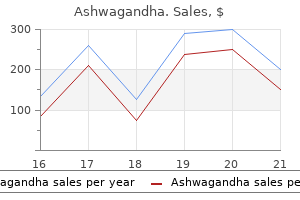
Ashwagandha
Ashwagandha dosages: 60 caps
Ashwagandha packs: 1 bottles, 2 bottles, 3 bottles, 4 bottles, 5 bottles, 6 bottles, 7 bottles, 8 bottles, 9 bottles, 10 bottles

Trusted ashwagandha 60 caps
In specific anxiety symptoms red blotches buy discount ashwagandha 60 caps line, the detection of antiemetics might help the hypothesis of suicide anxiety symptoms breathing problems discount 60 caps ashwagandha mastercard. Fatalities solely occur at excessive altitudes, for example whereas mountaineering, in cable cars, scorching air balloons or aeroplanes. Isolated fatalities have been reported, which can apparently be attributed to a failure of the pressure control system in plane. In the event of a sudden drop in cabin stress at an altitude of roughly 10,700 m, the passengers are only able to self-rescue for 30-60 seconds. Incapacitation and subsequent lack of consciousness may occur inside a very brief time. Perpetrators have been recognized to kill their victims utilizing one other type of traumatic violence after which place a plastic bag over their heads within the ultimate stages of the murder. Homicide also includes cases of human trafficking when individuals die of asphyxiation whereas being transported in containers. Cases of murder have been recorded by which victims are shut in cupboards or bins, which are then buried. Accident Accidents with plastic luggage happen in affiliation with autoerotic actions. Accidents may occur without any sexual aspect the place individuals sniff solvents, for example, from plastic baggage to attain a state of intoxication. In rare instances, small children asphyxiate after pulling a plastic bag over their own heads. Deaths in silos, accessible tanks and airtight pieces of furniture virtually invariably represent unintentional asphyxiation. This discovering should be current in General findings the symptoms embrace liquid cadaver blood, acute hyperaemia of the interior organs and mind oedema. For example, these conditions could lead to cardiac deaths or deadly status epilepticus. As a common rule, the traditional ligature is tied into a noose, which is passed a couple of times, extra rarely in a quantity of loops, around the neck. The latter is discovered, for example, in cases of hanging on taut ropes, mounted bathe hoses or bicycle helmets [26]. In these instances, the strangulation gadget only constricts the area located instantly beneath the chin and/or the front and lateral areas of the neck. Frequency/occurrence Throughout the world, hanging is one of the most common strategies of suicide and is favoured by males. Accidents are comparatively rare [19,80] and primarily involve incidents of autoerotic asphyxiation. Near-hanging to improve sexual stimulation is evidently nearly always practised by men [2]. For example, there have been cases of small children falling onto low, taut ropes leading to demise by hanging [22]. Extremely uncommon circumstances have been reported of homicidal hangings as foul play [23,24]. In a really small variety of cases, the sufferer was killed by another methodology and strung up in a dangling position in order to simulate a suicide for the purpose of concealing the crime. Classification of the circumstances Essentially, there are two distinct situations in which the physique could additionally be found, particularly incomplete and complete hanging. Strangulation units For the most part, ligature materials with a round cross part are used. These types of object could produce attribute indentations within the mark of the ligature. Occasionally, hanging is carried out utilizing objects of clothing, bed linen or towels. Occasionally, victims are additionally found in other positions, together with sitting, crouching, kneeling or mendacity down. In roughly 70 per cent of all suicides by hanging, the physique has direct contact of some sort with the surface under [27]. A lengthy drop is utilized in executions, known as judicial hangings, when the condemned prisoner falls down through a trapdoor and then hangs freely. In drops of greater than 1 metre into the tightening noose, fractures of the second cervical vertebra are widespread in adults. There have been several reports of suicides plunging from a peak with the noose round their necks. Depending on the peak of the drop, the physique weight and the elasticity of the ligature materials, this can outcome in decapitation [32,33]. In suicide hanging, a climbing aid will at all times be discovered in the instant vicinity of the deceased, otherwise there could be no explanation for the free suspension. In some instances, they could merely be current within the conjunctiva and/or in the pores and skin of the eyelids. In excessive instances, the congestion syndrome produced by incomplete hanging might resemble the appearance of a ligature strangulation [34]; thus, a differentiation is problematic at finest or might even prove inconceivable. This develops because of abrasion or compression of the pores and skin by the strangulation device, which usually has a tough surface. With time, the furrow dries out, taking over a brownish hue like other pores and skin abrasions. If a appreciable quantity of material is used or if Ligature mark gentle padding is placed under the noose, the ligature mark could merely be an impression of the folds of the material, or there could also be no mark at all. The varied ligature marks in deaths by hanging not often present related gentle tissue haematomas [27]. In suicide hangings, the marks usually run above or around the thyroid cartilage, in instances of suicidal complete hanging, the figure is much more than 70 per cent [27]. Where the ligature encircles the neck, will in all probability be attainable to establish or reconstruct the best level in practically every case. In closed nooses, the very best level, also referred to as the purpose of suspension, is usually at the site of the knot. In order to decide the highest point, it could be advantageous to measure the gap between the mark and the decrease edge of the auricles. Very typically, the ligature furrow is most distinguished reverse the point of suspension. If the noose slips upwards through the hanging, this will likely cause a quantity of parallel markings and broad abrasion zones that slope upwards. A band of purple skin, noticed sometimes above the ligature furrow only, is an incontrovertible postmortem finding. This is a results of the blood draining from the top and pooling across the ligature.
60 caps ashwagandha trusted
However anxiety back pain order 60 caps ashwagandha, actually in England anxiety symptoms or heart problems cheap 60 caps ashwagandha, the number of scenes to which pathologists are being known as is declining and the choice as to whether or not a death is suspicious is often being undertaken by individuals with limited experience and even much less coaching in pathology. Jumping to erroneous conclusions can result in the lack of evidence and, potentially, a missed murder. For a wider view on crime scene administration and investigation, the reader should seek the advice of extra specialist, dedicated crime scene reference sources [2,3]. They should also refer to their own native, regional or national protocols and pointers. It is past the scope of this chapter to present a comprehensive review of crime scene investigation on a country by country basis. For example, a person may be stabbed in a property but then manage to walk and even run away from the address solely to collapse far from the scene of the stabbing. In these cases, the physique is often discovered at the place the place the mechanism resulting within the demise was initiated. That is to not say that the death needed to have occurred on the web site of physique discovery: for example, an individual might have been ligature strangled at one scene and their body then moved to a separate deposition site. However, in cases of mechanical or chemical asphyxia, consciousness is commonly lost so rapidly that, without help from others, there may be steady progression to cardiorespiratory arrest and then the point of irreversibility with out further body movement throughout the scene [10]. For example, when coping with a case of hanging in a jail cell, although the scene stays confined to the cell itself, the ligature is usually reduce, and the body moved during resuscitation attempts. For example, in the course of the coaching of one of the authors in his position as a pre-hospital medical responder he was knowledgeable that, if an oropharyngeal airway is blocked with blood, this can be quickly eliminated, flicked to clear the obstruction and changed. In doing this, blood spatter could be produced on a nearby surface which may later confuse the investigating crime scene team. Blood patterns may additionally be produced by responding personnel walking by way of blood on the ground of the scene, creating extra footwear marks. This might trigger further disturbance of the scene as paths are cleared to remove the individual for medical therapy. Every scene is completely different and the strategy to every scene, regardless of how simple or advanced, is subsequently unique. Natural dying and suicides, once realised as such, might require relatively little scene evaluation. For scenes of suspicious and homicide deaths there are generic approaches to scene investigation that will be thought-about and utilized. These include methods for scene safety, internal and outer cordons, frequent paths of method, single factors of entry and exit, flooring plating and the sporting of generalized private protective tools (so-called scene suites). In certain circumstances - for example, hypothermia, drug and alcohol intoxication, diabetic coma and compression asphyxia - an individual may be found in a low-cardiac output state with shallow, rare breathing. Cases have been described of people being pronounced lifeless or taken to mortuaries and positioned into refrigeration solely to subsequently be found to be alive [7,8]. The pathologist ought to therefore by no means presume life is extinct and may, if necessary and the place attainable, comply with national guidelines when making this willpower. This expertise could be invaluable for scene examinations, notably in relation to analyzing deaths from natural or unnatural, non-suspicious causes. They may have a role to play at a scene after an post-mortem has been undertaken, when establishing what object or surface may have brought on an, as but, unexplained harm. It is also important to keep in thoughts that the health and safety of those attending the scene is paramount at all times. There are a quantity of roles which the pathologist can undertake at a scene of against the law. Establishing the precise fact of death If the body has not been pronounced life extinct, the pathologist, as a medical practitioner, may be referred to as upon to do this. The mark was recognized on examination by a forensic pathologist on the mortuary and had not been detected by these current on the scene. By using easy generic calculations, such because the Rule of Thumb, a pathologist could possibly provide an early rough estimate of the postmortem interval. However, it is extremely important to make it explicitly clear to the police that any scene-based estimation of postmortem interval is not extra than that and that investigations outdoors any estimated timeframe must not be overlooked or discounted at this stage. Establishing the place of death It could seem obvious that an individual died the place their physique was found. However, as with the pronouncement of life extinct, this could never be presumed. Through examination of the body at the scene, the pathologist could determine postmortem changes to the body, or marks or hint proof on the body that present an early indication that the scene of discovery is a deposition web site and never the site of dying. Is the lividity in a distribution that would be expected of the body position at the scene Loose fibres on the clothes may be left when a body is wrapped in a rug or carpet and then transported to the scene of physique discovery. Evidence recovery One of an important elements of scene examination is the event of a forensic strategy for the recovery of trace evidence. These strategies are adapted as the investigation progresses, however an initial plan should be in place prior to any examination or movement of the physique. Pathologists can play a part in helping develop the strategies and therefore must pay consideration to the principal types of bodily proof that can exist at a scene [3]. On a physique, this might embody blood spatter, clothing patterns, gunshot residue patterns, and the ante- and postmortem accidents. This is evidence ensuing from a specific action or occasion and can assist with reconstruction of events at a scene. While serving to with the recovery of swabs, tapings, clothes and personal artefacts, a pathologist can build up an preliminary impression of the death. This initial exterior examination and manipulation of the body allows Establishing the time of demise Pathologists should concentrate on the different strategies that can be employed on the scene of a dying to estimate the time since death. A review of such strategies is beyond the scope of this chapter and we propose that the reader considers a dedicated reference supply such as that of Madea [4]. Initial examination of the physique ought to embody identifying the presence of hypostasis and rigor mortis, recording body temperatures and assessing the stage of putrefaction. Even the scent of the body can assist with building up an image of the death, providing clues as to what the individual could have ingested, utilized to their physique, had utilized to their physique or been mendacity in. This helps to seize any proof that will fall from the physique, and it protects the physique from contamination from filth, soil or blood that could be in the quick neighborhood. It is changing into less widespread to use the normal approach of placing baggage over the heads, arms and ft in order to undertake sampling on the mortuary. There are a quantity of assets that may be accessed to help in these situations, together with national well being protection our bodies and internet-based sources such as ToxBase. These can typically be easily accessed on the scene on smart phones or pill computer systems. It is therefore necessary that anyone transferring a body at a scene has undertaken an appropriate handbook handling course. Even with essentially the most cautious body recovery course of, additional damage or disturbance of the physique can happen. By staying on the scene for the body restoration section, pathologists can be sure that any such occurrence is documented, making it easier to interpret any injuries identified at the following autopsy. Recording the scene Resource planning Another cause for a pathologist to attend a scene is to plan for the next stage of their examination.
Diseases
- Daish Hardman Lamont syndrome
- Aniridia type 2
- Blepharo facio skeletal syndrome
- Appendicitis
- Hereditary carnitine deficiency syndrome
- Abnormal systemic venous return
- Camurati Engelmann disease
60 caps ashwagandha purchase otc
They may also present cerebral seizures anxiety symptoms cold hands buy ashwagandha 60 caps otc, and even status epilepticus induced by the hanging has been observed [21 anxiety meds 60 caps ashwagandha buy with visa,27]. In the post-comatose stage, mind dysfunction manifests as cognitive and motor deficits in addition to memory and attention disorders [1], which can persist for a protracted time period [7]. Only in exceptional circumstances is reminiscence access to the events immediately earlier than hanging maintained. Among those that survive the hanging try, the likelihood of full neurological recovery is excessive [18]. Medicolegal examination and photo-documentation of the relevant findings occurred 6 hours after rescuing the sufferer by eradicating the noose (single loop formed by an electrical cable hooked up to the balcony wall). Reddish, streak-like hanging mark, pronounced signs of blood congestion with petechial haemorrhages within the facial pores and skin and hyposphagma (box within the left lower corner) could be seen. Two potential mechanisms are to be considered: � Thrombotic or embolic vascular occlusion [5]. Thromboembolic issues most regularly develop if the carotid artery intima has torn due to overstretching. Fractures of the cervical spine are not often seen in survived makes an attempt of hanging [2,three,17,35]. The frequency of accidents to the hyoid bone and laryngeal cartilages is dependent upon the modalities of hanging (freely suspended or supported), the course of the noose, and the age and sex of the victim. Abrading relative movement between the ligature and the pores and skin (as the noose tightens). In surviving victims, subcutaneous bleeding may occur underneath the hanging mark [5,9,29], while in primarily fatal acts of hanging such bleeding is prevented by persistent compression of the tissue. The hanging mark is stage with the encompassing pores and skin or even protrudes from it (in the presence of tissue oedema and/or haematoma). Both lethal acts and survived attempts of hanging could be related to blood congestion above the strangulation level. Pathophysiological circumstances for the formation of a congestion syndrome are occlusion of the cervical veins with a minimum of partial patency of the carotid and/or vertebral arteries. In survivors, the conjunctival petechiae are likely to kind a confluent haematoma (hyposphagma), which can remain visible for a number of days or even weeks. Andriuskeviciute G, Chmieliauskas S, Jasulaitis A, Laima S, Fomin D, Stasiuniene J. Types of injuries and interrelated conditions of victims and assailants in tried and homicidal strangulation. Beweisthema todesurs�chliche/lebensgef�hrliche Ha lskompression: pat hophysiologische Aspekte der Interpretation. Outcome of suicidal hanging sufferers and the function of focused temperature management in hanging-induced cardiac arrest. Retrospective research on skin reddenings and petechiae in the eyelids and the conjunctivae in forensic physical examinations. Cervical arterial harm after strangulation � different sorts of arterial lesions. Patterns of injury and useful end result after hanging: analysis of the National Trauma Data Bank. Epidemiology and prognostic elements in circumstances of close to hanging presenting to a referral hospital in Arak, Iran. In older victims, the haemorrhagicdysoric syndrome has been characterized as a morphologic correlate of mechanical asphyxiation [3]. Apart from clear instances with marked bruising and abrasion on the neck, the external (and sometimes also internal) evidence could additionally be extremely scanty. If the neck belongs to the dependent surfaces of the physique, postmortem hypostasis sometimes makes it tough to distinguish any further bruises. Fingernail marks and scratches are sometimes inconspicuous shortly after infliction and solely become seen as a end result of drying. It is therefore not uncommon that abrasions on the neck are overlooked during the first inspection, but simply identified when re-examining the corpse at a later date. For that reason, it has been recommended to perform a second external examination of the physique, a minimal of in critical circumstances [8]. Especially in victims recovered from water, it takes a while until the moistened abrasions assume a brownish colour as a result of drying [19]. In fatal strangulations by ligature, one may count on a minimal of one mark roughly corresponding with the width of the device utilized to constrict the neck. However, incessantly the mark is seen on solely part of the neck circumference quite than completely encircling it. Abrasions typically turn out to be clearly seen solely after drying has produced a brownish discolouration. Consequently, the long-acting stress on the neck may depart a deeply embedded groove. In different circumstances, the local findings are restricted to a band-shaped pale area without this chapter considers the potential sources of error within the assessment of demise from suffocation and pressure on the neck. An exhaustive enumeration of all deceptive findings and consequent misinterpretation is, of course, not achievable. According to the principle causes of error, the case examples compiled in this chapter are classed in three classes: � Minor manifestation or absence of indicative clues. In some instances, the crime is elucidated only because of a later confession of the perpetrator [2,12]. Blockage of the nostrils and mouth may be induced by hand, a delicate fabric or by urgent a cushion on the face. In some situations, the exterior look of the neck is by no means indicative of preceding ligature strangulation. This applies significantly to victims whose ligature is removed both by the perpetrator or by one other individual arriving on the scene earlier than the investigators. In hanging deaths, the local findings on the neck skin are topic to similar elements as discussed in the context of manual and ligature strangulation. It is due to this fact not shocking that deaths by hanging may even be ignored by the medical expert [11,19], particularly if the physique is not suspended and the noose has been removed. A vague hanging mark could be seen in victims suspended just for a quick time and/or with the physique being supported Broad hanging units made of sentimental materials and cushioned nooses additionally produce marks that are less impressive than those brought on by tough ropes and cords. Considering the good variability of the exterior findings in deaths from smothering and strangulation, a holistic method seems recommendable when examining relevant instances. This means taking into account the individual background, the scenario on the scene, the clothes, any concomitant accidents, any signs of congestion and, in fact, the post-mortem outcomes. In the early hours after death, the preliminary stage of hypostasis typically types blotchy purplish discolourations resembling grip marks if situated on the neck. In most cases, the colour of hypostasis is initially bluish due to the dearth of oxyhaemoglobin.
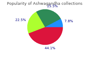
Ashwagandha 60 caps cheap otc
Since lymphatic vessels are often wrapped with an artery in a common connective tissue sheath anxiety symptoms out of nowhere ashwagandha 60 caps buy discount line, arterial pulsation additionally rhythmically squeezes the adjoining lymphatic vessels and contributes to lymph move anxiety symptoms checklist pdf ashwagandha 60 caps discount overnight delivery. A thoracic (respiratory) pump promotes the circulate of lymph from the belly to the thoracic cavity as one inhales, simply as it does in venous return. Finally, at the point the place the accumulating ducts empty into the subclavian veins, the quickly flowing bloodstream attracts the lymph into it. These tissues are composed of quite lots of lymphocytes and other cells with various roles in protection and immunity: 1. Neutrophils are aggressively antibacterial leukocytes and are described in section 18. T lymphocytes (T cells) are lymphocytes that mature within the thymus and later depend on thymic hormones; the T stands for thymus-dependent. B lymphocytes (B cells) are lymphocytes that differentiate into plasma cells-connective tissue cells that secrete antibodies. They are named for an organ in chickens (the bursa of Fabricius1) in which they had been first found. However, you could discover it more helpful to consider B for bone marrow, the site the place these cells mature in people. They come up from monocytes which have emigrated from the bloodstream and from division of preexisting tissue macrophages. They phagocytize tissue debris, useless neutrophils, micro organism, and different foreign matter (fig. They also course of foreign matter and display antigenic fragments of it to certain T cells, thus alerting the immune system to the presence of an enemy. They play an important role in alerting the immune system to pathogens that have breached the physique surfaces. They engulf foreign matter by receptor-mediated endocytosis somewhat than phagocytosis, but otherwise operate like macrophages. After internalizing an antigen, they migrate to a close-by lymph node and activate an immune reaction to it. The easiest type is diffuse lymphatic tissue, by which the lymphocytes are scattered somewhat than densely clustered. In some locations, lymphocytes and macrophages congregate in dense plenty referred to as lymphatic nodules (follicles) (fig. Abundant lymphatic nodules are, however, a relatively fixed function of the lymph nodes (see fig. In the ileum, the distal portion of the small gut, they form clusters called aggregated lymphoid nodules (formerly named Peyer patches). These organs embrace the red bone marrow, thymus, lymph nodes, tonsils, and spleen. The lymph nodes, tonsils, and spleen are known as secondary lymphatic organs as a outcome of immunocompetent lymphocytes migrate to these organs only after they mature in the primary lymphatic organs. Sinusoid Capillary Adipose cell Artery Endothelial cells Sinusoid Platelets and blood cell getting into circulation Megakaryocyte Reticular cells Red Bone Marrow Red bone marrow might not seem to be an organ; when aspirated from the bones for the purpose of biopsy or transfusion, it simply appears like extra-thick blood. Yet a cautious microscopic examination of much less disturbed marrow shows that it has a shocking degree of structure and consists of a number of tissues, assembly the standards of an organ, even if a really gentle one. Yellow bone marrow is mainly adipose tissue and may be disregarded for current functions, but red bone marrow is involved in hematopoiesis (blood formation) and immunity. Red bone marrow is delicate, loosely organized, extremely vascular materials, separated from osseous tissue by the endosteum of the bone. It produces all lessons of formed parts of the blood; its purple shade comes from the abundance of erythrocytes. Numerous small arteries enter nutrient foramina on the bone surface, penetrate the bone, and empty into large sinusoids (45 to eighty �m wide) in the marrow (fig. The sinusoids drain into a central longitudinal vein that exits the bone via the identical route that the arteries entered. The sinusoids are lined by endothelial cells, like other blood vessels, and are surrounded by reticular cells and reticular fibers. The blood-forming cells are connected in varied methods to the reticular cells and different elements of the framework (stroma) of the marrow. The reticular cells secrete colony-stimulating factors and other indicators that induce stem cells to become leukocytes, erythrocytes, and platelet-forming megakaryocytes. In the long bones of the limbs, aging reticular cells accumulate fat and remodel into adipose cells, eventually changing pink bone marrow with yellow bone marrow. The fashioned components of blood squeeze by way of the endothelial cells into the sinuses, which converge on a central longitudinal vein on the decrease left. The areas between the sinusoids are occupied by islands (cords) of hematopoietic tissue, composed of macrophages and blood cells in all levels of growth. The macrophages destroy malformed blood cells and the nuclei discarded by growing erythrocytes. As blood cells mature, they push their way through the reticular and endothelial cells to enter the sinusoid and circulate away within the bloodstream. It homes growing lymphocytes and secretes hormones that regulate their later activity. It is a bilobed organ located between the sternum and aortic arch within the superior mediastinum. The thymus exhibits a exceptional diploma of degeneration (involution) with age, as described and illustrated earlier (see fig. The fibrous capsule of the thymus offers off trabeculae (septa) that divide the gland into several angular lobules. Each lobule has a light central medulla populated by T lymphocytes, surrounded by a dense, darker cortex (fig. After growing within the cortex, T cells migrate to the medulla, the place they spend another 3 weeks. Epithelial cells of the thymus secrete a number of signaling molecules that promote the event and action of T cells both domestically (as paracrines) and systemically (as hormones); these include thymosin, thymopoietin, thymulin, interleukins, and interferon. If the thymus is removed from new child mammals, they waste away and never develop immunity. Other lymphatic organs also appear to depend on thymosins or T cells and develop poorly in thymectomized animals. They serve two features: to cleanse the lymph and to act as a website of T and B cell activation. A lymph node is an elongated or bean-shaped construction, normally lower than three cm long, typically with an indentation referred to as the hilum on one facet. It is enclosed in a fibrous capsule with trabeculae that partially divide the interior of the node into compartments. Between the capsule and parenchyma is a slim, relatively clear house called the subcapsular sinus, which accommodates reticular fibers, macrophages, and dendritic cells.
60 caps ashwagandha best
Electrocution with anal insertion of wire Causes and manners of dying of victims of autoerotic dying Natural 6 anxiety 4th breeders ashwagandha 60 caps purchase without prescription. Asphyxia via ligature encircling neck Autopsy and toxicological findings the pathological findings in circumstances of autoerotic asphyxia are often non-specific and are just like anxiety symptoms ruining my life ashwagandha 60 caps effective those encountered in asphyxial deaths [28,66, 76]. Several gross pathological options could also be observed singularly or in combination, or could additionally be absent in asphyxial deaths (Table 28. Investigators have postulated that petechiae result from a mechanical vascular process, specifically impaired or obstructed venous return regardless of persistent arterial enter [25,28]. Microscopic evidence of an asphyxial death may embrace hypoxic/ischaemic damage to tissues if the survival time is delayed. Six decedents had sustained fractures of the thyroid cartilage and one had subluxation of a cervical vertebra. In our research of 16 cases of autoerotic asphyxia [66], petechiae of the conjunctivae, skin of the eyelids and extremities, epicardium and pleura have been current in 12 victims (75%). In our examine of sixteen victims, the postmortem blood ethanol level was adverse in 11 subjects (68. Filmed non-lethal autoerotic ligature strangulations and hangings Sauvageau and colleagues scrutinized non-lethal ligature strangulations and hangings filmed by an autoerotic participant [56,57]. They reported loss of consciousness in eleven seconds, onset of convulsions in 7�11 seconds, and regaining of consciousness in 16�18 seconds in ligature strangulations [57]. Similar early agonal findings had been observed in hangings, together with decerebrate rigidity noticed at 20 seconds in an interrupted hanging [56]. These investigations into a behaviour of a living autoerotic practitioner may elucidate the timing of irreversible mind injury induced by ligature strangulations and hangings. Myths and actuality of autoerotic asphyxia Reality All autoerotic asphyxial deaths are unintentional, by definition. The lethal paraphiliac syndrome: Accidental autoerotic deaths in 4 ladies and a evaluate of the literature. Age, transvestism, bondage, and concurrent paraphilic activities in 117 deadly instances of autoerotic asphyxia. A comparison of typical death scene features in cases of fatal male and autoerotic asphyxia with a review of the literature. Searching for the G spot within the urinary bladder: Autoerotism and potential complications. Penile strangulation: An uncommon sexual apply that always presents an urological emergency. Conclusion A fantastic line exists between the attainment of cerebral hypoxia for sexual pleasure and unconsciousness which will quickly ensue and lead to inevitable dying. As autoerotic deaths are innately enigmatic, awareness that this follow might lead to a fatality is critical. Emergency drugs physicians who could encounter members of autoerotic asphyxia should be educated about the danger factors associated with this behaviour and must be encouraged to determine, report and counsel families and pals dealing with autoerotic asphyxia [75]. Several myths pertaining to autoerotic asphyxia persist, and this chapter has strived to dispel these myths (Table 28. The typical autoerotic asphyxia participant acts alone and uses a ligature for the only objective of attaining heightened sexual arousal with no intention of ending their life. Variations from the classic autoerotic case may be encountered, relying on the gender of the participant and specific sexual technique chosen. These alterations from the standard autoerotic demise scene warrant particular attention to determine the correct manner of death. Accidental dying as a outcome of complete autoerotic asphyxia associated with transvestic fetishism and anal self-stimulation: Case report. Letter to the editor: A revisitation of the commonest methods of autoerotic activity resulting in demise based on the new standardized classification of asphyxia. Autoerotic nonlethal filmed hangings: A case series and comments on the estimation of the time to irreversibility in hanging. Three nonlethal ligature strangulations filmed by an autoerotic practitioner: Comparison of early agonal responses in strangulation by ligature, hanging, and handbook strangulation. Aqua-eroticum: An unusual autoerotic fatality in a lake involving a home-made diving apparatus. Generalized subcutaneous emphysema caused by injection of air into the penis for autoerotic functions. It has been postulated that the sympathetic nervous system could also be stimulated when a plastic bag is placed over the pinnacle which may induce arrhythmias similar to ventricular fibrillation [32,51]. This deadly cardiac occasion could explain the everyday absence of postmortem findings in plastic bag asphyxial deaths, including cutaneous and conjunctival petechial haemorrhages, facial congestion, oedema and cyanosis [51,29]. Individuals with a debilitating medical illness may choose suicide utilizing the strategies outlined in Final Exit. This chapter discusses the impression of Final Exit on the suicide rate, with particular consideration to plastic bag asphyxial deaths with or without the supplementation of inert gases. We also delve into the surreptitious act of autoerotic asphyxia the place a participant may use a plastic bag to heighten sexual arousal with no intention of inflicting deadly penalties. This chapter classifies the mechanisms of asphyxia and mentions the findings that may be noticed throughout autopsy and toxicological evaluation of plastic bag asphyxia. The demise scene could additionally be ambiguous and deceptive to investigators of plastic bag asphyxia instances, especially if the evidence has been removed from the scene. A thorough scene analysis, historical investigation and autopsy examination could shed light on the circumstances in equivocal circumstances of plastic bag asphyxial deaths. It grew in recognition as individuals embraced the direct message set forth in the guide amid the controversy of euthanasia and assisted suicide internationally. Assisted suicide refers to receiving lethal drugs from a doctor and swallowing them to cause dying [26]. This method of death is legal in solely a small number of nations worldwide and in a couple of states in the United States [14]. Switzerland is the only country that permits foreigners to use medically assisted suicide to die. From methods of dying ranging from cyanide ingestion to self-starvation to helium inhalation to carbon monoxide intoxication [10], the step-by-step manual Final Exit particulars the way to acquire provides and execs and cons of various elements of each method [26]. While this e-book has been accused of encouraging suicide in individuals with psychological problems, primarily melancholy [49], adolescents [9] and non-terminal aged people [35], Humphry emphasised that his primary objective for writing the book was to provide a straightforward guide to those suffering from a terminal illness to finish their lives by the means of self-deliverance [26]. In 2000, Supplement to Final Exit: the Latest How-To and Why of Euthanasia/Hastened Death instructed including an inert fuel corresponding to helium to the plastic bag, as a outcome of the benefit of obtaining it [25]. Humphry based the Hemlock Society in 1980 which served as an end-of-life care group, providing information about voluntary euthanasia and ethical help with out direct help [26]. Similar to the New York City outcomes, suicidal asphyxiations using a plastic bag elevated by 30. These authors were unable to decide whether the victims had impulsively dedicated suicide after studying Final Exit or whether or not the suicidal choice had already been determined and the strategy was changed as a outcome of this guide. Plastic bag asphyxia: Suicide While plastic bags are available, a paucity of individuals select a plastic bag as a means to end their life. Plastic bag asphyxia could involve solely a plastic bag or may embody inhalation of a gasoline, such as chloroform [69], ether [3,68], helium [16,17,45,56,58], natural gas (high methane-content mixture) [39], nitrogen [38], butane [1], propane [13,15], exogenous carbon dioxide (dry ice) [13] and toluene [46].
Ashwagandha 60 caps buy lowest price
Neck dissection For anterior neck strap muscle dissection anxiety symptoms natural remedies purchase ashwagandha 60 caps mastercard, ordinary indications are exterior evidence of neck trauma anxiety guru 60 caps ashwagandha buy otc, strangulation, sexual assault with potential neck trauma or subcutaneous neck haemorrhage upon initial examination. Summary and opinion It is important to perceive that the death investigation is a stepwise course of. It is important to follow an evidencebased approach in providing the trigger of demise and the opinion. A comprehensive demise investigation together with a thorough evaluate of the historical past, circumstances and scene examination, and thorough post-mortem together with layered dissection of the neck and face and incorporating each external and inner examination findings are needed before concluding an opinion in a case of neck injury. This is often difficult within the presence of a competing alternative reason for death such as a head injury or a stab injury. In the presence of a competing explanation for demise, the troublesome query is to find out the exact contribution of the neck damage to the final reason for death. A forensic pathologist is commonly questioned in court about the potential of incapacitation by the neck injury before the death happens. Prerequisites There are numerous conditions earlier than the anterior neck dissection is began. It is necessary to take away the thoracic content under the level of the clavicles, remove the mind, and wait for about 10 minutes for the blood to drain from the neck constructions. Technique Knowing the anatomy of every layer is critical for performing this procedure. It is advisable to take stepwise images and mark the presence of any injury in appropriate diagrams at each step of the procedure. Layered dry neck dissection after elimination of the thoracic and stomach organs and the ten mind allows any congested blood which may be confused with true haemorrhage to drain from the neck area. The anterior neck constructions should be examined layer by layer and any injury ought to be documented with nature of damage, dimension and distribution and correlation ought to be made to any injury observed on the pores and skin floor. The incision from the tops of the shoulders could be further prolonged up to the mastoid areas making a rhomboid-shaped flap. The sternocleidomastoid muscle tissue run along the perimeters of the neck with the carotid and jugular sheath just underneath. The deep layer of muscle tissue consists of the sternothyroid and thyrohyoid muscular tissues. The sternothyroid muscle is minimize on the sternum and mirrored upwards, visualizing the underlying thyroid gland and cricothyroid muscles. The neck organs and attached strap muscle tissue are eliminated en bloc with the connected tongue, hyoid bone, larynx and trachea for detailed examination. The content material of the carotid sheath including the carotid arteries, inner jugular vein and vagus nerve should be examined for any injury [5]. The laryngeal block with the tongue, hyoid bone and larynx ought to be eliminated using forceps and a bigger knife The incision should be made as shut as attainable to the inferior floor of the mandible. After liberating the tongue, the tip of the tongue can be used to hold the neck buildings and a horizontal incision made in the soft palate simply above the uvula into oropharynx. Next, the incision is continued into the musculature surrounding the oropharynx, along the prevertebral fascia and the tissue block containing the tongue, hyoid bone, laryngeal structures and pharynx, and the upper oesophagus could be removed after incising the gentle tissue inferior to suprasternal notch and inferior surfaces of the medial ends of the clavicles. The neck dissection could be extended to the face to detect and doc facial accidents. The sternocleidomastoid muscles could be seen along the perimeters of the neck and the paired sternohyoid muscle tissue located centrally. Postmortem artefacts There are five main artefacts/pitfalls in interpretation of postmortem findings [3]: 1. Note the fractured left superior horn (lower a part of image) with associated hemorrhage. Developmental segments of the hyoid bone the hyoid bone is part of the hyoid-laryngeal complex which varieties the inner hard buildings of the throat. Discontinuities of the hyoid bone may be interpreted as fractures by an untrained person. The greater cornua and the body of the hyoid bone in early development stage are current as three separate bony structures. Due to variable developmental development, these bony structures may not be symmetrical in some individuals. In such instances, the synchondrodic joints may be unfused or partially unfused on one side of the hyoid bone. In the presence of artefactual haemorrhages, dysmobility may give rise to misinterpretation as fractures. Presence of triticeous cartilages Triticeous cartilages are small items of fibrocartilage which are linear or spherical and a few millimetres in dimension. The presence of triticeous cartilage could be misinterpreted as a fracture of a bone in this location. Another unusual postmortem artefact is extravasation of blood inside the strap muscles and the platysma in instances of drowning or due to immersion in water. Blunt-force neck trauma also needs to be differentiated from injuries brought on by neck compression. Prinsloo and Gordon haemorrhages these artefactual haemorrhages were named after the nice South African forensic pathologists Prinsloo and Gordon [4]. They can lead to misinterpretation of extravasation of blood into the gentle tissues of the neck as a mimic of bruising due to trauma. Postmortem handling of the neck structures similar to blunt dissection and incisions made during the neck dissection are the true trigger of those haemorrhages. The congested blood on this space may cause blood monitoring between and throughout the strap muscles. This artefact may be prevented by dissection of the neck after vascular decompression and systemic layered dry neck dissection. Interpretation of errors Interpretation errors can occur as a outcome of improper recording of data/findings, lack of know-how of postmortem artefacts, lack of expertise of anatomical variations and never following the established stepwise approach in systemic demise investigation. Awareness of the anatomical variations, postmortem artefacts and other pitfalls within the neck is crucial. The neck represents a critically essential anatomical construction in forensic medicine. Surface artefacts, such as a neck fold of an infant or an obese particular person, could also be mistaken for a ligature mark. Injury brought on by tight clothes across the neck, in particular when the body is decomposed, can even mimic a ligature mark. Postmortem hypostatic haemorrhages Postmortem hypostatic haemorrhages are because of extravasation of blood into the interstitial tissue. This occurs because of congestion of the venous plexus and distention of the venous plexus because of gravitational hypostasis. As a results of postmortem adjustments, the vascular integrity can be breached and the blood can be extravasated from the vessels into the soft tissues. Post-mortem dissection artifacts of the neck; their differentiation from ante-mortem bruises. Resuscitation-related neck artefacts There are three major resuscitation-related neck injuries that can result in interpretation issue. Injury to sternomastoid muscle on account of placement of a cannula in the internal jugular vein.
N-Acetylcysteine (N-Acetyl Cysteine). Ashwagandha.
- Reducing mucus and helping with breathing in various lung conditions.
- Preventing kidney problems with dyes used during some X-ray exams.
- Acetaminophen (Tylenol) poisoning.
- Preventing side effects of doxorubicin (used for certain types of cancer).
- Cystic fibrosis.
- Preventing side effects of ifosfamide (Ifex, used for certain types of cancer).
- Treating a lung disease called fibrosing alveolitis.
- How does N-acetyl Cysteine work?
- Treating organ failure.
Source: http://www.rxlist.com/script/main/art.asp?articlekey=96979

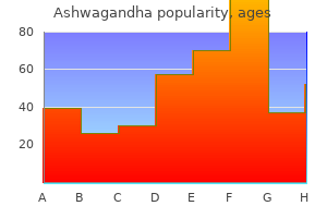
Ashwagandha 60 caps buy lowest price
You ought to familiarize yourself with these earlier than proceeding anxiety chest tightness generic 60 caps ashwagandha visa, as a outcome of later discussions on this chapter assume a working data of a few of these terms anxiety symptoms postpartum trusted 60 caps ashwagandha. To cough, we close the glottis and contract the respiratory and stomach muscles, producing high pressure within the decrease respiratory tract. We then abruptly open the glottis and release an explosive burst of air at speeds over 900 km/h (600 mi. Its mechanism is just like coughing besides that the glottis is regularly open, the taste bud and tongue block the move of air whereas thoracic strain builds, after which the soft palate is depressed to direct a half of the airstream via the nose. These actions are coordinated by coughing and sneezing centers within the medulla oblongata. Explain why contraction of the diaphragm causes inspiration but contraction of the transverse stomach muscle causes expiration. Identify a benefit and an obstacle of normal (nonpathological) bronchoconstriction. Suppose a wholesome person has a tidal volume of 650 mL, an anatomical useless house of one hundred sixty mL, and a respiratory price of 14 breaths per minute. Suppose an individual has a total lung capacity of 5,800 mL, a residual quantity of 1,200 mL, an inspiratory reserve quantity of 2,400 mL, and an expiratory reserve volume of 1,four hundred mL. As we are now concerned with atmospheric pressures and how they affect the partial pressures of blood gases, we return to mm Hg as our unit of measurement (not cm H2O as when we were considering pulmonary ventilation). If we assume the typical sea-level atmospheric pressure of 760 mm Hg and oxygen is 20. Alveolar air may be sampled with an equipment that collects the final 10 mL of expired air. We will flip our attention now to the behavior of those gases within the human body: how oxygen is obtained from inspired air and delivered to the tissues, and the way carbon dioxide is faraway from the tissues and launched into the expired air. This was all too sadly realized by French doctor Paul Bert (1833�86), who is often acknowledged because the founder of aerospace medication. He invented the primary stress chamber able to simulating the results of excessive altitude, and undertook a variety of experiments on human and animal topics to take a look at the consequences of variation in oxygen partial stress. Their aim was to investigate the consequences of the low oxygen pressures attainable solely by ascending to a really high altitude. But after passing 24,000 toes, they experienced stupefaction, muscular paralysis, euphoria, and at last unconsciousness. The balloon finally descended on its own with two of the three men useless; only Tissandier lived to write about it. If the partial stress of the gasoline is bigger within the air, it diffuses into the water. Thus, the Po2 of alveolar air is about 65% that of inhaled air, and its Pco2 is greater than 130 occasions larger. For oxygen to get into the blood, it must dissolve in this water and pass by way of the respiratory membrane separating the air from the bloodstream. For carbon dioxide to depart the blood, it should cross the opposite method and diffuse out of the water film into the alveolar air. Whenever air and water are in contact with one another, gases diffuse down their gradients until the partial stress of each gas within the air is equal to its partial stress within the water. Even in vigorous exercise, when the blood flows quicker, an erythrocyte is within the alveolar capillary for about 0. Several variables affect the efficiency of alveolar gas trade, and under abnormal conditions, some of these can prevent the complete loading and unloading of gases: Pressure gradients of the gases. Po2 is about 104 mm Hg in alveolar air and forty mm Hg in blood arriving at an alveolus. Oxygen subsequently diffuses from the air into the blood, the place it reaches a Po2 of 104 mm Hg. Pco2 is about 46 mm Hg in blood arriving at the alveolus and 40 mm Hg in alveolar air. Blood entering lungs Po2 40 mm Hg Pco2 46 mm Hg Blood leaving lungs Po2 ninety five mm Hg Pco2 forty mm Hg these gradients differ beneath particular circumstances such as high elevation and hyperbaric oxygen therapy (treatment with oxygen at >1 atm of pressure) (fig. Atmospheric Po2, for instance, is 159 mm Hg at sea stage however solely 110 mm Hg at 3,000 m (10,000 feet). In a hyperbaric oxygen chamber, by contrast, a patient is uncovered to three to four atm of oxygen to treat such situations as gangrene (to kill anaerobic bacteria) and carbon monoxide poisoning (to displace the carbon monoxide from hemoglobin). Carbon dioxide is about 20 instances as soluble as oxygen, and oxygen is about twice as soluble as nitrogen. In such coronary heart conditions as left ventricular failure, nonetheless, blood strain builds up within the lungs and promotes capillary filtration into the connective tissues, causing the respiratory membranes to turn out to be edematous and thickened (similar to their condition in pneumonia; fig. Under these circumstances, blood leaving the lungs has an unusually low Po2 and high Pco2. In good health, each lung has about 70 m2 of respiratory membrane out there for gasoline exchange. Since the alveolar capillaries comprise a total of solely a hundred mL of blood at any one time, this blood is spread very thinly. Several pulmonary illnesses, nonetheless, lower the alveolar surface space and thus lead to low blood Po2-for example, emphysema (fig. Gas exchange requires not only good air flow of the alveoli but in addition good perfusion of their capillaries. Ventilation�perfusion coupling refers to physiological responses that match airflow to blood circulate and vice versa. For example, if a part of a lung have been poorly ventilated due to tissue destruction or an airway obstruction, it will be pointless to direct a lot blood to that tissue. This stimulates local vasoconstriction, rerouting the blood to better-ventilated areas of the lung the place it can choose up extra oxygen (left facet of fig. In distinction, increased air flow raises the local blood Po2 and this stimulates vasodilation, rising blood circulate to that region to reap the benefits of the oxygen availability (right facet of similar figure). These reactions of the pulmonary arteries are reverse from the reactions of systemic arteries, which dilate in response to hypoxia. Furthermore, adjustments within the blood move to a area of a lung stimulate bronchoconstriction or dilation, adjusting ventilation so that air is directed to the best-perfused components of the lung (fig. Trace the partial strain of oxygen from impressed air to expired air and explain each change in Po2 alongside the greatest way. The poisonous effect of carbon monoxide stems from its competition for the O2 binding site (see Deeper Insight 22. The rate of loading is dependent upon the steepness of the gradient from alveolar air to the venous blood arriving on the alveolar capillaries. Compared with the oxygen gradient at sea level (blue line), the gradient is less steep at excessive elevation (red line) because the Po2 of the atmosphere is decrease. In a hyperbaric chamber with 100% oxygen, the gradient from air to blood is very steep (green line), and oxygen loading is correspondingly rapid.
Discount ashwagandha 60 caps otc
However anxiety 4 year old boy buy 60 caps ashwagandha overnight delivery, these findings are unspecific with regard to the actual mechanism of dying anxiety symptoms jaw pain order ashwagandha 60 caps without a prescription. Axon injury could be observed not only after hypoxic-ischaemic alterations, but additionally after craniocerebral trauma because of blunt violent effects and in circumstances of a number of sclerosis. It is usually assumed right now that changes within the neurons are detectable about 7 hours after a relevant cerebral O2 deficit [3,8]. Astrocytes accumulate and proliferate, and neutrophil granulocytes and macrophages migrate, ingesting the apoptotic cell materials. Within 12-24 hours the variety of activated microglia and macrophages will increase considerably. Immunohistochemical investigations on the brains of deceased newborns and infants have been printed by Fineschi et al. The aim of the research was to estimate the time of onset of perinatal hypoxic-ischaemic cerebral O2 deficiency. Depending on the sample of immunohistochemically positive findings, it was potential to make rough estimates of the time at which the brain harm developed. Furthermore, the authors have successfully used the pattern of different markers current within the serum and cerebrospinal fluid A controversial medicolegal issue: Timing the onset of perinatal hypoxic- ischemic brain injury. Immunohistochemical investigation of hypoxic/ ischemic mind damage in forensic post-mortem circumstances. Morphological evaluation of astrocytes in the hippocampus in mechanical asphyxiation. Pontine axonal harm after mind trauma und nontraumatic hypoxicischemic mind harm. Methodical strategy to mind hypoxia/ischemia as a basic drawback in forensic neuropathology. In addition, frozen sections of fastened tissue should be prepared for the detection of fats parts. The microscopic analysis of instances follows with regard to general morphological changes (alveolar and interstitial oedema, hyperaemia, alveolar and interstitial haemorrhages, emphysema, dystelectasis) and particularly alterations of lung blood vessel contents. These may be regarded as positive, if 20 per cent of the cells in a vessel are non-erythrocytic. The percentage of constructive vessels is then noted and graduated (<5%, 5%�20%, 21%�50%, >50%). For special purposes, immunohistochemical methods for the detection and characterization of alveolar macrophages and pulmonary big cells could additionally be carried out. Introduction In the medicolegal investigation of deadly instances of asphyxiation, marked external indicators of vitality corresponding to cyanosis/congestion or petechiae may be missing. For these reasons the histological and immunohistochemical examination of pulmonary tissue has been evaluated by a quantity of authors. As a central organ within the pathophysiology of asphyxiation, the lung might exhibit alterations of its microstructure and/or cell content. In mixture with microembolism syndrome, they regarded it as a software to differentiate death by obstructive asphyxia from different causes of demise with similarly short periods of agony. Janssen, in addition, observed the appearance of quite a few alveolar macrophages and intraalveolar big cells in cases of protracted oxygen deficiency (throttling, smothering, thoracic compression), an agonal mobilization and proliferation of alveolar cells with the formation of multinuclear giant cells [9,10]. A semi-quantitative rely of constructive cells seems to be sufficient (negative, weak, reasonable, sturdy reactions; variety of cells per microscopic field). Main results and which means In accordance with macroscopic findings, general structural changes may be observed in all kinds of strangulations more or less regularly. Apart from emphysema (present in about 10%�40% of cases) the Methods For finding out routine lung histopathology, no much less than one sample of every lobe ought to be taken during post-mortem. Microscopic alterations of the pulmonary blood vessel contents consist of three major phenomena: fats embolism, bone marrow tissue embolism and intravascular cell accumulations. In specific, bone marrow tissue embolism happens primarily sporadically (once or twice a case). The cell accumulations consider medium-sized and smaller arteries and appear partly as aggregates filling the entire vessel and partly in a disseminated manner. On the whole, these modifications are principally restricted to a quantity of sections of a case or even to a couple of vessels within one part. Most regularly, less than 5 per cent or actually lower than 20 per cent of all vessels show optimistic reactions on this sense. The phenomenon is extra frequent in non-hanging cases (approximately one-third vs one-tenth). The phenomenon of intravascular cell accumulation is present in control circumstances as properly when (longer) resuscitation is documented/probable. In circumstances with out indicators of blunt pressure, they level to protracted agony programs in the sense of shock equivalents. The prevalence of quite a few alveolar macrophages and pulmonary big cells has been reported in both deadly asphyxia and different causes of demise. Oedema could be found in practically 100 per cent of instances (among these in one-quarter extreme oedema involving greater than 50% of alveoli). In a minority of instances with beginning intra-alveolar oedema, perivascular and interstitial oedema may be distinctly differentiated. In about one-third of deadly strangulations, small air bubbles throughout the alveolar oedema fluid are present. Alveolar and interstitial haemorrhages also occur frequently, but mainly in a very discrete manner. On the entire, these basic structural changes of the lung are non-specific for asphyxia/strangulation/suffocation, 122 Table 13. The German forensic pathologist Janssen described a mobilization and proliferation of alveolar cells with the formation of multinuclear giant cells in fatalities with protracted oxygen deficiency [9,10]. Further investigations together with our personal outcomes [5,7,eight,11] result in totally different conclusions as to the function of pulmonary macrophages in asphyxia/suffocation. Giant cells occurred even more than twice as regularly in opiateinvolved deaths (group with prefinal oxygen lack) in comparison with strangulations. For these causes, there have just lately been many attempts to identify pathomorphological criteria that are absent under physiological conditions and seem particularly in the context of asphyxiation, increasing of their expression with the length of the hypoxic agony [8]. Thus, the main target of the research has been on the lung, a major effector organ of the dysregulation of respiration and circulation as a end result of a fatal suffocation. Acute alveolar hypoxia, characterizing suffocation, is a bodily state observed in numerous medical conditions and illnesses, together with anaphylactic shock, mind damage, intoxication and acute cardiac insufficiency. Clinical analysis subsequently provides significant perception into physiological mechanisms, mobile modifications and additional asphyxiation markers within the lung, all of that are additionally helpful in forensic pathology. Microstructure of the lung and the position of alveolar macrophages and large cells Pathophysiologically, suffocation is characterized by decreased alveolar pO2, which induces vasoconstriction of the pulmonary arteries within the lung periphery and a redistribution of blood from basal to apical lung segments in accordance with the Euler-Liljestrand mechanism. As oxygen is the primary requirement for the secretion of needed vasoactive substances, a gentle lung injury results in inflammatory adjustments as a outcome of acute hypoxia [27]. Consequently, the permeability of blood vessels, generated by a high number of pores and vacuoles in endothelial cells, is elevated, adopted by rising pulmonary extravasation of fluid, electrolytes and albumin. However, their comparative study in 50 rats and 15 rabbits of strangulation vs varied various causes of dying This research merely underlined the higher frequency and the higher degree of the lung alterations described above within the instances of deadly asphyxia compared to the controls.
Ashwagandha 60 caps buy otc
In some studies anxiety symptoms 6 months buy generic ashwagandha 60 caps line, the bodily disfigurement attributable to depigmented patches emphasised the significance of bodily appearance in psychological adjustment [45] anxiety symptoms 4 days buy 60 caps ashwagandha with amex. Decreased photodamage and low incidence of non-melanoma pores and skin most cancers in 136 sun-exposed Caucasian sufferers with vitiligo. Decreased threat of melanoma and nonmelanoma pores and skin cancer in sufferers with vitiligo: A survey amongst 1307 patients and their partners. A higher understanding of the disease and analysis of psychosocial impression may assist the treating doctor to deal with sufferers in a holistic method. Introduction, epidemiology, quality of life, analysis, differential analysis, associations, histopathology, etiology, and work-up. The evaluation of genetics and associated autoimmune diseases in Chinese vitiligo sufferers. Vogt-Koyanagi-Harada illness: Extensive vitiligo with prodromal generalized erythroderma. Both might initially appear as focal or localized vitiligo, which entails a small area [14]. Dermatomal sample is believed to be the commonest, and an area roughly associated to the trigeminal dermatome is essentially the most commonly affected. However, a quantity of hypotheses have been proposed, including neuronal mechanisms and a mosaicism speculation [7�9]. The restricted nature of the disease and restriction of the depigmentation to a well-defined phase should be emphasised while counseling patients. Various therapeutic modalities and superior outcomes of surgical intervention ought to be discussed intimately with each patient before starting any remedy. The coexistence of generalized and segmental vitiligo is seen in some patients and it might point out that mixed vitiligo represents type 2 mosaicism of Happle [19�21]. This is in accordance with the speculation that the segmental lesions of sort 2 mosaicism are more severe than the nonsegmental sort [19�21]. Facial lesions, shorter length, and earlier preliminary response to therapy seem to be the favorable elements for medical therapy [22]. Revised classification/ nomenclature of vitiligo and associated points: the Vitiligo Global Issues Consensus Conference. Segmental vitiligo because the potential expression of cutaneous somatic mosaicism: Implications for widespread nonsegmental vitiligo. Relevance of thyroiditis and of different autoimmune ailments in kids with vitiligo. Topical calcineurin inhibitors have shown good results due to their melanocyte-stimulating impact along with their immunomodulator motion. A therapeutic trial of minimum 6 months may be given with topical remedy earlier than declaring it as ineffective [25]. Surgical treatment should be thought of as first-line therapy in sufferers with leukotrichia as a outcome of medical management is sure to fail in these circumstances. This consists of punch graft, Thiersch graft, blister graft, and full-thickness skin graft. This is a novel process primarily based on the concept of the presence of undifferentiated stem cells in the hair follicle. These cells form a superb reservoir of melanocytes responsible for repigmentation. Segmental vitiligo related to generalized vitiligo (mixed vitiligo): A retrospective case series of 19 patients. Segmental vitiligo: A randomized controlled trial to consider efficacy and safety of 0. Since there was a scarcity of consensus relating to the nomenclature and classification used across the globe till now, a new classification system of vitiligo was proposed in 2012 by Vitiligo Global Issues Consensus Conference [2]. The major groups described on this classification are segmental and nonsegmental, of which nonsegmental vitiligo is a group comprising acrofacial, mucosal, generalized, common, or blended varieties. Generalized and acrofacial are probably the most generally reported patterns of vitiligo from India [3,4]. Speeckaert and van Geel evaluated the association of age of onset and affected sites in seven-hundred vitiligo sufferers [8]. Predominant involvement of the beard area in males and the periocular space in girls was noted. An earlier age of onset, periocular vitiligo was noticed when compared to the perioral space, and of lower extremities in comparability with upper extremities. Lesions in the genital areas are also included within the acrofacial group of vitiligo [5]. A latest research from China confirmed a big affiliation of mucosal vitiligo with lesions on the acrofacial areas and proposed that mucosal vitiligo is probably a special form of acrofacial vitiligo [6]. Dermal findings can range from a mild-to-dense lichenoid band-like inflammatory infiltrate [12]. The discount in practical melanocytes can be confirmed by special stains similar to Fontana-Masson, a stain for melanin, or dihydroxyphenylalanine alanine for tyrosinase. This approach has been efficiently used in the administration of eyelid and lip vitiligo as properly [22,23]. It has proven superior efficacy over blister grafting within the administration of secure and treatment-resistant vitiligo [24]. Various research have proven a lower response of medical therapies in these cases; hence surgical choices have extra commonly been opted for on this variant of vitiligo. In a meta-analysis of nonsurgical repigmentation therapies in vitiligo, topical class 3 and 4 had the highest success rates (56% and 55%, respectively) [14]. Younger and darker-skin sufferers with head and neck involvement have proven a relatively better response [15]. Children have shown a better response over acrofacial areas in comparability with adults [17]. In a potential case examine of generalized (338), segmental (170), and localized (166) instances of vitiligo, topical vitamin D3 remedy was considerably effective for the localized kind. Combination with topical corticosteroids, calcineurin inhibitors, and vitamin D3 analog has resulted in a greater response compared to monotherapy [18]. Revised classification/nomenclature of vitiligo and associated issues: the Vitiligo Global Issues Consensus Conference. A sample of affiliation between medical form of vitiligo and disease-related variables in a Brazilian population. Vitiligo in North-Eastern China: An affiliation between mucosal and acrofacial lesions. Ohguchi R, Kato H, Furuhashi T, Nakamura M, Nishida E, Watanabe S, Shintani Y, and Morita A. Risk elements and therapy responses in patients with vitiligo in Japan-A retrospective large-scale research. Lichenoid inflammation in vitiligo-A scientific and histopathologic review of 210 instances. Presence or absence of melanocytes in vitiligo lesions: An immunohistochemical investigation. Vitiligo treated with topical corticosteroids: Children with head and neck involvement reply well.
Order 60 caps ashwagandha with mastercard
When oxygen becomes obtainable again anxiety loss of appetite discount ashwagandha 60 caps visa, the liver oxidizes lactate back to pyruvate anxiety at night discount 60 caps ashwagandha with visa, which might then enter the aerobic pathway described shortly. The liver can even convert lactate again to G6P and can do both of two issues with that: (1) Polymerize it to form glycogen for storage, or (2) take away the phosphate group and release free glucose into the blood. Although anaerobic fermentation retains glycolysis running slightly longer, it has some drawbacks. Skeletal muscle is comparatively tolerant of anaerobic fermentation, and cardiac muscle is much less so. In the presence of oxygen, pyruvate enters the mitochondria and is oxidized by aerobic respiration. This reaction generates oxaloacetic acid, which is out there to start the cycle another time. The citric acid cycle not only oxidizes glucose metabolites however is also a pathway and supply of intermediates for the synthesis of fat and nonessential amino acids. The connections between the citric acid cycle and the metabolism of different nutrients are mentioned later. The membrane reactions are carried out by a collection of compounds known as the mitochondrial electrontransport chain (fig. Iron�sulfur (Fe�S) centers, complexes of iron and sulfur atoms certain to membrane proteins. Unlike the other members, this is a relatively small, cell molecule that moves about within the membrane. Hydrogen atoms are split aside as they Cyt a3 transfer from coenzymes to the chain. Transport molecules are reduced when it receives an electron pair and grouped into three enzyme complexes, each of which acts as a proton pump. Molecules on the upper left of the determine have a comparatively excessive free power content, and molecules oxidized again when it passes the electrons on the decrease proper are comparatively low in power. Each oxygen atom (half of an O2 molecule) accepts two electrons (2 e�) from cytochrome a3 and two protons (2 H+) from the mitochondrial matrix. Without it, this response stops and, like a visitors jam, stops all the opposite processes leading to it. The Chemiosmotic Mechanism Of major importance is what happens to the vitality liberated by the electrons as they cross along the chain. Each advanced collectively acts as a proton pump that removes H+ from the mitochondrial matrix and pumps it into the house between the inside and outer mitochondrial membranes. Coenzyme Q is a shuttle that transfers electrons from the primary pump to the second, and cytochrome c shuttles electrons from the second pump to the third. That is, they create a steep electrochemical gradient across the inside mitochondrial membrane. If the internal membrane have been freely permeable to H+, these ions would have a powerful tendency to diffuse down this gradient and back into the matrix. As H+ flows by way of these channels, it creates an electrical present (which, you might recall, is just shifting charged particles). This process is known as the chemiosmotic7 mechanism, which suggests the "push" created by the electrochemical H+ gradient. Each enzyme complex pumps hydrogen ions into the house between the mitochondrial membranes. The common adult body accommodates about four hundred to 450 g of glycogen: almost one-quarter of it in the liver, three-quarters of it within the skeletal muscular tissues, and small quantities in cardiac muscle and different tissues. The enzyme glycogen synthase then cleaves off the phosphate group and attaches the glucose to a rising polysaccharide chain, thus assembling glycogen one glucose at a time. The enzyme glycogen phosphorylase begins by phosphorylating a glucose residue and splitting it off the glycogen molecule as G1P. Liver cells, however, have an enzyme referred to as glucose 6-phosphatase, which removes the phosphate group and produces free glucose. Gluconeogenesis8 is the synthesis of glucose from noncarbohydrates such as glycerol and amino acids. Explain the origin of the word glycolysis and why this is an appropriate name for the perform of that response pathway. What essential enzyme is found in the inside mitochondrial membrane aside from those of the electron-transport chain Describe how the liver responds to (a) an excess and (b) a deficiency of blood glucose. Glucose 6-phosphate Glycogen synthase Glycogenesis Glycogenolysis Glycolysis Glucose 1-phosphate Glycogen Pi phosphorylase Pi Glycogen Key 26. In most cells, the glucose 1-phosphate generated by glycogenolysis can bear solely glycolysis. In liver, kidney, and intestinal cells, it can be converted again to free glucose and launched into circulation. These pathways additionally serve for the oxidation of proteins and lipids as gasoline and as a source of metabolic intermediates that can be used for protein and lipid synthesis. Glycogenolysis this course of and eventually produce simply as much glucose because the liver does. Lipogenesis employs compounds such as sugars and amino acids to synthesize glycerol and fatty acids, the triglyceride precursors. As glucose and amino acids enter the citric acid cycle by means of acetyl-CoA, the acetyl-CoA can be diverted to make fatty acids. The glycerol and fatty acids can then be condensed to form a triglyceride, which may be stored within the adipose tissue or converted to other lipids. When carbohydrate is unavailable, oxaloacetic acid is converted to glucose and becomes unavailable to the citric acid cycle. Fat oxidation then produces extra ketones, leading to elevated blood ketones (ketosis) and doubtlessly to a ensuing pH imbalance (ketoacidosis). Name the acid�base imbalance that outcomes from the buildup of the ketone our bodies shown within the oval. The fatty acid component is catabolized within the mitochondrial matrix by a course of known as beta oxidation, which removes 2 carbon atoms at a time. The ensuing acetyl (C2) groups are bonded to coenzyme A to make acetyl-CoA-the entry level into the citric acid cycle. Excess acetyl teams could be metabolized by the liver in a course of known as ketogenesis. Two acetyl teams are condensed to form acetoacetic acid, and some of that is additional transformed to -hydroxybutyric acid and acetone. Some cells convert acetoacetic acid back to acetylCoA and thus feed the C2 fragments into the citric acid cycle to extract their energy. When the physique is rapidly oxidizing fat, however, extra ketone bodies accumulate. These combine with the amino acids from the diet to kind an amino acid pool that cells can draw upon to make new proteins. The fastest fee of tissue protein turnover is in the intestinal mucosa, the place epithelial cells are replaced at a really high fee. Dead cells are digested together with the food and thus contribute to the amino acid pool.