
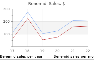
Benemid
Benemid dosages: 500 mg
Benemid packs: 60 pills, 90 pills

Buy cheap benemid 500mg
Conversely valley pain treatment center buy benemid 500mg without a prescription, inflammation of a tube (salpingitis) could result from infections that spread from the peritoneal cavity pain treatment for rheumatoid arthritis generic benemid 500 mg with visa. Accumulation of radiopaque fluid or the looks of gas bubbles within the pararectal fossae region of the peritoneal cavity signifies that the tubes are patent. Arrowheads, uterine tubes; c, catheter in the cervical canal, vs, vaginal speculum. Ectopic Tubal Pregnancy In some girls, collections of pus may develop in a uterine tube (pyosalpinx) and the tube could additionally be partly occluded by adhesions. Tubal pregnancy is the commonest kind of ectopic gestation; it happens in approximately 1 of every 250 pregnancies in North America (Moore et al. In both cases, the parietal peritoneum is inflamed in the identical basic space, and the pain is referred to the right decrease quadrant of the abdomen. The scenario is exacerbated within the presence of a disrupted perineal body or with atrophic ("relaxed") pelvic floor ligaments and muscles (see the blue box "Disruption of Perineal Body," p. The size and other traits of the uterus may be decided on this means. Because of the small dimension of the pelvic cavity during infancy, the uterus is especially an stomach organ. The cervix remains relatively massive (approximately 50% of complete uterus) all through childhood. All these phases characterize normal anatomy for the actual age and reproductive status of the girl. Because no peritoneum intervenes between the anterior cervix and the base of the bladder, cervical cancer may spread by contiguity to the bladder. Hysterectomy Owing to the frequency of uterine and cervical cancer, hysterectomy, excision of the uterus (G. Cervical Cancer, Cervical Examination and Pap Smear Until 1940, cervical cancer was the main cause of dying in North American women (Krebs, 2000). Lateral distension is proscribed by the ischial spines, which project posteromedially, and the sacrospinous ligaments extending from these spines to the lateral margins of the sacrum and coccyx. Urine enters the vagina from each vesicovaginal and urethrovaginal fistulas, however the move is steady from the former and happens solely throughout micturition from the latter. Digital Pelvic Examination Because of its comparatively skinny, distensible walls and central location inside the pelvis, the cervix, ischial spines, and sacral promontory can be palpated with the gloved digits within the vagina and/or rectum (manual pelvic examination). Chapter 3 � Pelvis and Perineum Forceps 397 Culdoscopy and Culdocentesis An endoscopic instrument (culdoscope) may be inserted by way of the posterior part of the vaginal fornix to examine the ovaries or uterine tubes. Although it involves less disruption of tissue, culdoscopy has been largely replaced by laparoscopy, which, nonetheless, supplies greater flexibility for operative procedures and better visualization of pelvic organs (see the blue field "Laparoscopic Examination of Pelvic Viscera," p. Anesthesia for Childbirth Laparoscopic Examination of Pelvic Viscera Visual examination of the pelvic viscera is particularly useful in diagnosing many conditions affecting the pelvic viscera, corresponding to ovarian cysts and tumors, endometriosis (the presence of functioning endometrial tissue outdoors the uterus), and ectopic pregnancies. Insufflation of inert gas creates a pneumoperitoneum to present house to visualize, and the pelvis is elevated so that gravity will pull the intestines into the stomach. The uterus can be externally manipulated to facilitate visualization, or extra openings (ports) can be made to introduce other instruments for manipulation or to allow therapeutic procedures. General anesthesia has advantages for emergency procedures and for women who select it over regional anesthesia. Clinicians monitor and regulate maternal respiration and each maternal and fetal cardiac operate. Childbirth occurs passively beneath the control of maternal hormones with the help of an obstetrician. Women who choose regional anesthesia, such as a spinal, pudendal nerve, or caudal epidural block, often want to participate actively. The anatomical basis of the administration of a pudendal block is provided in the blue field "Pudendal and Ilio-inguinal Nerve Blocks," p. The anesthetic agent circulates into the cerebral subarachnoid area within the cranial cavity when the patient lies flat following the delivery. Pudendal nerve Key Intraperitoneal viscera Subperitoneal viscera Somatic structures Chapter 3 � Pelvis and Perineum 399 anesthesia bathes the S2�S4 spinal nerve roots, including the ache fibers from the uterine cervix and superior vagina, and the afferent fibers from the pudendal nerve. � Coursing in a peritoneal fold (mesosalpinx) that makes up the superior margin of the broad ligament, each uterine tube has a fimbriated, funnel-like infundibulum, a wide ampulla, a narrow isthmus, and a short uterine half that traverses the uterine wall to enter the cavity. The ovaries and uterine tubes receive a double (collateral) blood supply from the belly aorta by way of the ovarian arteries and from the inner iliac arteries via the uterine arteries. � this collateral circulation permits the ovaries to be spared to provide estrogen when a hysterectomy necessitates ligation of the uterine arteries. Uterus: Shaped like an inverted pear, the uterus is the organ by which the blastocyst (early embryo) implants and develops into a mature embryo and then a fetus. The uterus is normally anteverted and anteflexed so that its weight is borne largely by the urinary bladder, although it also receives vital passive help from the cardinal ligaments and active help from the muscular tissues of the pelvic flooring. Vagina: the vagina is a musculomembranous passage connecting the uterine cavity to the outside, permitting the entrance/insertion of the penis, ejaculate, tampons, or analyzing digits and the exit of a fetus or menstrual fluid. The vagina is indented (invaginated) anterosuperiorly by the uterine cervix in order that an encircling pocket or vaginal fornix is shaped around it. � the vagina is capable of exceptional distension, enabling handbook examination (palpation) of pelvic landmarks and viscera (especially the ovaries) in addition to of pathology. Innervation of uterus and vagina: the inferiormost (perineal) portion of the vagina receives somatic innervation by way of the pudendal nerve (S2�S4) and is, subsequently, sensitive to touch and temperature. Lymphatic vessels from the superolateral elements of the bladder pass to the exterior iliac lymph nodes, whereas these from the fundus and neck cross to the internal iliac lymph nodes. However, a number of vessels from the female urethra may drain into the sacral nodes and, from the distal female urethra, to the inguinal lymph nodes. Lymphatic vessels from the inferior half of the rectum drain on to sacral lymph nodes or, especially from the distal ampulla, comply with the center rectal vessels to drain into the interior iliac lymph nodes. Boundaries and floor options of the perineal region with projections of the osseous boundaries and muscular tissues of the superficial muscle tissue of the perineum. The osseofibrous buildings bounding the pelvic outlet and perineum are identified. The airplane between the bladder and rectum is occupied by inner genitalia and a septum formed throughout embryonic development because the urogenital sinus was partitioned into the urinary bladder and urethra anteriorly and the anorectum posteriorly. In males, the fatty layer is tremendously diminished within the urogenital triangle, being changed altogether within the penis and scrotum with easy (dartos) muscle. The anal canal and its orifice, the anus, represent the most important deep and superficial features of the triangle, mendacity centrally surrounded by ischioanal fats. The subcutaneous tissue of the perineum, like that of the inferior anterior stomach wall consists of a superficial fatty layer and a deep membranous layer, the (superficial) perineal fascia (Colles fascia). The external urethral sphincter and deep transverse perineal muscle span the area of the urogenital hiatus, which is closed inferiorly by the perineal membrane extending between the ischiopubic rami. This coronal part of the male urogenital triangle is within the plane of the prostatic urethra. This coronal section demonstrates the subcutaneous tissue of the proximal penis and scrotum.
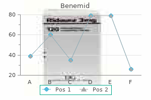
Cheap benemid 500mg with mastercard
The investigators additionally discovered a barely decrease prevalence in the Chinese inhabitants when in comparability with wrist pain treatment stretches cheap benemid 500mg mastercard Caucasians unifour pain treatment center nc 500 mg benemid cheap with amex. However, it does happen sparingly within the second and third decade of life in developing lesions. It is thought that ruptured atherosclerotic plaques are adopted by calcification as a way of stabilizing the unstable plaque. It should be famous that only roughly 20% of the entire plaque quantity is calcified. The Agatston rating prevails because of the availability of the database for these scores that allows risk stratification. One of the limitations of the Agatston and other scoring strategies is that calcium scoring is conventionally carried out at 2. Calcium scoring could underestimate the amount of macroscopically seen plaque and fail to detect such plaque in some instances, which can have implications for danger stratification. This methodology is thought to have resolved the difficulty of slice thickness, though portability of this method is affected in a way just like that of the Agatston methodology. For example, if two lesions of equal area differ in calcium content material, extra area above the nominal threshold shall be detected in the higher content material lesion. Additionally, linear interpolation will unfold the sign of the higher content lesion contained inside one slice to other slices, thereby rising the quantity artificially. Mass measurements are computed on the basis of the sum of the integrals of all pixels above a given threshold, multiplied by the voxel volume in mm3, and factored to an acceptable scale. The computed worth expresses the entire mineral content unbiased of slice thickness and spatial decision, however only within the absence of noise. Threshold settings that maximize constructive predictive worth (minimize false-positive pixels) alter the measurement itself. Nevertheless, this technique most intently approaches portability when the brink setting is appropriately low and the scaling issue is known. The Agatston method has stratified this patient as belonging to the 98th percentile inside his age group. Is coronary artery calcium the key to assessment of cardiovascular danger in asymptomatic adults Lesion- and vessel-specific coronary artery calcium scores are superior to whole-heart Agatston and quantity scores within the prognosis of obstructive coronary artery disease. A rosetta stone for coronary calcium threat stratification: Agatston, quantity, and mass scores in eleven,490 people. Numerous cardiac danger elements have been identified in epidemiological studies, similar to hypertension, diabetes, smoking, hypercholesterolemia, weight problems, sedentary life-style, and family historical past. However, thrombosis superimposed on a ruptured atherosclerotic plaque can trigger an acute coronary event that might be probably fatal (Table 44. Factors that promote plaque vulnerability and rupture include a skinny fibrous cap overlying lipid pools and a necrotic core, increased macrophage activity, intraplaque hemorrhage, and plaque progression. This preserves a close to normal cross-sectional lumen area until the lesion occupies roughly 40% of the internal elastic lamina space. Negative remodeling is defined as a compensatory lower in the size of exterior elastic lamina. According to one study, patients with clinically unstable coronary lesions had extra optimistic transforming, whereas sufferers with secure lesions had more adverse reworking. Complex lesion Fibrous cap could also be ruptured and probably ulcerated Fibromuscular tissue deposition may be seen with repair efforts following repeated plaque rupture May slowly grow in measurement and produce important arterial narrowing macrophages and metalloproteinases which would possibly be liable for arterial reworking. Negative reworking is assumed to be related to fibrotic changes of the arterial wall, which stabilize the plaque, making it less susceptible to rupture. Calcium deposition may be detected by noninvasive methods in both vulnerable and sophisticated plaques, but classification of the histological sort of the detected plaque has but to be fully elucidated. Moreover, it should be famous that extensively calcified plaques ("hard plaques") are much less more doubtless to be related to acute coronary syndromes. Mineral deposition renders these plaques extra stable by uniformly including stress resistance. Conversely, delicate to average amounts of calcification destabilize plaques through a nonuniform stiffening impact, making them more susceptible to shear stress. Cone-down view of a frontal chest radiograph demonstrates a heavily calcified left main coronary artery (arrows). High sensitivity and specificity, constructive predictive value, and unfavorable predictive value of this modality within the detection of coronary stenoses in comparability to invasive angiography have been reported in a quantity of studies. In addition, the increased protection of 320-slice scanners eliminates oversampling and stair-step artifact on volumetric reconstruction by enabling acquisition of the entire heart in a single heartbeat/gantry rotation without heart-rate restrictions. Of specific scientific curiosity, noninvasive identification of probably vulnerable plaques is typically potential through the quantification of lesion attenuation values. However, characterization of plaque morphology and composition is imprecise, and its scientific value is unknown. A small ultrasound transducer mounted to a coronary angioplasty catheter is advanced by an endovascular route past the distal plaque segment of curiosity. A series of 360-degree cross-sectional images are obtained because the catheter is drawn proximally. Noninvasive evaluation of plaque morphology and composition in wrongdoer and secure lesions in acute coronary syndrome and stable lesions in secure angina by multidetector computed tomography. Evaluation of coronary artery calcification by multi-detector row computed tomography for the detection of coronary artery stenosis in Japanese sufferers. Role of electron-beam computed tomography and nuclear stress testing in cardiovascular danger assessment. Arterial remodeling and coronary artery illness: the concept of "dilated" versus "obstructive" coronary atherosclerosis. However, stenosis higher than 70% results in 41% survival over the same time span. The distal portion of the artery (the space of bifurcation into the left anterior descending and the left circumflex) is mostly involved in atherosclerosis growth due to low shear stress in that area. Left main equal illness is defined as a larger than 70% narrowing of both the left anterior descending and the left circumflex coronary arteries. These atherosclerotic lesions are usually advanced plaques, with characteristic internal surface ruptures, fissures, and hemorrhages. More advanced scanners may have dual-source know-how and wide z-coverage and allow for potential triggering and iterative reconstruction, thereby decreasing radiation publicity. Small vessel caliber and cardiac movement can result in suboptimal picture high quality and diagnostic accuracy in the evaluation of luminal stenosis as in contrast with that with typical angiography. Comparison could be made by acquiring photographs at relaxation and under adenosine-induced stress.

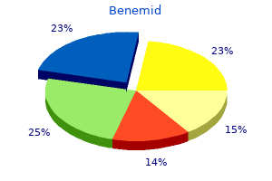
Benemid 500 mg buy lowest price
When the site of bleeding is identified throughout colonoscopy pain treatment machine purchase 500mg benemid mastercard, it might enable for therapeutic interventions to stop bleeding back pain treatment usa benemid 500 mg purchase line, however is unsuccess ful in 20% of cases. Mortality is greater in aged sufferers with comorbidities, and these features ought to immediate con sideration for admission to an intensive care setting. Uncross matched type 0 blood is ordered for patients with unstable vital indicators and important blood loss. Octreotide is useful in reducing the speed of bleeding, the incidence of rebleeding, and mortality by lowering portal hypertension. It is especially helpful in variceal bleeding, but can also cut back bleeding from nonvariceal sources. Surgical intervention may be required in sufferers with uncontrolled hemorrhage, perforation, or patients with liver disease and portal hypertension. Young steady patients with normal hemoglobin, no lively bleeding, evidence of hemorrhoids or fissures as a possible supply, and no evidence of portal hypertension, coagulopathy, or different significant comor bidities could additionally be discharged with close follow-up. Outpatient administration of sufferers with low-risk upper-gastrointestinal haemor rhage: Multicentre validation and prospective evaluation. H i ckey, M D Key Points � � Intesti nal obstruction presents with acute abdom inal pain, belly distension, and vomiting. Abdominal radiog raphs can demonstrate obstruction, however computed tomography is more sensitive. Strangulation is a compl ication of obstruction that may lead to bowel ischemia, peritonitis, and sepsis. Complete obstructions carry extra r isk of morbidity and may end up in strangulation. As bowel contents are prevented from forward circulate, elevated secretions result in overdistention, which causes bowel wall edema and lowered lymphatic and venous outflow. This is referred to as strangulation and can progress to bowel ischemia, necrosis, perforation, and peritonitis. Up to 40% of small bowel obstructions become strangulated, most commonly from volvulus, adhesions, and hernias. This ends in very excessive risk of strangulation as a result of bowel contents are prevented from each forward and retrograde move. Small bowel obstructions characterize lSo/o of hospital admissions for acute stomach ache. Approximately 300,000 operations are performed yearly within the United States for obstruction. Mortality fee general is approxi mately So/o, whereas the mortality fee from strangulated obstructions approaches 30%. In distinction to mechanical obstruction, practical obstruction (eg, adynamic ileus) occurs when intestinal contents fail to move because of disturbances in gut motility. It mostly happens instantly after surgery, however can also be seen in inflammatory conditions, electrolyte abnormalities, and from certain medicines (namely, narcotics). Unless famous in any other case, the remainder of this chapter refers to mechanical obstruction. If the obstruction is proximal, the affected person can also complain of nausea and vomiting. Fever, tachycardia, and hypotension are ominous indicators and will suggest peri tonitis or sepsis. Physical exam is significant for a distended, diffusely tender stomach, tympany to percussion, and hyperactive bowel sounds. Patients ought to be examined for evidence of prior abdominal sur geries (eg, incision scars) and examined for hernias. Third spacing of fluid and dehydration from vomiting may cause elevated blood urea nitrogen or creatinine. I ntestinal isch emia may cause an anion hole metabolic acidosis with an elevated lactic acid. Leukocytosis could additionally be current on a complete blood rely and in addition suggests ischemia or peri tonitis. An "obstructive sequence" classically consists of three radiographs: upright chest movie, supine belly film, and upright stomach movie. The upright chest movie is used to consider for proof of perforation (free air under the diaphragm). It represents a predominance of fluid in the bowel lumen with small quantities of air trapped between the valvulae conniventes of the bowel. I n adynamic ileus, radiographs will reveal dilation of the bowel without air-fluid levels. Determine which nostril is less congested by having the patient blow the nostril on either side. I nject viscous lidocaine into the nostril or alternatively spray benzo caine into the nostril and mouth. Check the placement ofthe tube by inserting 60 mL of air and listening over the abdomen f gurgling. Aspiration or of abdomen contents will also indicate that the t ube is in the correct location. Do not rule out obstruction based on the presence of flatus or bowel actions or the lack of vom iting, a s these findings may develop later. This results in decompression of the bowel lumen, offers symptomatic relief, and should avoid the necessity for surgical procedure. Broad-spectrum antibiotics that cowl gram-negative and anaerobic organisms (eg, piperacillin-tazobactam, ciprofloxacin plus metronida zole) ought to be given in the presence of fever, peritonitis, or evidence of strangulation. Surgical consultation should be obtained in case the affected person requires surgical interven tion. For sufferers with adynamic ileus, remedy involves cessation of any narcotic medicines and initiation of motility brokers (eg, metoclopramide). Urgent surgery is required in patients with peritoni tis, perforation, or strangulation. Acute mechanical bowel obstruction: clinical presentation, etiology, handle ment and outcome. Admission All patients with intestinal obstruction require admission, both to a surgical service or a drugs service with a surgeon on seek the guidance of. Intensive care unit admission is indicated for patients with unstable important signs (tachycardia, hypotension) or Mesente ric Ische mia Ross A. Over time, the hypoxemia ends in tissue break down with lack of bowel integrity. Delay in diag nosis is common, however with reviews that early intervention increases survival price, it is necessary to always have this prognosis within the differential for aged sufferers presenting with stomach pain. Four etiologies of mesenteric ischemia are described, and every has completely different r isk components and variation in presen tation. Arterial thrombosis at the narrowing of mesenteric arteries in patients with atherosclerosis is answerable for 20% of acute shows.

Benemid 500 mg discount online
Two pulmonary veins pain after zoom treatment benemid 500 mg safe, a superior and an inferior pulmonary vein on all sides treatment pain behind knee cheap benemid 500mg otc, carry oxygen-rich ("arterial") blood from corresponding lobes of each lung to the left atrium of the heart. Each terminal bronchiole offers rise to several generations of respiratory bronchioles, characterized by scattered, thin-walled outpocketings (alveoli) that reach from their lumens. Due to the presence of the alveoli, the respiratory bronchioles are concerned each in air transportation and gasoline trade. Alveolar ducts are elongated airways densely lined with alveoli, resulting in frequent spaces, the alveolar sacs, into which clusters of alveoli open. New alveoli continue to develop till about age eight years, by which era there are roughly 300 million alveoli. Note that the proper pulmonary artery passes underneath the arch of the aorta to reach the right lung and that the left pulmonary artery lies completely to the left of the arch. Except within the central, perihilar region of the lung, the veins from the visceral pleura and the bronchial venous circulation drain into the pulmonary veins, the relatively small quantity of low-oxygen blood getting into the large quantity of oxygen-rich blood returning to the heart. The single proper bronchial artery may also arise directly from the aorta; however it generally arises indirectly, either by means of the proximal a half of one of the higher posterior intercostal arteries (usually the best third posterior intercostal artery), or from a common trunk with the left superior bronchial artery. Then they usually pass alongside the posterior aspects of the principle bronchi, supplying them and their branches as far distally because the respiratory bronchioles. The deep bronchopulmonary lymphatic plexus is positioned in the submucosa of the bronchi and within the peribronchial connective tissue. It is largely involved with draining the constructions that type the foundation of the lung. Lymphatic vessels from these nodes continue to comply with the bronchi and pulmonary vessels to the hilum of the lung, where in addition they drain into the bronchopulmonary lymph nodes. From them, lymph from each the superficial and deep lymphatic plexuses drains to the superior and inferior tracheobronchial lymph nodes, superior and inferior to the bifurcation of the trachea and main bronchi, respectively. Many, but not all, of the lymphatics from the lower lobe of the left lung, nonetheless, drain to the proper superior tracheobronchial nodes; the lymph then continues to comply with the right-side pathway. The lymphatic vessels originate from superficial subpleural and deep lymphatic plexuses. All lymph from the lung leaves along the basis of the lung and drains to the inferior or superior tracheobronchial lymph nodes. From right here the lymph traverses a variable variety of paratracheal nodes and enters the bronchomediastinal trunks. Lymph from the parietal pleura drains into the lymph nodes of the thoracic wall (intercostal, parasternal, mediastinal, and phrenic). These nerve networks include parasympathetic, sympathetic, and visceral afferent fibers. Their cell our bodies (sympathetic ganglion cells) are within the paravertebral sympathetic ganglia of the sympathetic trunks. After contributing to the posterior pulmonary plexus, the vagus nerves continue inferiorly and turn out to be part of the esophageal plexus, typically dropping their identity after which reforming as anterior and posterior vagal trunks. Branches of the pulmonary plexuses accompany pulmonary arteries and particularly bronchi to and throughout the lungs. Thus the parietal pleura usually extends roughly two ribs inferior to the lung. The anterior borders of the lungs lie adjacent to the anterior line of reflection of the parietal pleura between the 2nd and 4th costal cartilages. Here, the margin of the left pleural reflection strikes laterally after which inferiorly on the cardiac notch to attain the sixth costal cartilage. The anterior border of the left lung is more deeply indented by its cardiac notch. On the right facet, the pleural reflection continues inferiorly from the 4th to the 6th costal cartilage, paralleled intently by the anterior border of the proper lung. Consequently, the cervical pleura is particularly weak to harm during infancy and early childhood. Normal lungs in situ remain distended even when the airway passages are open because the outer surfaces of the lungs (visceral pleura) adhere to the internal floor of the thoracic partitions (parietal pleura) as a outcome of the surface pressure offered by the pleural fluid. The elastic recoil Bullet punctures thoracic wall and parietal pleura, admitting air and causing lung to collapse. Chapter 1 � Thorax 121 of the lungs causes the stress in the pleural cavities to be sub-atmospheric. The stress is usually about -2 mm Hg; during inspiration, it drops to about -8 mm Hg. Laceration or rupture of the floor of a lung (and its visceral pleura) or penetration of the thoracic wall (and its parietal pleura) results in hemorrhage and the doorway of air into the pleural cavity. In open-chest surgical procedure, respiration and lung inflation should be maintained by intubating the trachea with a cuffed tube and utilizing a positive-pressure pump, various the strain to alternately inflate and deflate the lungs. The needle passes by way of the intercostal muscular tissues and costal parietal pleura into the pleural cavity. The needle must be angled upward, to keep away from penetrating the deep facet of the recess (a skinny layer of diaphragmatic parietal pleura and diaphragm overlying the liver). The inflamed surfaces of pleura can also cause the parietal and visceral layers of pleura to adhere (pleural adhesion). A brief incision is made in the 5th or sixth intercostal area in the midaxillary line (which is roughly at nipple level). In different procedures, adherence of the parietal and visceral layers of pleura is induced by overlaying the apposing layers of pleura with an irritating powder or sclerosing agent (pleurodesis). The indirect and horizontal fissures could also be incomplete or absent in some specimens, with consequent reductions in the number or distinctiveness of lobes. Consequently, the left lung generally has three lobes and the proper lung solely two. The commonest "accent" lobe is the azygos lobe, which appears in the proper lung in approximately 1% of people. Appearance of Lungs and Inhalation of Carbon Particles and Irritants the lungs are light pink in wholesome kids and people who discover themselves non-smokers and stay in a clear surroundings. However the lungs are capable of accumulating a considerable amount of carbon without being adversely affected. To preserve a extra sterile surroundings and avoid aspiration of overseas objects, some dentists insert a thin rubber dam into the oral cavity earlier than performing sure procedures. If the tracheobronchial lymph nodes within the angle between the principle bronchi are enlarged as a result of cancer cells have metastasized from a bronchogenic carcinoma, for instance, the carina is distorted, widened posteriorly, and motionless. Awareness of these segments can be important for surgical resection of diseased segments. An embolus in a pulmonary artery forms when a blood clot, fats globule, or air bubble travels in the blood to the lungs from a leg vein, for instance, after a compound fracture. Lymphatic Drainage and Pleural Adhesion If the parietal and visceral layers of pleura adhere (pleural adhesion), the lymphatic vessels in the lung and visceral pleura may anastomose (join) with parietal lymphatic vessels that drain into the axillary lymph nodes.
Benemid 500mg discount with visa
In this process pain treatment with heat 500 mg benemid order with visa, carried out during the first few days of life pain management for older dogs buy benemid 500 mg cheap, the aorta and major pulmonary artery are divided near their roots. The aorta is reimplanted into the left ventricle, and the main pulmonary artery is reimplanted into the proper ventricle. Because of D-looping, the ventricles are in normal position with the right ventricle to the right of the left ventricle. In the pulmonary circuit, oxygenated blood flows from the pulmonary veins into the left atrium, through the mitral valve, into the left ventricle, and at last out by way of the pulmonary artery. Blood then returns to the pulmonary veins, with out passing thought the systemic circuit. The pulmonary artery is ligated proximally and an extracardiac conduit is then constructed immediately from the right ventricle to the pulmonary artery. The conduit transmits deoxygenated blood from the proper ventricle to the pulmonary arteries. The disadvantage of this repair is that the conduit could require reoperation as the patient grows, to place a bigger conduit or to address conduit stenosis. The coronary arteries also must be excised from above the proper ventricle and reimplanted within the neoaorta originating from the left ventricle. Long-term data from the arterial change How to Approach the Image Much of the preoperative analysis of this disorder happens with echocardiography and, if essential, cardiac catheterization. Following a Jatene restore, the main pulmonary artery is situated anterior to the neoaortic root, with the proper and left main pulmonary arteries extending posteriorly "hugging" the neoaorta. The baffle separates the "neo-right atrium" (long thin arrow), which receive the systemic venous return, from the "neo-left atrium (thick arrow)," which receives the pulmonary venous return. After an arterial switch procedure, pulmonary arterial stenosis ought to be evaluated. There is stenosis of the left pulmonary artery (arrow), a known complication of this process. Quantitative move assessment of the proper and left pulmonary arteries demonstrated two-thirds of the pulmonary blood flow supplying the proper lung and one-third supplying the left lung. The neonatal targets of therapy are to hold shunts open to permit mixing between the parallel pulmonary and systemic circuits. This is completed medically with prostaglandin, to maintain the ductus arteriosus open, or interventionally with a balloon atrioseptostomy, known as the Rashkind procedure, which allows mixing on the atrial level. However, it is a momentary resolution and definitive surgical procedure must be undertaken; the arterial change operation is currently favored. The timing bolus exhibits early enhancement of the "neo-left atrial appendage" (arrow) earlier than pulmonary venous enhancement indicating a leak within the superior limb of the baffle. Some of these phrases have totally different shades of which means and ranges of precision, but they will be used interchangeably in this chapter. Key determinants of poor outcomes are tricuspid regurgitation and proper (systemic) ventricular failure. Anatomy and Physiology During the third week of embryonic growth, the heart tube loops, with the cranial end looping ventrally and caudally. In addition to the anatomic issues, there are additionally physiological considerations. How to Approach the Image During the neonatal interval and even later, echocardiography generally offers a whole evaluation of the cardiac chambers and great arteries. Still, there could additionally be loss of the aortic knob contour, narrowing of the mediastinum from the pulmonary artery and aorta being superimposed on the frontal projection, and mesocardia or dextrocardia. Common Variants Congenitally corrected transposition might exist in two types: S,L,L, the more widespread kind, where the atria are in situs solitus however the ventricles and great arteries are each inverted, and I,D,D, the place the atria are inverted and the ventricles and nice arteries are both inverted relative to these inverted atria. Note the parallel course of the pulmonary artery (black arrow) and the aorta (white arrow). Note the crossing sample of the aorta (white arrow) and the pulmonary artery (dotted black arrow), which crosses posterior to the aircraft of this picture. Before attempting surgical repair, coronary artery anatomy should be delineated to forestall catastrophic coronary artery harm. Cardiac venous anatomy, corresponding to absence of the coronary sinus, may occur and be clinically relevant when making an attempt to insert a pacing lead into an epicardial vein for biventricular pacing. A Mustard/Senning process can be utilized to reroute blood at the atrial stage, and a Jatene process can be used to reroute blood on the level of the nice arteries. The aorta is anterior and to the left of the pulmonary artery, best seen on axial photographs. The nomenclature, definition and classification of discordant atrioventricular connections. Congenitally corrected transposition of the great arteries presenting in a nonagenarian. Congenital Heart Surgery Nomenclature and Database Project: corrected (discordant) transposition of the great arteries (and related malformations). Burns and Hugo Spindola-Franco Definition Ebstein anomaly is congenital displacement of the posterior and septal leaflets of the tricuspid valve towards the right ventricular apex. More than 90% of neonates present with cyanosis secondary to an associated proper to left atrial level shunt or in the presence of pulmonary valve stenosis or atresia. Older adults with mild forms of Ebstein anomaly might current with arrhythmias or right coronary heart failure from long-standing tricuspid insufficiency. Supraventricular tachycardia and Wolff-Parkinson-White syndrome are commonly related to Ebstein anomaly. There is a strong affiliation of Ebstein anomaly with maternal oral lithium remedy during pregnancy. Severe displacement of the tricuspid valve leaflets presents early in the neonatal and childhood interval, whereas less severe displacement could be tolerated into adulthood. Neonates with extreme Ebstein anomaly and an associated right-to-left atrial degree shunt normally current on the primary day of life with cyanosis and a tricuspid insufficiency murmur. Older youngsters or young adults might current with Anatomy and Physiology the traditional tricuspid valve has three leaflets: septal, anterior, and posterior. The anterior portion of the septal leaflet inserts into the membranous septum, whereas the mid and posterior parts insert into the muscular septum. In Ebstein anomaly (b), the septal leaflet of the tricuspid valve is displaced toward the right ventricular apex. There is atrialization of a half of the best ventricle (white asterisk) with abnormal thinning of the affected portion of the septal wall. The black asterisk is within the remaining practical portion of the best ventricle distal to the insertions of the leaflet. The presence of noncompaction of the left ventricular apex and adjoining lateral wall is by the way famous. For this cause, the anterior tricuspid valve leaflet is commonly described as "sail-like. The vast majority of sufferers with Ebstein anomaly may have tricuspid insufficiency. Rarely, redundancy of the anterior leaflet could cause obstruction of the right ventricular outflow tract during systole, whereas an imperforate valve ends in tricuspid atresia. Displacement of the posterior and septal tricuspid leaflets toward the proper ventricular apex also results in atrialization of a half of the best ventricle.
Discount benemid 500 mg overnight delivery
Rupture of the vessel remains a rare complication as a outcome of neuropathic pain treatment guidelines benemid 500mg buy otc early detection back pain treatment youtube benemid 500mg with amex, however may end up in cardiac tamponade. Clinical Features Aneurysms are a common incidental discovering on coronary angiography. Patients with coronary artery aneurysms could be asymptomatic or current with angina, congestive heart failure, myocardial infarction, or sudden dying. In one examine, the vast majority of patients with coronary artery aneurysms also had vital coronary artery stenoses. The most typical etiology of coronary artery aneurysm formation in the United States is atherosclerosis, accounting for 50% of circumstances. Aneurysm formation is assumed to result from intimal plaque formation leading to an adjacent degenerated tunica media causing plaque rupture and wall dilatation. Other causes embody congenital aneurysms (20�30% of cases), connective tissue disease. A disadvantage of this modality is underestimation of aneurysm measurement when thrombus is present. However, its inferior spatial resolution and inability to reveal peripheral linear calcifications within the aneurysm makes it a less favorable choice. Differential Diagnosis Cardiac chamber aneurysm Post-traumatic pseudoaneurysm of the ascending aorta or pulmonary trunk Coronary artery fistula Cardiac or pericardial tumor Mediastinal mass. Medical treatment of coronary artery aneurysms involves the use of anticoagulants and antiplatelet brokers. Coronary artery aneurysms: a review of the natural history, pathophysiology, and management. Key Points Coronary artery aneurysm, or ectasia, is defined as a segment greater than 1. The overall annual incidence within the United States is 17 to 27 per one hundred,000 children youthful than 5 years of age. Other cardiac manifestations embody myocarditis with depressed cardiac function, coronary arteritis without aneurysm formation, valvulitis, and pericardial effusion. Echocardiography, nevertheless, is much less sensitive for detecting distal lesions and worsening coronary stenoses in arterial segments affected by aneurysms. Both methods are optimized with heart rates of lower than 70 beats per minute, which can necessitate the use of beta-blockers. Patients with aneurysms may endure periodic nuclear stress testing for inducible ischemia. As previously noted, no distinction is seen within and distal to the second aneurysm, signifying full vascular occlusion. However, contrast is seen distal to the third aneurysm, probably owing to retrograde move from collateral vessels. Coronary artery aneurysms may be related to thrombosis or stenoses with resulting ischemia, arrhythmias, or sudden dying. Fate of coronary aneurysms in Kawasaki illness: serial coronary angiography and long-term follow-up study. Muta H, Ishii M, Egami K, et al: Early intravenous gamma-globulin therapy for Kawasaki illness: the nationwide surveys in Japan. Diagnosis, therapy, and long-term management of Kawasaki illness: a statement for well being professionals from the Committee on Rheumatic Fever, Endocarditis, and Kawasaki Disease, Council on Cardiovascular Disease in the Young, American Heart Association. Detection of coronary artery aneurysms, stenoses and occlusions by multislice spiral computed tomography in adolescents with kawasaki disease. Stunning could be the result of a single ischemic occasion, as in "acute gorgeous," or of multiple episodes of ischemia, termed "repetitive stunning. A second state of myocardial dysfunction, generally known as "hibernating myocardium," is the end result of chronically lowered resting perfusion, which can be reversed with restoration of sufficient perfusion. When hibernating myocardium is recognized in sufferers with poor left ventricular perform, revascularization has been demonstrated to markedly scale back morbidity and mortality. Therefore, clinicians must determine patients in whom revascularization can be expected to enhance cardiac function. Once myocardial dysfunction is identified, it have to be decided whether or not the area of dysfunctional myocardium is viable or scarred, as solely viable myocardium will profit from revascularization. Symptoms and indicators are these of ischemic cardiomyopathy and congestive coronary heart failure and embody the following: Anatomy, Physiology, and Pathophysiology In each surprised and hibernating myocardium, ischemia is the cause. It is the timing of the ischemia, however, that differentiates the pathophysiology. Stunned myocardium results from a single acute occasion or repetitive episodes of ischemia. Following these ischemic occasions, high intracellular calcium ranges cause myofilament desensitization and lysis. Reperfusion leads to generation of oxygen free radicals that disturbs ion pump and mitochondrial function. This leads to reworking of myocytes, together with dedifferentiation, lowered production of contractile proteins, lowered measurement of mitochondria, interstitial fibrosis, and glycogen accumulation. While normally perfused myocardium preferentially utilizes fatty acids for vitality, hibernating myocardium demonstrates comparatively elevated uptake of glucose, as glucose could be metabolized anaerobically. Dyspnea on exertion Exercise intolerance Angina Fluid retention Reduced ejection fraction/cardiac output Infarct Arrhythmia How to Approach the Image Identification of hibernating and viable myocardium by imaging is made possible by the physiological differences between well-perfused, poorly perfused, and infarcted and scarred myocardium. A variety of imaging modalities have been used to successfully differentiate hibernating and viable myocardium from infarcted and scarred myocardium. In the higher row, blood move imaging utilizing rubidium eighty two in short-axis view shows markedly decreased perfusion, extending from distal to basal slices in the apical, inferior, inferolateral, and septal regions of the left ventricle at relaxation. Reproduced with permission from Macmillan Publishers Ltd: Nature Clinical Practice Cardiovascular Medicine, 2008. In the first-pass/blood pool phase, regular and stunned myocardium is nicely perfused, whereas infarcted and scarred and hibernating and viable myocardium will show lack of perfusion. Because infarcted and scarred myocardium has an expanded extracellular space relative to viable and shocked myocardium, tracer will accumulate in infarcted tissue in the steady-state phase, leading to delayed contrast enhancement. Myocardium that demonstrates lack of each perfusion and metabolism is taken into account to have a flow�metabolism match, indicating infarcted and scarred myocardium. This necessitates identification of patients who will probably profit from revascularization or from medical administration. On the other hand, anterior and lateral infarctions are often related to improvement of ventricular dysrhythmias. The coronary artery circulation is divided right into a right-, left- or co-dominant circulation. Microscopic findings primarily comprise eosinophilic coagulative necrosis, which can later evolve into granulation tissue and a fibrous scar. Additionally, nuclear scintigraphy can detect a reduced or absent tracer uptake during stress and relaxation images.
500 mg benemid purchase
Physical Examination the bodily examination ought to focus on excluding other abdominal or thoracic causes of pain and figuring out the degree of pain in the proper higher quadrant fibromyalgia treatment guidelines american pain society purchase 500mg benemid. The Murphy sign is essentially the most particular bodily exam finding for cholecystitis and is described because the patient halting inspiration when the examiner is palpating deeply in the proper upper quadrant fibroid pain treatment relief buy cheap benemid 500 mg online. The examiner must also assess for costovertebral angle tenderness and right decrease quadrant tenderness. La boratory Studies It is hard to discriminate biliary colic from early cholecys titis, and laboratory evaluation is sort of at all times indicated. It is essential to think about different situations which will masquerade as gall bladder ache. Right lower lobe pneu monia can also present with right upper quadrant pain and vomiting. Patients with choledocholithiasis are often misdiagnosed as having pancreatitis or gastritis. In aged sufferers or those with coronary illness, it could be very important contemplate the risk of an inferior myocardial infarc tion. Patients who seem septic or with peritoneal signs could have perforation or ascending cholangitis. Surgical session is important when the analysis of acute cholecystitis is established. If there are indicators of an infection (leukocytosis, fever), then antimicrobial protection with a second-generation cephalosporin or quinolone with metronidazole is appro priate. If the patient presents with sepsis or is in danger to develop sepsis (elderly, immune system c ompromise, excessive danger presentation such as ascending cholangitis, emphyse matous cholecystitis), then broad-spectrum antibiotics should be began promptly. The antibiotic routine ought to cover both gram-positive and gram-negative organisms. Patients with sepsis or severe illness ought to be admitted to an intensive care unit setting. Admission must also be strongly considered for sufferers with persistent symptoms but without definitive proof of acute cholecystitis, as testing could be regular early in the course of the illness. Discharge Patients with biliary colic may be discharged house if their pain has resolved, testing is regular, they usually can tolerate oral fluids. They ought to be advised to return for persistent signs, more severe pain, or fever. The price of enlargement and danger of rupture are associated to rigidity on the wall of the aneurysm, which in flip is expounded to the diameter of the aneurysm and to the underneath lying stress. Rupture of aneurysms smaller than four em is uncommon, whereas the annual threat of rupture for aneurysms bigger than eight em has been estimated at 30-50%. It is a common explanation for sudden demise and is liable for 1-2% of all deaths in males older than sixty five years. The mortality price for elective open operative restore is 2-7%; current advances in endovascu lar technique have mitigated early morbidity and mortality. Rarely, patients with rupture can current with syncope alone or with nonspecific signs such as vomiting, diarrhea, or dizziness. Transient hypotension can also occur and could be erroneously attributed to a vaso vagal etiology. Abdominal examination could detect a pulsa tile mass, however this can be tough with small aneurysms or overweight sufferers and is subject to vital interobserver variability. I n addi tion to the aneurysm, ultrasound might reveal intraperito neal free fluid in circumstances of rupture. Other emergent causes of abdominal, again, and flank pain ought to be considered and evaluated concurrently. These sufferers could profit from early elective restore, relying on the scale of the aneurysm. Patients with by the way found asymptomatic aneu rysms ought to be referred for surveillance or elective repair. Consider smoking cessation counseling, beta blockers, antihyperlipid brokers, and low-dose aspirin, as applicable. B a ke r, M D Key Points � � � Aggressive resuscitative measures (intravenous entry, crysta lloid bol us, and blood products) a re needed in unsta ble patie nts with gastrointesti nal (G I) bleeding. Laboratory Complete blood count, electrolytes, renal function, and coagulation studies must be obtained. Blood merchandise should be ordered for patients with unstable very important indicators or important blood loss. Less common causes embody pseudomembranous colitis, infectious diar rhea, aortoenteric fistula, radiation colitis, mesenteric ischemia, and Meckel diverticulum. The presence of free air underneath the diaphragm is diagnostic of perforation and is a surgical emergency. History In most cases, patients will report hematemesis, coffee ground emesis, hematochezia, or melena. For hematemesis, you will need to determine whether or not blood was current initially or appeared after a quantity of episodes of vomiting. These patients may have a coagulopathy, making control of hemor rhage tougher. Patients with pep tic ulcer illness may report epigastric abdominal ache associated to consuming. Elderly sufferers with acute hemorrhage could initially current with syncope or near-syncope. The stomach may then be lavaged with 200-300 mL saline to see if the aspirate clears. Note that false negatives could occur with bleeding distal to the pylorus, and false positives could happen from nasal trauma. When abnor malities are current, therapy is frequently necessary earlier than obtaining a radical history. Tachycardia and hypotension indicate hypovolemic shock and require instant resuscitation. The abdomen should be totally examined, noting areas of tenderness or perito nitis. Examination should also elicit any evi dence of the stigmata of cirrhosis together with ascites, spider angioma, j aundice, or palmar erythema. In emergent cases, colonoscopy misses the analysis in 40% of circumstances due to poor bowel preparation. These patients regularly have other types of atherosclerosis similar to coronary artery disease. Mesenteric venous thrombosis, which may be related to peripheral deep vein thrombosis, accounts for 5-lOo/o of shows.