
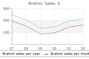
Brahmi
Brahmi dosages: 60 caps
Brahmi packs: 1 packs, 2 packs, 3 packs, 4 packs, 5 packs, 6 packs, 7 packs, 8 packs, 9 packs, 10 packs

Brahmi 60 caps low cost
The tumor is associated with fibrosis treatment gonorrhea generic brahmi 60 caps, hyalinization medicine 79 brahmi 60 caps generic mastercard, and irritation, however the patient received neoadjuvant chemotherapy previous to nephroureterectomy. Identifying this part confirms the urothelial origin of the sarcomatoid carcinoma. These tumors are rare however in the same morphologic spectrum of tumors which are seen within the bladder that might be encountered in the renal pelvis. Renal Cell Carcinoma: Unclassified Type Metastatic Squamous Cell Carcinoma of Kidney (Left) this tumor displays complicated cystic and papillary infiltrating morphology. Cut Section of Renal Pelvic Fibroepithelial Polyp Fibroepithelial Polyp of Renal Pelvis: Papillary Projections (Left) Fibroepithelial polyps are comparatively common benign lesions of the renal pelvis and ureter within the pediatric inhabitants, but most happen in adults. They predominantly consist of proliferation and growth of subepithelial stroma, often accompanied by prominent vascularity and inflammatory infiltrate. A outstanding basement membrane surrounding the epithelial nests is a characteristic function. Many lesions seem polypoid or papillary, raising the suspicion of a papillary neoplasm. Typically, the liner cells in nephrogenic metaplasia are cuboidal and single layered. Inverted Papilloma von Brunn Nest Hyperplasia (Left) Inverted papilloma may be very uncommon within the higher urinary tract. It reveals interconnecting cords/trabeculae invaginated into the lamina propria, with a flat surface urothelium. When florid, this finding may be confused with malignancy corresponding to nested variant of urothelial carcinoma. Villous Adenoma: Renal Pelvis Clear Cell Renal Cell Carcinoma Involving Renal Pelvis (Left) Villous adenoma is morphologically identical to its more common colorectal counterpart. This neoplasm is characterized by distinguished thin papillae, lined by dysplastic mucin-producing columnar epithelium. This tumor offered as a extra centrally located mass that obliterated the medullary portion of the kidney, concerned the renal sinus, and protruded into the renal pelvis. Small Cell/Neuroendocrine Carcinoma and Urothelial Carcinoma In Situ Schwannoma (Left) Benign mesenchymal tumors involving, or arising from, the pelvis are very uncommon. These embody schwannoma (as proven here), hemangioma, lipoma, myxoma, and leiomyoma, among others. For any spindle cell neoplasm of the pelvis, the potential for a sarcomatoid urothelial carcinoma must be excluded. Schwannoma: S100 Immunostain Inflammatory Myofibroblastic Tumor: Renal Pelvis (Left) Inflammatory myofibroblastic tumor of the renal pelvis is rare and morphologically similar to that occurring at other websites, including the bladder. Notice alternating loose and hypercellular areas, with extravasated purple blood cells, and scattered inflammatory cell infiltrate. Adenocarcinoma may be pure but usually seems in affiliation with a urothelial part. Squamous Cell Carcinoma: Renal Pelvis Urothelial Carcinoma: Renal Pelvis (Left) Squamous cell carcinoma, seen here invading the renal parenchyma, is the 2nd commonest carcinoma in the pelvis. Most instances happen as aberrant differentiation in urothelial carcinoma, however pure varieties could also be seen. This picture reveals a noninvasive micropapillary element in otherwise typical urothelial carcinoma. Sarcomatoid Carcinoma of Renal Pelvis: Panoramic View Sarcomatoid Carcinoma With Heterologous Elements (Chondrosarcoma) (Left) this whole-mount shows a big exophytic tumor mass, completely filling a markedly dilated pelvicalyceal system. Sarcomatoid carcinoma and sarcomas could present with large cumbersome and polypoidal intraluminal lots. In all sarcomatous tumors of the pelvis, the risk of sarcomatoid carcinoma must always be excluded. A patent urachus is a uncommon anomaly, showing a very open lumen connecting the umbilicus via the urachus to the bladder. Clinical Anomalies: Sinuses Histology: Incidental Urachal Remnant (Left) Incidental urachal remnant was encountered at cystectomy. The lumen may be solitary/central or tortuous or clustered as here, lined by urothelium with inspissated or calcified luminal secretions. Outside the periluminal, dense connective tissue are easy muscle bundles & adventitia. Lining could also be urothelial (shown) or present areas of squamous or glandular metaplasia. The perilesional stroma is chronically infected and fibrotic, as is characteristic. Busto Mart�n L et al: Urachal adenocarcinoma of the bladder, our expertise in 20 years. Urachal Adenocarcinoma, Not Otherwise Specified Urachal Adenocarcinoma: Mucinous Type (Left) Within the not otherwise specified variants of urachal adenocarcinoma are examples showing cribriform architecture that may mimic endometrioid-type endometrial adenocarcinoma. Correlation with clinical/imaging analysis is crucial to set up a urachal origin. Such cases might morphologically mimic metastases from a selection of anatomic places. This instance of a carcinoma arising in the urachus is a noninvasive, highgrade papillary urothelial carcinoma. The distinction from a urinary bladder primary is predicated completely on the anatomic location of the tumor. Generous sampling is recommended as higher invasion is designated as frank invasion. A urothelium with reactive atypia is at upper left, whereas urothelial carcinoma in situ is seen decrease proper. The differential consists of pseudosarcomatous stromal reaction and first bladder sarcoma. The nuclear positivity for p63 is most useful given its practically absent expression in mesenchymal lesions. Spindle Cell Lesions: Rhabdomyosarcoma Spindle Cell Lesions: Rhabdomyosarcoma (Left) A spindle cell sarcoma with fibrosarcomatous morphology from the bladder dome and urachus of a pediatric affected person is shown. MyoD1 confirmed a similar sample, while a smaller subset of cells additionally confirmed induction of desmin expression. Urothelial-Associated Markers Urothelial-Associated Markers (Left) S100p, or placental S100, is an rising marker utilized in urothelial histogenesis, optimistic in urothelial and pancreaticobiliary primaries. Urothelial-Associated Markers Uroplakins: Highly Specific (Left) Uroplakin-3 is exquisitely particular for urothelial neoplasms when scored for plaque-like membranous positivity. Helpfully, keratins and p63 ought to be adverse, while chromogranin and synaptophysin optimistic, with S100 optimistic sustentacular cells. Enteric-Type Adenocarcinoma Enteric-Type Adenocarcinoma (Left) A moderately differentiated enteric-type adenocarcinoma is seen within the bladder dome. In a bladder/urachal major, membranous accentuation with relative nuclear sparing (pictured) is anticipated. The remaining prostate is coronally sectioned at 3- to 5-mm intervals and submitted sequentially.
Diseases
- Shellfish poisoning, neurotoxic (NSP)
- Choroiditis, serpiginous
- Epiphyseal dysplasia hearing loss dysmorphism
- Proximal spinal muscular atrophy
- Hypomelanotic disorder
- Lymphangiomatosis, pulmonary
- Hand foot uterus syndrome
Brahmi 60 caps fast delivery
However medicine game 60 caps brahmi buy amex, the potential for a mucosaassociated lymphoid tissue lymphoma needs to medications 1 brahmi 60 caps generic online be excluded, and therefore immunohistochemical confirmation is a should. Therefore, morphologic as nicely as the expression of different immunohistochemical options are necessary for proper classification. Secondary malignant lymphomas of the kidneys are extra usually bilateral than main renal lymphomas. Plasmacytoma Plasmacytoma (Left) H&E exhibits unfastened sheets of neoplastic plasma cells from a case of a number of myeloma involving the kidney. Many cells are atypical, with distinguished nucleoli, a common characteristic in plasma cell neoplasms. Langerhans Cell Histiocytosis Langerhans Cell Histiocytosis (Left) this greater magnification view depicts the everyday nuclear options of Langerhans cell histiocytosis, including pale chromatin, marked nuclear angulations, and frequent grooves. Such lymphomas are extremely rare, and predominant involvement of glomeruli is reported. Involvement of many more extraglomerular lymphovascul ar channels than is identified on H&E sections is highlighted on immunohistochemistry. Posttransplant Lymphoproliferative Disorder Posttransplant Lymphoproliferative Disorder (Left) H&E shows posttransplant lymphoproliferative dysfunction, polymorphic type, involving the kidney. The infiltrate is polymorphous, however ample, massive atypical lymphoid cells are present. Cut surface is usually light tan to yellow, mostly homogeneous, but may be hemorrhagic and partially cystic. Tumor Circumscription Glomus Tumor-Like Features (Left) Juxtaglomerular cell tumors often present relative uniformity of nuclear dimension and form. Cells typically contain a moderate quantity of eosinophilic cytoplasm and cell membranes are normally distinguished. Kuroda N et al: Juxtaglomerular cell tumor: a morphological, immunohistochemical and genetic study of six cases. The cells are in small clusters and will raise the potential for a renal epithelial neoplasm. Cytoplasmic Eosinophilia Endocrine-Like Nuclear Atypia (Left) Focal, marked nuclear atypia could also be current in juxtaglomerular cell tumor. Some could present thinwalled edematous cystic foci as a outstanding sample in the tumor. Others might have antler-like vascularity, similar to that seen in solitary fibrous tumor/hemangiopericytoma. In this image, a papillary configuration is seen with a number of tubular constructions occupying the cores of the papillae. However, many tumors show plentiful mast cell infiltration, associated with lymphoplasmacytic infiltrates. This instance exhibits some cystic areas that on microscopy seem as cystically dilated entrapped tubules. An occasional case might compress the pelvicalyceal system and end in hydronephrosis. These are the most common renal tumors, and thorough sampling of the kidneys reveals an incidence of > 40%. This H&E image exhibits a wellcircumscribed renomedullary interstitial tumor imperceptibly merging with the encircling renal medullary tissue. Notice the adjacent renal medulla, during which virtually all of these tumors are located. Loose Myxoid Stroma Sclerotic Stroma (Left) Some renomedullary interstitial cell tumors present sclerotic stroma and comparatively scant spindle or stellate cells. However, the stroma is extra free and faintly basophilic, at least focally, generally. Keloid-Like Bands Cystic Entrapped Tubules (Left) Rarely, renomedullary interstitial cell tumors might show cystic dilatation of the entrapped tubules. Spindle/Stellate Cells 228 Renomedullary Interstitial Cell Tumor Kidney Tumors and Tumor-Like Conditions Myxoid Stroma Cellular Stroma (Left) In some renomedullary interstitial tumors, the stroma may be completely myxoid or basophilic. In many tumors, nevertheless, both the sclerotic and basophilic-type stroma are current in combination, in variable proportions. However, circumscription and different typical myxoid areas in the tumor exclude that possibility. Cellular Stroma Bland Cytology (Left) Some renomedullary interstitial cell tumors are fairly cellular. Glomus tumors of the kidney are quite uncommon, and at both the gross and microscopic ranges want distinction from juxtaglomerular cell tumor. This example reveals sheets of glomus cells within a background of compressed capillary framework. Glomus Tumor: Glomangioma Pattern Glomus Tumor: Glomangiomyoma Pattern (Left) Some glomus tumors are highly vascular, with dilated vascular channels and roundto-oval glomus cells dispersed within the stroma between the vessels. Actin-sm Laminin: Pericellular Staining (Left) Glomus tumors present diffuse immunoreactivity for actin-sm and common muscle actin. However, staining for desmin is negative, even in tumors with glomangiomyoma features. Leiomyoma, as depicted right here, could come up throughout the renal parenchyma, or from the renal capsule or partitions of hilar vessels. Most of the previously reported renal leiomyomas, particularly those arising from the capsule, presumably symbolize leiomyomatous angiomyolipomas. Most leiomyosarcomas involving the kidney come up within the retroperitoneal delicate tissue or renal vein, and due to marked compression and distortion of the kidney, are clinically thought to come up from it. Leiomyosarcoma 234 Other Rare Tumors Kidney Tumors and Tumor-Like Conditions Leiomyosarcoma: Necrosis Small Cell Carcinoma (Left) this picture of a leiomyosarcoma reveals a big space of tumor necrosis. Immunohistochemically, nearly all of tumors are unfavorable for cytokeratin and epithelial membrane antigen. Hemangioma Cavernous Hemangioma (Left) After angiomyolipoma and leiomyoma, hemangioma and lipoma are the other most frequent benign mesenchymal renal tumors, though both are extremely uncommon. Both capillary and cavernous hemangiomas are described, and most described circumstances have been of the cavernous type, as proven here. The diagnosis was revealed by sufficient sampling of perirenal gentle tissue that confirmed its well-differentiated liposarcomatous component. Angiosarcoma: Histological Features Angiosarcoma: Histological Features (Left) this major angiosarcoma of the kidney reveals an infiltrative tumor within the renal parenchyma. In this subject, the tumor is predominantly composed of nondiagnostic pleomorphic massive cells, necessitating immunostaining to verify the prognosis. Malignant Spindle Cell Tumor: Sarcomatoid Carcinoma Renal-Adrenal Fusion: Gross Features (Left) Adequate sampling and ancillary research. Most of those are sarcomatoid carcinomas, though rare circumstances might symbolize true sarcoma.
60 caps brahmi generic mastercard
The characteristic feature is the presence of prominent nucleoli with perinucleolar halos medications like zovirax and valtrex brahmi 60 caps buy generic on-line. It has virtually exclusive clear cell cytology and nuclei aligned in a linear fashion away from the basal facet medications during pregnancy chart brahmi 60 caps with visa. However, they often have clear cells, show excessive nuclear grade, and some cells with voluminous cytoplasm. They are often multifocal, with completely different tumors usually displaying variable histologies, and are incessantly associated with the presence of angiomyolipomas and cysts within the surrounding renal parenchyma. Microscopic Features Gross Features (Left) the brown minimize surface of this chromophobe renal cell carcinoma suggests prominence of cells with eosinophilic cytoplasm in this tumor. Perinuclear halos round irregular, raisinoid nuclei seen in this eosinophilic variant are the most diagnostic characteristic of chromophobe renal cell carcinoma. The apparent thick, plant-like cell wall appearance results from ample microvesicles pushing other organelles to the periphery. The clear cells are inclined to focus along the septa, and the eosinophilic cells are toward the center. In spite of cytologic differences, both the classic and eosinophilic variants share many cytogenetic options and, to a fantastic extent, immunophenotypic profiles. Microscopic Features Microscopic Features (Left) Architectural sample in some tumors is often totally acinar. Such structure is especially more widespread in tumors composed virtually solely of cells with eosinophilic cytoplasm (eosinophilic variants). Uncommon Microscopic Features Uncommon Microscopic Features (Left) Rhabdoid and sarcomatoid features could typically coexist in a tumor. While intuitively brisk mitotic rates could be expected as a marker of aggressive habits, none of the recent giant studies have been capable of verify this. Their predominant perinuclear location results in the sunshine microscopic perinuclear halos. Microvesicles additionally displace different organelles to the cell periphery, resulting in the apparently distinguished cell membranes. Cytology 96 Chromophobe Renal Cell Carcinoma Kidney Tumors and Tumor-Like Conditions Uncommon Microscopic Features Uncommon Microscopic Features (Left) this image shows an space with markedly pleomorphic, hyperchromatic, and polyploid nuclei. Biopsy Diagnosis Biopsy Diagnosis (Left) Classical and most circumstances of eosinophilic variants can easily be identified on needle core biopsies if the attribute morphological features are current. This multinodular tumor exhibits a mix of strong, tan, and hemorrhagic necrotic areas. The epithelial part in this tumor reveals a fine, sieve-like microcystic progress sample. In giant tumors, it could be difficult to confirm whether the tumor is centered in the renal medulla. Notice the outstanding desmoplastic stroma and lymphocytic response, each characteristic features of the the tumor. Such architecture makes distinction from invasive urothelial carcinoma difficult in some instances. The intervening fibrotic renal parenchyma in this image incorporates sclerotic glomeruli, in addition to atrophic renal tubules. Most tumors show perinephric or sinus fat invasion, or invasion into the venous system. Other frequent websites of metastasis at presentation embrace lungs and skeletal system. However, cribriform structure, and intratumoral neutrophils, as an alternative of lymphocytes, are extra widespread within the latter. However, these characteristically show very outstanding nucleoli with perinucleolar halos. The tumors, notably the smaller ones, are renal medulla based mostly, with infiltrative borders. Irrespective of the architectural patterns, stromal desmoplasia is a consistent discovering. Alvarez O et al: Renal medullary carcinoma and sickle cell trait: A systematic review. Intratumoral inflammatory infiltrate is typical and is often neutrophilic but typically may be predominantly lympho-plasmacytic. Papillary and micropapillary options are sometimes current focally, usually together with other architectural patterns. Cribriform structure with small lumina could sometimes give an impression of adenoid cystic carcinoma-like areas. This feature, along with the excessive nuclear grade and extensively infiltrative margins, are in maintaining with high metastatic potential for this tumor. The presence of crenated/sickled red cells in a metastatic focus, and in the surrounding tissues, might deliver to discover the presence of the hemoglobinopathy for the 1st time in some cases. Notice the retained reactivity within the stromal cells and lymphocytes, in addition to a benign renal tubule. Argani P et al: Xp11 translocation renal cell carcinoma in adults: expanded scientific, pathologic, and genetic spectrum. Argani P et al: Translocation carcinomas of the kidney after chemotherapy in childhood. The solid alveoli typically show central cell dyscohesion and morphologically mimic alveolar soft half sarcoma. The voluminous cytoplasm in some of the cells is a pointer toward translocationassociated carcinoma. Intraand inter-cellular hyaline globules are additionally sometimes seen, and these could act as nidus for psammomatous calcifications. Dispersed among the many bigger cells are clusters and islands of smaller cells with high nuclear/cytoplasmic ratio and considerably denser-appearing nuclear chromatin. The smaller cells are arranged round small nodules of hyaline materials ultrastructurally shown to be basement membrane material. The tumor entrance in a few of such tumors seems multi-nodular and could also be related to lymphoid aggregates. The cells lining the tubules are low columnar or cuboidal, with uniform, lowgrade, spherical nuclei. While the tubules show some branching typically, in some mucinous tubular and spindle cell carcinomas this branching is exquisite and could also be associated with marked interconnecting tubular architecture. Microscopic Features Microscopic Features (Left) the banal-appearing spindle cells seem to be intimately admixed with the tubular part. The juxtaposition or the imperceptible mixing of these 2 options is typical in mucinous tubular and spindle cell carcinoma. Rarely, foci with more plentiful and clear cytoplasm may be observed, and such cells are often carefully related to the other extra typical cell varieties. Microscopic Features Microscopic Features (Left) Some mucinous tubular and spindle cell carcinomas present marked paucity of epithelial part. Such spindle cell predominant tumors may be mistaken for a sarcoma, but the cytologic options are low grade, necrosis is absent, and a few epithelial element is invariably seen on cautious search. Typical histology in the the rest of the tumor ought to point in course of the true analysis.
Generic brahmi 60 caps amex
Drug-induced or pill esophagitis could be seen in younger or older grownup patients (see Practice Note 5-1) symptoms pink eye purchase brahmi 60 caps with amex. Typically symptoms and diagnosis order brahmi 60 caps on-line, commonly administered medicines that are bigger in measurement (tetracycline, potassium, quinidine) turn into lodged at the stage of the aortic arch and dissolve, inflicting irritation and stricture. Symptoms of chest ache, odynophagia, heartburn, and dysphagia could additionally be present, normally extra acutely in youthful patients. The stricture usually is smooth and progressively tapering, with a symmetric lumen that follows the anticipated path of the conventional esophagus. The lateral view exhibits a long, tapered look of a stricture within the esophagus. Proper administration requires each therapy of the underlying irritation and dilation of the stricture. Although reflux is the most typical cause of esophagitis, different possibilities should be considered, especially in patients with atypical histories, an uncommon distribution of inflammation, or failure to reply to reflux remedy. However, the stricture could additionally be relatively unyielding and require stiffer dilator techniques. Even when ongoing inflammation completely ceases, periodic dilatation could additionally be necessary, especially in the course of the first 12 months after preliminary remedy, when maturation of the fibrotic reaction continues on the expense of luminal diameter. Malignant Stricture Although benign tumors may come up from the esophagus, the majority of clinically significant tumors of the esophagus are malignant. In the previous, most esophageal malignancies have been squamous cell carcinomas, although current research recommend a dramatic improve in adenocarcinoma of the distal esophagus. As with different kinds of stenotic lesions, dysphagia initially occurs for solids only. However, it usually progresses rapidly, with dysphagia for delicate foods and even liquids creating within a few months of the onset of signs. Radiographically, esophageal malignancies appear as strictures of variable length. By the time of presentation, the cancerous tumor or area is normally many centimeters long and entails the complete circumference of the esophageal lumen, producing a stricture. However, not all esophageal cancers are clearly malignant on barium radiography, and occasional malignant-looking strictures could also be benign. Curative remedy is primarily surgical, though apparent cures by radiotherapy have been reported. Unfortunately, by the point signs develop, the most cancers is usually very advanced and incurable. Among these sufferers, survival was improved fourfold over charges reported for surgical procedure alone and twofold over those with proof of residual tumor at surgical resection. Characteristics distinguishing it from a benign stricture embody the sharp, shelflike proximal margin and the extra irregular configuration of the stenotic segment. Unlike some malignant strictures, this stricture follows the anticipated path of the esophageal lumen. Each of these approaches is directed at opening the esophageal lumen to allow eating, in recognition that the most important explanation for early death in patients with esophageal cancer is malnutrition and aspiration pneumonia. Dilatation typically offers limited and short-lived relief however is beneficial in preparing for other forms of remedy. The selection between other modalities depends on particular features of the tumor and native technical expertise and resources. Endoscopic laser therapy and bipolar electrocautery can be utilized to destroy tumor tissue that blocks the esophageal lumen; this will present a quantity of months of aid, allowing continuing oral intake. The stent permits ingestion of a modified food plan, concentrating on delicate, simply chewed meals and purees. The use of stents for palliation has decreased dramatically since the development of thermal strategies of remedy. However, stents proceed to be useful in certain situations, particularly within the presence of a tracheoesophageal fistula that always complicates the natural historical past or remedy of esophageal cancer. In this situation, a properly placed stent can maintain the esophageal lumen while overlaying the opening to the airway. The recent introduction of expandable steel stents has made insertion easier and offers a bigger inside luminal diameter, allowing patients to eat a less-restrictive food plan. Although endoscopic treatment with laser, bipolar electrocautery, or stent placement may be highly successful in reestablishing luminal patency, a substantial proportion of patients with esophageal most cancers have poor appetites and are unable to acquire weight. Luminal Deformities Extrinsic Compression Some diploma of luminal deformity caused by extrinsic compression by normal mediastinal structures (the aortic knob, the left mainstem bronchus, and the left atrium of the heart) is generally seen on barium studies and rarely, if ever, causes symptoms. More pronounced compression can happen with mediastinal situations, such as aortic aneurysm, cardiomegaly, congenital abnormalities of the massive mediastinal arteries. Video 5-4 on the Evolve web site shows a patient with cardiomegaly and lowered bolus circulate. The enlarged coronary heart is seen as a large shadow (note heartbeat) in the center of the video picture. The elasticity of the contralateral esophageal wall usually tends to reduce symptoms until compression is way superior. Dilatation is often ineffective because the drive of dilatation is absorbed by the elastic, uninvolved wall. Effective therapy, when essential, requires shrinking or removing the mass producing the compression. Unfortunately, this is usually not practical in patients in whom compression produces important symptoms. When symptoms do happen, they include dysphagia for liquids and solids, regurgitation of previously swallowed food again into the mouth, or both. Most often, esophageal diverticula are a consequence of obstruction distal to the region of bolus collection. Increased strain in the esophagus leads to bulging at a point of relative weak point. Less commonly, diverticula can result from periesophageal irritation, which causes traction on the esophageal wall (traction diverticulum). Although most traction diverticula happen within the midesophagus, most midesophageal diverticula, like their distal esophageal counterparts, are caused by pulsion. Video 5-5 reveals a diverticulum that fills and causes a momentary obstruction to bolus flow. Treatment of pulsion-type diverticula is necessary only if a diverticulum is symptomatic. Because they frequently give rise to motor or structural issues, it is essential to look for pulsion-type abnormalities as causes for the development of the diverticulum. It may be tough to distinguish between the underlying obstructive disorder and the diverticulum as a reason for signs. It is appropriate to attempt to deal with the underlying explanation for elevated pressure with dilatation within the case of structural obstruction or with medicine for dysmotility. In some patients signs initially believed to be a consequence of the diverticulum enhance considerably or resolve totally with such conservative remedy. Surgery limited to diverticulectomy, nevertheless, is associated with a high incidence of early anastomotic leakage or late recurrence, in all probability because it fails to take care of the underlying reason for increased intraesophageal pressure and creates an space of relative esophageal wall weak spot.
Wild Celandine (Jewelweed). Brahmi.
- Mild digestive disorders, rash from poison ivy, and other conditions.
- Are there safety concerns?
- What is Jewelweed?
- How does Jewelweed work?
- Dosing considerations for Jewelweed.
Source: http://www.rxlist.com/script/main/art.asp?articlekey=96535
Brahmi 60 caps fast delivery
The causes for scoring <100% are prone to medications used to treat depression buy 60 caps brahmi visa be numerous and should embody an incomplete representation and measurement of the relevant cardiac biology walmart 9 medications 60 caps brahmi buy mastercard, differences due to the temperature used during information technology, binding kinetics of medication to channels, pharmacokinetic properties, and uncertainty in the experimental measurements. The ensemble approaches have since been utilized in increasing frequencies moving away from the concept of a single variant per single experimental remark to a mess of equally legitimate variants per observed measurement (Britton et al. Another issue impacting model accuracy is understanding tips on how to account for the role of kinases, Gproteincoupled receptors, adrenergic receptor stimulation, drug accumulation in cardiac tissues, and trafficking of ion channel proteins to the cell floor. While these approaches increase complexity in the fashions, different approaches have attempted to simplify the parameters by figuring out the mannequin features which might be most impactful in predicting a set of outcomes (Mistry et al. These extra empirical and pragmatic approaches supply an opportunity to mirror on the important thing biological mechanisms that drive the noticed cardiac effects, and provide one of the best probability for an early drug discovery virtual display (Kramer et al. Inclusion of the drug pharmacokinetic properties would moreover enable users the flexibility to contemplate the relevant drug publicity in the coronary heart. It is due to this fact important to have a technique that, in the context of drug discovery, permits technology of knowledge sets that account for as many endpoints as attainable and help in selecting molecules with the lowest likelihood of cardiac liabilities for development to human studies (Pollard et al. Therefore, the event of human derived in vitro models could help enhance human security predictivity. Furthermore, an increasing give attention to the Replacement, Refinement, and Reduction (3Rs) in relation to animal research supplies an additional incentive to investigate the appliance of a simplified in vitro humanized platform to prioritize compounds before enterprise animal research. The limited availability of adult human cardiac tissue/ cells from donors for investigation of illness mechanisms and cardiac effects of medicine stays a major challenge within the subject. Calcium transient measurements: the detection of Ca2+ transients is widely used in highthroughput screening assays in cells preloaded with a fluorescent Although preclinical security assessment approaches of some pharmaceutical corporations presently present adequate evaluation of druginduced cardiac threat, analysis is often advanced and requires integration of findings from a number of in vitro and in vivo assays using nonhuman animal species, which can not all translate properly to people. In many of the above assays, other parameters together with beat price, amplitude and slope of the upstroke, and incidence of arrhythmias could be additionally measured. However, the translational value of those parameters to in vivo or man nonetheless must be further investigated. The effectivity and reproducibility of the differentiation procedures used to generate these cells should be improved to allow costeffective production suitable for the pharmaceutical trade. Advantages of telemetry embody remote acquisition of highfidelity alerts for extended periods from acutely aware, unrestrained animals, where autonomic homeostatic reflexes and circadian rhythms remain intact, permitting the conduct of preclinical research beneath situations that intently resemble a scientific scenario. Once data are acquired, software packages enable automated extraction of particular parameters from the physiological alerts using refined algorithms. These advances not only increase scientific quality, but additionally they benefit animal welfare by refinement of experimental strategies and discount of using animals, according to the 3Rs. All of these potential arrhythmia danger markers may be readily addressed utilizing information acquired and analyzed utilizing telemetry methods and statistical methodologies. Nevertheless, use of telemetry to consider hemodynamics stays an important part of both efficacy and safety evaluations throughout drug discovery and development. The telemetric strategy to safety science is much broader than for cardiovascular functions alone, and some further opportunities on this field are talked about within the following. Some examples include: Gastrointestinal motility: Biotelemetry capsules ("good capsule"; wi-fi motility capsule) have been developed for 24h noninvasive measurement of gut intraluminal pressure and motility, as nicely as monitoring temperature and pH gradients in the gastrointestinal tract, which can be used to consider gastrointestinal dysfunction. Negative inotropic results may be contraindicated in sufferers with established cardiac insufficiency and will, in the worst case, drive them into acute heart failure. In this case, an imbalance between oxygen supply and demand results in the development of myocardial ischemia, notably within the deeper layers of the myocardium distal to the coronary stenosis. Thus, both positive and unfavorable modulation of myocardial contractility may be clinically related, and medicines with these actions should be detected to assess their potential limitations in particular affected person populations. As such, having preclinical assays to determine these activities early within the drug research and improvement process may be critically important. Output is modulated by each neuronal and humoral inputs that may modify the cardiac output through either modifications within the coronary heart fee or contractile pressure, generally both. There can additionally be a dependency of the myocardial contractile force on the speed, offering a further mechanism to finely tune the relationship between oxygen consumption and oxygen supply. It is subsequently not surprising that drug exposure may have an impact on myocardial contractility. Examples of those embrace sympathomimetics and adrenoreceptor blockers that either mimic or block, respectively, the endogenous sympathetic input to the heart. Other pharmacological mechanisms can also have substantial effects on myocardial contractility. Itraconazole, an antifungal medicine, is a good example of a compound that was found to have a considerable adverse inotropic impact on the center through a mechanism that has not yet been elucidated (Qu et al. Due to the recognition that the loading situations of the center, that means the strain within the ventricle prior to contraction or "preload" as nicely as the strain towards which the guts pump is making an attempt to expel blood or "afterload," the quantitative assessment of myocardial contractility is challenging. A further consideration is that the inotropic state of the guts is intrinsically linked to the speed of contraction, such that changes within the coronary heart rate must be thought-about when making such determinations. The presently favored approach to decide the myocardial contractile state was proposed some years ago by Suga et al. Plotting stress versus volume over time leads to a "pressure�volume loop" with which a strain and coronary heart rateindependent assessment of the inotropic state of the guts can be made. Despite its theoretical attractiveness, using this method for screening has not found much software in drug discovery and improvement because of its technical challenges and quite low throughput. For the early drug discovery process, in vitro approaches with a medium to excessive throughput would be preferable. Some of those objectives can be achieved with the use of isolated myocardial tissue from varied sources and isolated hearts from varied animal species and, extra recently, by way of the use of cells or tissues engineered from human stem cells. Each of these approaches offers advantages and drawbacks which are summarized within the following. Isolated atria, papillary muscles, trabeculae, or strips taken from papillary muscles or the ventricular wall have been used for this kind of examine (Toda, 1969; Brown and Erdmann, 1985; Brown et al. Isolated myocardial tissue must be maintained in a temperaturecontrolled physiological buffer system and suspended in an equipment that allows for both an isometric (contraction at fixed length) or isotonic (muscle shortening in opposition to a defined load) contraction of the muscle. Small quantities of the test article can be added to the buffer to take a look at for changes in the contractile performance of the muscle at completely different drug concentrations. The research require technical expertise, are quite low throughput, and require animals as the source of the tissue. One advantage of this approach is that the druginduced results can be assessed in the absence of secondary elements that will come into play in an in vivo system, such that direct results on myocardial contractility may be detected and immediately associated to the drug concentration. Novel approaches to the efficiency of such studies and their analysis have been suggested. For instance, the usage of a workloop evaluation seems to improve the predictivity of druginduced results seen in man (Gharanei et al. This strategy shares most of the characteristics of the isolated tissue approach, including the necessity for technical expertise to run the examine, low quantities of compound required, and the power to examine effects over a spread of drug concentrations. Contraction of cardiomyocyte monolayers or threedimensional cultures utilizing these cells has emerged as a novel approach to detect druginduced results on myocardial contractility (Ramade et al. Particularly, contractility and excitation�contraction coupling are very complex physiological processes that depend upon each subcellular microstructures and their adequate physiological operate. A perfect match of structure and performance is tough to achieve in an ex vivo surroundings (Rao et al.
Discount 60 caps brahmi
For instance treatment yeast in urine order 60 caps brahmi otc, the tail of the rodent pancreas has a larger density of islets and may be a more common site for lesions originating in or near islets treatment hyponatremia discount brahmi 60 caps fast delivery. In the case of enormous animals, consultant sections may be evalu ated from completely different anatomical regions in addition to from sites of gross abnormalities. Immunohistochemistry together with histomorphometry via picture analysis in addition to stereological approaches can be utilized to establish and quantify adjustments in specific cell populations or other tissue options relative to the tissue space or quantity, respectively. Frozen tissue samples could additionally be used for molecular analyses to assess exposurerelated modifications in gene expres sion or protein levels as well as drug tissue focus to determine if the drug candidate is selectively taken up by and concentrated in the pancreas. In regard to exocrine toxicity, probably the most widely used conventional biomarkers are digestive enzymes similar to amylase and lipase, which originate from the exocrine pancreas. Although these bio markers may be useful and indicative of druginduced results, they often lack adequate specificity and/or sensitivity. To address these biomarker gaps, a cross industry working group is being established (c. The biomarker part will give attention to exocrine injury bio markers and endocrine useful biomarkers which might be rele vant to drug discovery and development. For exocrine biomarkers, the part is divided into conventional, explor atory, and rising pancreatic biomarkers. Traditional bio markers, as outlined in this manuscript, are these parameters for which assay methodologies are presently out there and can be added to a preclinical security examine protocol if pan creas is recognized or suspected as a probable goal organ of toxicity. Exploratory biomarkers are these which are sparingly used in human and veterinary diagnostic medication and are adapted in preclinical drug discovery settings with addi tional fitforpurpose assay improvement. Emerging bio markers are these not at present used in veterinary or human drugs, however which are being explored in the pharmaceutical business as a outcome of particular pancreatic harm biomarker wants. While serum cytokines, chemokines, acutephase proteins, and complement products may provide added worth for detecting pancreatitis, the speci ficity of those biomarkers to the pancreas is questionable and thus past the scope of this discussion. For every of those biomarker sections, background, biomarker value, and present gaps might be discussed. In terms of assay methodology, serum amylase and lipase use an activitybased endpoint, which makes use of the substrate. The assays have been used for over a decade in diagnostic and experimental settings. Shortduration elevations in serum amylase and lipase can be noticed following acute acinar damage in humans and preclinical species, and these assays are generally utilized in a diagnostic setting to evaluate for pancreatic injury. Current Gaps Although commonly utilized in a diagnostic setting to test for pancreatic damage in people, serum amylase and lipase have limited utility for detecting drug candidate induced pancreatic damage because of their poor sensitivity, lack of specificity, and quick duration of clinically relevant ele vations even within the context of acute widespread acinar cell necrosis (Banks and Freeman, 2006). The sensitivity of those biomarkers may be additional decreased inside the context of persistent illness or decreased food consumption and/or physique weight loss, which may reduce pancreatic shops of enzymes and the likelihood that enzyme leakage into the serum might be detected if acinar injury should occur. In basic, lipase is taken into account to be extra delicate for detecting pancreatic injury than amylase, especially in people. It is price noting that there are interspecies variations in regard to the tissue dis tribution of the isozymes of amylase. The presence of amylase in a number of tissues in preclinical species is also mirrored by the big normal reference range. Similar to amylase iso zymes, will increase in serum complete lipase isoforms are also not pancreas particular and may be associated to injury to the gastric mucosa or decreased renal clearance (Lassen, 2006a). However, in suspected circumstances of pancreatic damage the place elevations in amylase or lipase do happen, the shortage of tissue specificity could be addressed by the use of assays specific for the pancreatic type of each enzyme. While the operate of pancreatic lipase is similar as some other lipase within the physique, the molecular structure is exclusive. Under physiologic circumstances, trypsinogen activation happens within the intestinal lumen and is mediated by enteropeptidase. The idea is predicated on the increased release of peptidases (carboxypeptidase A, chymotrypsin, and elastase) throughout pancreatitis. Additional baseline knowledge and proof of specificity are wanted beneath a wider range of druginduced harm and illness states as properly as in different species including people. Available isletspecific biomarkers routinely used to beneath stand islet well being in general embrace parameters that take a look at the functionality of islets relative to the synthesis of insulin and concurrent regulation of blood glucose concentrations. A extended eleva tion (>120 min) in each plasma glucose and insulin consti tutes impaired glucose tolerance and insulin resistance. In addition to measurement of insulin, byproducts of insulin synthesis, similar to Cpeptide, may be assessed. As such, each Cpeptide and insulin can be used to evaluate endocrine operate (Wu et al. When evaluating potential islet pancreatic toxicants, con siderations for timing of glucose problem must be primarily based on peak focus of the toxicant achieved after single or a number of doses. Concurrent measurement of pancreatic hormones corresponding to insulin, Cpeptide, and glucagon along with serum glucose could additionally be helpful in the interpretation of the power of the endocrine pancreas to maintain glucose homeostasis. In the absence of concurrent vascular harm in non pancreatic tissues, changes in vascular damage biomarkers within the blood may be interpreted as likely originating from the pancreas. This expertise continues to be within the explor atory phases and needs further assay validation and quali fication (linkage to the vascular damage) earlier than this assay is on the market for prospective biomarker applications. Testing species speci ficity, when reagents can be found, in addition to developing new speciesspecific reagents for these assays is an approach that might rapidly result in prioritization of assays using wellannotated pancreatic damage samples. Practically speaking, there are bottlenecks in the growth of reli ready immunoassay reagents across species. A broad protein biomarker discovery approach could additionally be challenging as a end result of reproducibility points because of important proteolysis within the pancreas compared to different organs, such because the liver or kidney. However, testing wellannotated samples with the serum peptide biomarkers already reported within the literature is fea sible and can be executed within reasonable time and prices. Investigators could have the option to utilize newer platforms available for broad biomarker profiling and discovery. In addition, a small set of biomarkers is what is required to handle acute pancreatic damage quite than a larger panel of exploratory biomarker candidates typically used to perceive novel toxicity of a drug candidate. The main advantage of in vitro work is that it allows one to mannequin drug candidate exposure and mobile response and explore attainable mechanisms of toxicity within a simplified, outlined, and con trolled surroundings. However, if the mecha nism of toxicity involves greater than a single tissue element or has a more complicated pathogenesis, as it might in the pancreas, given its complex anatomy and physiology, this approach may be inadequate to mannequin the in vivo toxicity. However, there are restricted knowledge and literature pertaining to the isolation of reside pancreatic cell subsets, mechanistic modeling, and dependable high throughput screening for in vitro assessments of toxicity mainly because of the complicated homeostatic regulatory systems involved. Acini and islets can each be isolated by perfusion of the frequent bile duct or direct infiltration of the pancreatic lobules with collagenase and a trypsin inhibitor (to prevent autodigestion), followed by handbook dissociation and measurement separation into the smaller acinar cells and larger complete islets that comprise multiple subtypes of hormonesecreting cells. Unlike the mechanical isolation of many other paren chymal cell populations, a 3day incubation period is recom mended to permit for recovery of the cells from the isolation process. After isolation, the entire islets can be further sub divided into particular person cells by additional dissociation and move cytometric cell purification. Autophagy is a fundamental catabolic mechanism that involves the uptake into vacuoles and degradation of pointless or dysfunctional mobile elements, including zymogen granules, via the actions of lysosomes. Autophagy could have a physiological role in that it promotes cell survival during starvation by maintaining cellular vitality levels; nonetheless, autophagy can be an indicator of sublethal damage to acinar cells and may precede acinar cell necrosis in drug candidateinduced damage (Wallig and Sullivan, 2013). The cells can be lysed to probe via Western blot for these autophagic markers (Meyer et al. Acinar cell degranulation outcomes from sign transduction within the cells throughout a stress response that releases previously inactive proenzymes as secretory enzymes (such as trypsin, chymotrypsin, amylase, and lipase) from zymogen granules (Ji et al.
Brahmi 60 caps purchase with mastercard
For occasion treatment bulging disc brahmi 60 caps order with mastercard, dysphagia of esophageal origin is more likely when solids are robust or fibrous medicine in the 1800s order brahmi 60 caps. Softer, more simply chewed meals are much much less more probably to trigger symptoms of esophageal dysphagia. An exception to this powerful food�soft meals dichotomy is that many patients even have explicit bother with gentle, absorbent meals corresponding to bread or pasta, which swell when combined with saliva throughout mastication. Once bolus impaction happens, the affected person could have issue with liquids as nicely, obscuring the characteristic solids-only nature of esophageal stenosis. However, a careful history usually reveals that liquid dysphagia begins with ingestion of solids (see Differential Diagnosis later on this chapter). The widespread knowledge that patients accurately localize symptoms to the site of obstruction is usually inaccurate. In fact, roughly one third of patients with obstructing lesions of the distal esophagus level to the neck as the location of obstruction. Between these extremes (20 and 10 mm), symptoms range each in frequency and severity depending on the presence of related motor dysfunction and the selection and preparation of meals. Stenosis is handled by opening or removing the narrowed section, depending on the particular trigger. This is usually completed with Maloney (bougie) dilators or with balloon dilatation. Common intrinsic structural abnormalities that slender the esophagus embody mucosal rings, benign strictures, and malignant tumors. Rings and Webs the esophagus may be narrowed by a band of tissue composed of mucosa and submucosa. By custom, this kind of lesion known as a hoop when positioned at the esophagogastric junction and a web when located elsewhere in the esophagus or hypopharynx. A suspected web on the cervical level additionally may be seen in Video 5-1 on the Evolve website accompanying this text. Patients usually report that symptoms are intermittent and less doubtless in the occasion that they select their meals correctly and chew fastidiously (see the section on Differential Diagnosis). Conversely, symptoms are more doubtless if the patient eats away from house or carries on a dialog while eating; in these situations the selection of food is more restricted and correct preparation of meals earlier than swallowing is harder. Once the food is dislodged, the affected person typically can return to the meal without additional issue. The extent to which consideration to the mechanics of chopping and chewing controls signs is restricted. When the lumen is severely compromised, the patient could find it inconceivable to maintain the level of attention required to stay symptom free without avoiding solids totally. The affected person might describe symptoms with none apparent progression in frequency or severity that date back for a number of years. Radiographically, rings and webs appear as skinny (2 to four mm) bands that type shelflike constrictions anyplace alongside the esophagus. Although radiologists occasionally refer to thicker lesions as webs or rings, these are in all probability quick strictures or irregular muscular contractions. Treatment of webs or rings includes dilatation or rupture of the ring by any considered one of quite a lot of esophageal dilator systems. Dilatation might present everlasting aid, though a big proportion of patients need periodic redilatation at variable intervals. The majority of benign esophageal strictures are acquired in adulthood as a consequence of esophagitis. In a circular construction such because the esophagus, edema ensuing from ongoing irritation and fibrosis as part of the therapeutic course of happens at the expense of luminal diameter. However, dysphagia is progressive, with episodes turning into extra frequent and extreme over a period of months or years. As luminal narrowing increases, the patient reviews trouble swallowing food that beforehand brought on no problem. Stenosis sometimes can turn into so severe that even thick liquids trigger dysphagia. Even then, however, dysphagia is just about always higher for solids than liquids. Esophagitis might vary in severity from microscopic inflammation to mucosal edema to erosion, ulcerations, and stricture. Gastroesophageal reflux Infections (Candida, viral) Trauma (prolonged nasogastric intubation) Acute chemical ingestion (lye, industrial acids) Drug-induced esophagitis (tetracycline, iron, potassium, quinidine, nonsteroidal antiinflammatory drugs) 6. Skin conditions (pemphigus, cicatricial pemphigoid, epidermolysis bullosa dystrophica, lichen planus, poisonous epidermal necrolysis, Stevens-Johnson syndrome) eight. Because sudden onset of a swallowing dysfunction is rare in youthful individuals, the only potential cause that came to mind was pillinduced esophagitis. When I requested whether or not he was taking medicine, she reported that he had just started taking tetracycline for his acne and the day before he had forgotten to take his medicine at home utilizing the conventional amount of water. The barium within the narrowed lumen has a considerably irregular look due to the erosions. In some sufferers the esophagus appears to be comparatively insensitive to acid publicity. These people by no means experience important reflux signs regardless of extreme esophagitis and progression to stricture formation. Although most benign esophageal strictures are a result of reflux esophagitis, any source of esophagitis can cause stricture formation (Box 5-1). Therefore diverticulectomy should be combined with treatment of the underlying disorder-motor (with a surgical myotomy) or structural (with dilatation). The dividing line between normal and pathologic levels of dysmotility is poorly outlined. The incidence of abnormal contractions modifications with bolus kind (it is elevated with dry swallows), though not with age. In abnormalities of esophageal peristalsis, contraction amplitude could also be too excessive or low, contraction duration extended, or the orderly progression of the contractile wave down the size of the esophagus uncoordinated. Disorders of Peristalsis Motor dysfunction of the physique of the esophagus could trigger signs of dysphagia, chest pain, or regurgitation. Dysphagia is usually for liquids as well as solids, though not necessarily in equal measure. Chest ache may mimic that of cardiac disease and trigger appreciable concern on the part of each affected person and physician. Although pain initiated or exacerbated by swallowing strongly implicates the esophagus as the location of origin, a clear relation to eating is often absent. Similarly, the presence of other signs implicating the swallowing mechanism supports the likelihood that the esophagus is the cause of chest ache. However, cardiac disease is sufficiently common, especially in older sufferers, to justify a cardiology analysis. Diffuse Esophageal Spasm Esophageal spasm is a graphic term with an imprecise meaning. The prognosis of esophageal spasm is used quite freely among physicians, including gastroenterologists.

Buy brahmi 60 caps line
Adipsin: a biomarker of gastrointestinal toxicity mediated by a practical gsecretase inhibitor lanza ultimate treatment generic brahmi 60 caps overnight delivery. Functional evaluation of multiple genomic signatures demonstrates that classification algorithms choose phenotyperelated genes adhd medications 6 year old brahmi 60 caps overnight delivery. Genomics, Transcriptomics, and Proteomics: Novel Detection Technologies and Drug Discovery. Interpreting patterns of gene expression with selforganizing maps: methods and software to hematopoietic differentiation. The Ah locus and the metabolism of chemical carcinogens and different overseas com kilos. Glutathione and glutathione Stransferases in clones of cultured rat liver epithelial cells that categorical varying exercise of gammaglutamyl transpeptidase. Effects of arsenic, cadmium, chromium, and lead on gene expression regulated by a battery of thirteen different promoters in recombinant HepG2 cells. Microarray evaluation of hepatotoxins in vitro reveals a correlation between gene expression profiles and mechanisms of toxicity. Qualitative and quantitative features of the biosynthesis of ribonucleic acid and of protein in the liver and the lung of the Syrian golden hamster. Mechanism of benzo(a)pyrene induction of alphahuman chorionic gonadotropin gene expression in human lung tumor cells. Integrated pathway analysis of rat urine metabolic profiles and kidney transcriptomic profiles to elucidate the methods toxicology of model nephrotoxicants. Development of a toxicogenomics in vitro assay for the efficient characterization of compounds. Identification of similar transcript adjustments in liver and entire blood throughout acetaminophen toxicity. This is achieved by early involvement (target selection and goal derisking), deciding on the absolute best chemical matter (lead optimization; screens and counterscreens), and security characterization and issue investigation. Discovery toxicologists must understand the biology of the target/pathway and predict the following toxicology that may happen when the system is modulated by a pharmaceu tical agent. In addition, the discovery toxicologist must possess a working data of chemical motifs that can result in off course actions that could end in chemicalrelated poisonous ities. Throughout the drug discovery phase, the aim should be to enable rational decisionmaking, together with molecule development and molecule modification to mitigate toxicity and/or molecule/program termination for intractable targets as soon as attainable. Until comparatively recently, toxicology was viewed as a drug growth exercise, and toxicologists were typically not concerned in early discovery. These setbacks prompted pharma ceutical firms to explore and characterize the toxicity of pharmaceutical agents a lot earlier in a program. As such, toxicological limitations of a lead molecule are assessed within the discovery part, where the expectation is that intractable targets may be identified early and discontinued and that rational lead optimization and investigative efforts would deliver candidate drugs with a higher probability of scientific success. Discovery toxicologists can evaluation avail able information, both printed or inside, to assess the safety of the chosen goal, put this information into the context of the desired indication and patient inhabitants, and suggest a derisking plan for the program. This info may be provided in the format of a security target assessment and must be part of an entire and customized lead optimization strategy. During the early stages of drug discovery, previous to the provision of the primary chemical matter (hittolead phase), Drug Discovery Toxicology: From Target Assessment to Translational Biomarkers, First Edition. In addition, certain humanacquired illnesses may inform the organic phenotype. In addition, certain human genetic illnesses also can aid in predicting biological phenotype. Overall, the assessment of the geno typic to phenotypic relationship of a target might help gauge the security of the goal, and it can provide a benchmark for inter pretation of mechanisms of toxicity (on vs. If there are particular security considerations related to the biology of the target, tool molecules can show useful in interrogating these issues. Tool molecules are by defini tion imperfect, sometimes missing potency or selectivity and different instances exhibiting poor oral bioavailability, precluding using in vivo fashions, though different dosing methods, corresponding to intraperitoneal dosing, can overcome excessive firstpass metabolism and/or low oral absorption of device molecules. However, with a good understanding of their limitations, they can be utilized to rationally get hold of helpful security information. An instance of early safety exploration utilizing a tool molecule is provided by Lee et al. In general, early data of poten tial goal liabilities obtained using device molecules can be very helpful in interpreting toxicity findings and informing decisionmaking for a discovery program. Early leads are also not best molecules to discover the security of the goal, but if the potential confounder of chemicalrelated toxicities is balanced with fastidiously chosen chemical variety. Alternatively, active and inactive enantiomers could be utilized to characterize pharmacologically versus nonphar macologically mediated toxicities although these chemical instruments are hardly ever obtainable throughout earlystage hittolead campaigns. Ideally, these experiments may be done via testing specific speculation in in vitro mechanistic studies or in shortterm toxicology studies (compound availability is usually limited at this early stage). At termination of the research, gross nec ropsy observations or scientific pathology samples and tissue samples for microscopic examination may be helpful in understanding the toxicology of the molecule. Caveats to this approach embody the comparatively low doses sometimes used in these studies, which only enable for a restricted exploration of the protection dose� response, and the reality that safety indicators might be challeng ing to learn within the background of a disease mannequin. At some point within the lead optimization part, corporations sometimes have a critical investment milestone (latestage research transition, lead identification, or candidate nomina tion) after which security failure is considerably extra expensive. Safety info at this stage should provide stable derisking of the target, and an inexpensive derisking of offtargets for the chemical sequence, notably if only a single chemical sequence is being pursued. Although sure general tips could be established for drug discovery pro grams. Human translation: When adverse findings emerge in nonclinical animal studies, the relevance and impli cations for human safety are oftentimes unclear, however the default assumption is that a toxicity ought to be con sidered related to people in the absence of robust evidence to the contrary. In some cases, in vitro systems that use human cells or tissues can present a degree of beneath standing concerning the potential human relevance of a selected toxicity. For instance, if a molecule causes liver or bone marrow toxicity in preclinical fashions and if the toxicity could be recapitulated with a reason in a position diploma of confidence in vitro. Although the case for lower human sensitivity, or lack of human rel evance, for a selected toxicity is tough to make in the absence of sturdy mechanistic data, these data can at least be used for inside decisionmaking. However, there are numerous examples the place offtarget exercise may be mitigated inside the chemical series. In this regard, it may be very important have a balanced approach including efforts to clear up the toxicity empirically, even with restricted understanding of the molecular mechanism, and in parallel investigating the molecular mecha nism, since this knowledge might help with screen growth, understanding of human relevance, and monitoring strategies. Knowing the actual molecular offtarget could be very helpful in mitigating the toxicity. On the other hand, for severe, irreversible, and/or nonmonitor able preclinical toxicities, caution should be exercised by the sponsor and the regulators to guarantee the protection of the clinical trial topics and patients. In this effort, it could be very important rigorously contemplate a rational technique for the particular program and avoid a "onesizefitsall" strategy.
Brahmi 60 caps for sale
Traditional cutaneous safety testing of prescribed drugs and cosmetics has relied on the usage of animal fashions medications rapid atrial fibrillation 60 caps brahmi discount with mastercard. In addition treatment diabetes generic brahmi 60 caps overnight delivery, questions regarding the ethics of utilizing animals for safety testing of cosmetics have led to worldwide efforts to develop humanbased in vitro cutaneous tests. This chapter will cover the stateoftheart of in vitro human pores and skin fashions and the utility of those models for preclinical drug development purposes together with screening for skin sensitization, phototoxicity, irritation, genotoxicity, pigmenta tion results, and percutaneous absorption. The validation status of the various fashions and applications and areas where further improvement is needed are also highlighted. Although a variety of medication could cause adverse pores and skin reactions, antibiotics and nonsteroidal antiinflammatory medicine are among the extra regularly reported causes (Svensson et al. Thus, immunogenic drugs should possess reactive electrophilic properties or endure bioactivation to form reactive electro philic metabolites beneath organic situations. In order for antagonistic immunologic reactions to happen throughout the skin, the protein conjugation occasion additionally doubtless occurs inside the pores and skin, with antigen presentation to immune cells by epidermal kera tinocytes or resident dendritic cells. Photochemical reactions leading to phototoxicity or pho toallergy are additionally answerable for a significant proportion of antagonistic cutaneous drug reactions. Following initial absorption of pho tons, phototoxicity could be produced by the formation of free radicals, which may type covalent adducts or lead to the oxidation of cellular molecules (type I reaction). Drugs whose mechanism of therapeutic motion includes interference with particular cellular molecules or signaling path methods can also produce mechanismbased adverse unwanted facet effects in tissues including pores and skin (Rudmann, 2013). Viral infections can affect and increase the incidence of those cutaneous adverse drug reactions (Svensson et al. Pharmacogenomics play an essential function in particular person susceptibility to antagonistic pores and skin reactions as nicely (Lonjou et al. Primary and immortalized fibroblast and melanocyte cultures are also now extensively out there for in vitro research and toxicology testing applications (Hsu and Herlyn, 1996; Costin and Hearing, 2007). However, monocytederived dendritic cells and dendritic cells derived from twine blood precursors are well established, as are a wide range of immortalized dendriticlike cell traces (Ayehunie et al. Methods for in vitro reconstruction of threedimensional (3D) differentiated human pores and skin have been developed during the Eighties (Bell et al. RhE may also be cocultured with dermal constructs containing viable fibroblasts. Systemic publicity is mimicked by addition of check brokers to the tradition medium under the constructs. When produced using cells obtained from diseased donors, these cultures supply alternatives to model human ailments such as psoriasis (Chamcheu et al. An advantage of excised skin is the presence of numerous cell types and appendages such as hair and sweat glands. Human explant pores and skin cultures and in vitro RhE models have each been proven to possess important drug metabolizing capabilities that may affect drug exercise and toxicity or promote hapten formation inside cutaneous tissue (Hu et al. The disadvantages of excised pores and skin or cadaver fashions are limited accessibility of the pores and skin tissue, reproducibility points, and lack of tissue viability and metabolism (in the case of cadaver skin). Deficiencies of current commercially out there in vitro RhE models embrace the shortage of fatty layer, lack of pores and skin appendages such as hair follicles and sweat glands, and lack of immune cells, innervation, or blood perfusion. However, (a) (b) hair folliclecontaining skin fashions have been reported in the literature (Michel et al. Due to the continuing excessive stage of research and growth activities in the area of 3D organotypic models including pores and skin models, further advances in complexity and functionality of in vitro pores and skin fashions are anticipated. A central cross part of each tissue culture compartment aligned along the interconnecting microchannel is depicted. A major limitation of these assays is an incapability to consider insoluble supplies or completed formulations. For waterinsoluble materials or formulations, strategies that make the most of 3D RhE models are in growth. A promising in vitro strategy for pores and skin sensitization check ing that utilizes 3D RhE fashions are assays that measure activation of assorted gene expression pathways such because the antioxidant response pathway (Natsch and Emter, 2008). A abstract of in vitro approaches to evaluation of immunemediated pores and skin results is proven in Table 12. Cytotoxicity (cell death) causes a discount in the uptake of Neutral Red (a dye) by the cells. These assays additionally measure tissue viability after exposure to a chemical in the presence and absence of light. The preliminary assessment considers the photochemical properties (290�700 nm absorption spectrum) and inherent photoreactivity. A sturdy scientific phototoxicity evaluation indicating no concern supersedes any optimistic non clinical results. A optimistic end in an in vitro phototoxicity check could also, on a case-by-case foundation, be negated by tissue distribution data (see text). In the United States, for merchandise utilized dermally, a dedicated scientific trial for phototoxicity on the to-be-marketed formulation may be warranted in assist of product approval. If these checks reveal an absence of phototoxic potential, no further testing is important. However, a optimistic outcome should be confirmed by further in vivo or medical testing. However, the sensi tivity of the in vitro 3D exams has in some circumstances been found to be lower than in vivo tests, so assay circumstances should consider using larger energy formulations or increased 12. Monolayer keratinocyte cultures have been successfully utilized to elucidate these mechanistic pathways and may also be helpful for screening candidate therapeutics for potential cutaneous results. Alternate methods have been developed to evaluate the severity of skin irritation for completed beauty formulations, and these may be adaptable for testing topical or systemic prescribed drugs. In this study, 22 formulations covering a broad vary of in vivo scores and totally different beauty product classes have been tested in humans and in vitro with three RhE fashions. This examine clearly demonstrated the usefulness and relevance of RhE fashions for the protection evaluation of topical formulations. Moreover, it demonstrated a high correlation of RhEderived in vitro information with outcomes obtained in people. Reconstructed human skin models have been evaluated and also used for in dermal penetration evaluation (Dreher et al. Substances had been tested in ten laboratories under carefully managed situations beneath each infinitedose and finitedose protocols. In basic, permeation of test chemical compounds through the RhE models exceeded that of human epidermis and pig pores and skin, but the ranking of substance permeation through the three RhE fashions and pig skin precisely reflected the permeation through human dermis. These research demonstrate that RhE fashions are helpful alternatives to human skin for the in vitro evaluation of chemical permeation and penetration. In vitro 3D RhE models that carefully mimic human epidermal barrier and metabolism provide a related approach for genotoxicity testing of topically applied chemicals and formulations or for evaluation of pores and skin particular genotoxicity.