
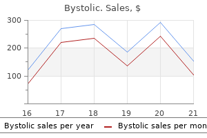
Bystolic
Bystolic dosages: 5 mg, 2.5 mg
Bystolic packs: 30 pills, 60 pills, 90 pills, 120 pills, 180 pills, 270 pills, 360 pills

Discount bystolic 5 mg free shipping
In macular translocation arrhythmia only at night bystolic 5 mg cheap on line, the retina is moved so that the fovea is relocated to an space away from submacular illness blood pressure goals purchase 2.5 mg bystolic fast delivery. In adults, multipotent stem cells exist that can differentiate to exchange misplaced or injured cells. Bone marrow cells, for instance, can undergo metaplasia to type skeletal muscle;57 neural stem cells can develop into muscle58 (Table 128. It seems that stem cells exhibit plastic behavior relying upon their surroundings and alerts from broken tissues. They exist in both embryonic and grownup tissues and can differentiate into glial and neuronal lineages. The capacities for self-renewal and pluripotency render stem cells candidates to substitute lost or injured cells. The results of such reprogrammed cells could possibly be the creation of genetically matched cell lines for autologous transplants. Indeed, in a single study, when embryonic stem cell-derived neural cell precursors were injected into mouse subretinal house, 50% of the eyes developed teratomata inside eight weeks. Thus far, research have described the differentiated cells at early time factors solely, and full characterization has not been reported. To be useful for cell-based alternative therapy, stem cells must have the next properties: 1. Proliferate extensively to generate sufficient portions of fabric to serve as a "universal donor" and preserve genetic stability after expansion. PolarizedSheet Delivery of a cell suspension to the subretinal area has the benefit of simplicity, i. Theoretically, cell suspension supply might cowl a bigger space whereas sheet supply is limited to the scale of a sheet that can be manipulated for successful placement. Potential benefits to transplanting sheets of cells on a scaffold are threefold. There may be an advantage to decreasing the antigen load with regard to stimulating immune surveillance of the transplanted cells. Patients had been immunosuppressed with low-dose tacrolimus and mycophenolate mofetil 1 week previous to surgical procedure, which was to be continued for several months postoperatively. It is feasible that other cells such as macrophages which have engulfed released pigment might account for pigmented cells observed within the subretinal space. The sheets were harvested from a porcine collagen I gel substrate using collagenase I to launch the sheet from the substrate. Toluidine blue-stained semi-thin sections of nondystrophic (A), and dystrophic (B,C) Royal College of Surgeons rat retina. Scale bars: 200 �m (top left), a hundred �m (bottom left, prime right), 50 �m (bottom right). Retinal pigment epithelial cell transplantation after subfoveal membranectomy in age-related macular degeneration: clinicopathologic correlation. Note the presence of a pigmented patch of transplanted cells (B,C, arrows) that turns into bigger and more pigmented by 6 months. Patches of pigmented cells are evident around the border of baseline atrophy in retinal pigment epithelium (E) that become more distinguished at 1 12 months (F, arrows). An space of transplanted retinal pigment epithelium cells is seen at the superior half of the atrophic lesion at 6 months (H) that becomes bigger and extra pigmented at 15 months (I). Immune privileged tissue can survive for prolonged durations in a nonimmune privileged site in comparison with nonimmune privileged tissue. Thus, resistance of an allograft to immune rejection may be due to properties of each the transplantation website and the transplanted tissue. Immune privilege in the eye is the end result of multiple anatomic and physiologic elements appearing on each the innate and adaptive immune systems. Subretinal allografts resist immune rejection and induce the event of systemic tolerance in the type of a suppressed antigen-specific delayed hypersensitivity response. That immune privilege is prolonged despite this disruption indicates the importance of the elements in the microenvironment. In summary, immune privilege may promote allograft survival within the subretinal house, but the changes due to illness and surgical intervention may significantly affect the degree of privilege. No signs of acute graft rejection have been famous within the 18 patients of this examine, indicating that immune suppression following allogeneic cell transplantation is possible. Another confounding factor in evaluating the animal research involving transplant rejection (reviewed in reference227) is that the transplantation surgical procedure in some studies resulted in harm to the blood retinal barrier, which may have promoted transplant rejection. Preoperative autofluorescence imaging exhibits hypoautofluorescence centrally with small satellite tv for pc lesions of hypo-autofluorescence, and a surrounding rim of hyperautofluorescence (A). There is speckled hyperautofluorescence in the higher half of the central atrophic lesion (B,C, arrows) that diminishes by 1 year (D). It is most likely going that cell choice, preparation, and supply technique will differ, relying upon the goal illness and the stage of illness progression. Regardless of the method of differentiation, rigorous testing at all stages of medical grade cell manufacturing should take place starting with the procurement of the starting cells through the final product to guarantee purity, safety, stability, and performance of transplanted cells. Independent of its effectiveness, systemic immune suppression may be sophisticated by side-effects such as nephrotoxicity, hypertension, risk of malignancy, susceptibility to an infection, hepatotoxicity, seizures, and even anaphylaxis, relying on the medications used. Sustained slow-release cyclosporine delivery was as efficient at promoting transplant survival as repeated intravitreal injections of a lot higher doses. High-dose dexamethasone remedy is known to be effective in stopping acute allograft rejection259,260 though lymphocyte resistance to prednisolone can develop. Use of the sustained dexamethasone supply system has prevented allogeneic corneal transplant rejection in rats. Another method has included enhancing the expression of cell-matrix adhesion molecules, i. Because cell-scaffold placement could require a larger retinotomy and a bigger retinal detachment, it might create extra surgical harm than dispersed cell subretinal injections. Human clinical studies point out that durations of macular detachment as much as 2 weeks are suitable with recovery of visible acuity of 20/50 or higher in a considerable number of sufferers. Cones are extra susceptible to apoptosis with detachment (versus rods)294 although in a single examine, roughly 75% of S- and M-cones survived a 1-day detachment (followed by retinal reattachment) in the floor squirrel. In addition to length of detachment, the peak of detachment influences photoreceptor survival. Transvitreal delivery is a much less traumatic and more pertinent technique to human retinal transplantation than the transscleral approach. Furthermore, the rod to cone ratio in rodent retina340 differs from that of the comparatively cone-rich human retina. To overcome these problems, animal fashions using larger eyes, such because the chemically ablated rabbit retina328 and the Abyssinian Rdy cat,341,342 have been employed in addition to regular rabbits,328,343,344 pigs,345,346 and primates. Photoreceptor microaggregates, that are retinal fragments during which tissue integrity is disrupted by gentle trituration with cells remaining connected to each other in small clusters (<0. Rosette formation prevents the reconstruction of the conventional retinal anatomy and interferes with establishing contacts between graft photoreceptor terminals and host second-order neurons. Sheet preparations preserve the organization and polarity of grafted photoreceptors and keep the densely packed arrangement of photoreceptors present in situ and thought to be important for good visible acuity.
5 mg bystolic discount visa
The cells stained optimistic for prostate-specific antigen phosphatase and prostate-specific antigen blood pressure guide nhs 2.5 mg bystolic sale, which is diagnostic of prostatic carcinoma hypertension webmd cheap 5 mg bystolic with visa, with the mass infiltrating the choroidal tissue. Chorioretinal biopsy offered useful data for figuring out the analysis (multifocal choroiditis with subretinal fibrosis, sarcoidosis, and viral retinitis) and led to change of remedy in 5 patients. Foulds and colleagues10,forty three,fifty three,54 reported on 34 transscleral biopsies of the choroid and retina for the analysis of choroidal melanoma, acute retinal necrosis, continual uveitis, and progressive retinal pigment epitheliopathy. An opposed occasion occurred in just one case: a retinal break developed, with related vitreous hemorrhage and resultant proliferative vitreoretinopathy. Echographic study showed a plaque on the surface of the mass similar to a beforehand reported benign fibrous tumor. A 25-gauge, 112-inch needle is inserted by way of the pars plana on this eye and into the tumor, which is located posteriorly. Once the needle is within the lesion to be biopsied, an assistant exerts forceful suction on a 10-mL syringe linked to the needle via tubing. The biopsy needle is linked to a 10-mL plastic disposable aspirating syringe via a standard plastic tubing. The large tumor cell within the decrease right field has pigment within the cytoplasm and a large round nucleus with outstanding nucleolus, characteristic of malignant melanoma. Results Transscleral fine-needle aspiration biopsy can be feasible in diagnosing choroidal melanoma. In addition, aspiration biopsy might assist in assessing cytogenetics of these tumors. In a prospective, interventional case sequence, 30G fine-needle aspiration biopsy recognized macular choroidal melanoma in 17 of 24 (71%) eyes. Chorioretinal biopsy for diagnostic purposes in cases of intraocular inflammatory illness. Cytomegalovirus retinitis after low-dose intravitreous triamcinolone acetonide in an immunocompetent patient: a warning for the widespread use of intravitreous corticosteroids. Postoperative complications and intraocular pressure in 943 consecutive cases of 23-gauge transconjunctival pars plana vitrectomy with 1-year follow-up. These embrace increased intraocular stress, cataract development, peripheral retinal tears, retinal detachment, choroidal hemorrhage, vitreous hemorrhage, endophthalmitis, exacerbation of the underlying inflammatory disease, and proliferative vitreoretinopathy. Noninvasive means of building a diagnosis are exhausted before surgical procedures are thought-about. Vitreous, Retinal, and Choroidal Biopsy supplement to typical microbiological diagnostic strategies. The vitreous entice: a easy, surgeon-controlled method for obtaining undiluted vitreous and subretinal specimens throughout pars plana vitrectomy. A 27-gauge sharp-tip short-shaft pneumatic vitreous cutter for transconjunctival sutureless vitreous biopsy. A 27-gauge instrument system for transconjunctival sutureless microincision vitrectomy surgery. Real-time polymerase chain reaction test to discriminate between contamination and intraocular infection after cataract surgical procedure. Polymerase chain response in pediatric post-traumatic fungal endophthalmitis amongst Egyptian kids. Polymerase chain reaction analysis of aqueous and vitreous specimens in the prognosis of posterior section infectious uveitis. An improved approach for the prognosis of viral retinitis from samples of aqueous humor and vitreous. Quantitative analysis of peripheral vasculitis, ischemia, and vascular leakage in uveitis using ultra-widefield fluorescein angiography. Retinal and choroidal biopsies are helpful in unclear uveitis or suspected infectious or malignant origin. An experimental approach to the tissue analysis and study of choroidal and retinal lesions. Intraocular biopsy using special forceps: a brand new instrument and refined surgical approach. Fine-needle aspiration biopsy of stable intraocular tumors: indications, instrumentation and techniques. Transvitreal retinochoroidal biopsy provides a representative sample from choroidal melanoma for detection of chromosome three aberrations. Spectral-domain optical coherence tomography analysis of choroidal melanoma and nevus fine-needle aspiration biopsy incision websites. Prolonged graft survival and a normal fee of graft growth and differentiation were noted. Expanding on this work, del Cerro and his colleagues3 transplanted embryonic rat retina into the anterior chamber of grownup eyes from various rat strains with comparable results. In principle, successful transplantation requires graft survival and integration with the host. Graft survival is determined by a selection of factors, both immunologic and non-immunologic,7 that will be discussed in this chapter. In contrast, synapse formation between retinal grafts and host retina is much more complex. Experiments in the central nervous system, nevertheless, have established the chance that synapse formation between donor and host neural tissue can occur. For instance, Lund and others8,9 demonstrated that embryonic retina transplanted into neonatal and grownup rat tectum develops the proper layering of the conventional retina and extends neuronal projections solely to the conventional retinorecipient structures, i. Similarly, intraocular retinal transplants set up with their targets functional connections that elicit light-driven visible response10 and mediate visual behaviors. Studies in neural retinal transplantation have utilized tissue from embryonic, early postnatal, late postnatal, and adult donors in addition to transplantation of homo- and heterotypic neural progenitor cells (stem cells, see below). In one examine, attempts to get hold of photoreceptor sheets from embryonic retina utilizing vibratome-sectioning resulted in irregular morphology and poor survival. Finally, acquiring embryonic or fetal tissue for transplantation has many logistical and ethical constraints. Adult retinal grafts seem to show normal morphology and organization within the host retina and are associated with minimal rosette formation, which could enable for better restoration of retinal anatomy,328,334,347 particularly if the outer blood�retina barrier remains intact. The strategies employed, together with the kind of cells used, in these paradigms depend on the primary derangement that causes photoreceptor cell death. For example, Schwann cells, derived from peripheral nerves, have been used as autologous grafts to rescue photoreceptors. However, in distinction to Schwann cells, olfactory ensheathing cells had been shown to phagocytose porcine retinal outer segments in an in vitro assay. Transplanted rods might rescue present cones that might be misplaced secondary to rod degeneration. This rescue effect was observed at some distance from the grafted cells, suggesting the existence of diffusible trophic issue launch by the transplant.
Diseases
- Thomas Jewett Raines syndrome
- Partial atrioventricular canal
- Kumar Levick syndrome
- Occipital horn syndrome
- DOOR syndrome
- Pterygium colli
- Lumbar malsegmentation short stature
- Bull Nixon syndrome
- B?b? Collodion syndrome
- Hypothermia
Generic 5 mg bystolic fast delivery
The distinction within the anatomy as teenagers and adults is putting when growth of irradiated orbits is in contrast with the expansion of nonirradiated orbits blood pressure 50 over 70 2.5 mg bystolic generic with mastercard. In a report from China high blood pressure medication valsartan order bystolic 5 mg amex, moderately good outcomes had been reported with a hydroxyapatite onlay bone graft substitute covered with a vascularized pedicle flap. The lacrimal gland, cornea, and conjunctiva can every tolerate as much as 50 Gy (5000 rad) of radiation, whereas the retina tolerates forty five Gy or less. The system of stabilizing the eye with a vacuum contact lens developed by Schipper and colleagues reduced the number of eyes that develop surgical radiation cataracts. One eye developed retinal detachment, 4 had amblyopia, and three developed tumor recurrence, one with extension of retinoblastoma into the subconjunctival house through the sclerotomy. Appropriate timing for cataract removing in retinoblastoma sufferers has not been documented or verified in the literature. Another necessary issue is age of therapy, with the very best threat for these patients who were handled within the first 12 months of life. In the July 1993 concern of the Journal of the National Cancer Institute, Eng and colleagues provided follow-up on 1000 sufferers with retinoblastoma treated in New York and Boston. Appropriate surveillance for second main tumors involves the dad and mom performing weekly examinations of their at-risk children searching for foci of ache, tenderness, or swelling. Such signs or signs should immediate a visit to the pediatrician or major care specialist with an accompanying assertion that the child is at high danger for bone cancer and should be examined. We have one affected person who has survived 17 years after prognosis of an osteosarcoma of the greater wing of the sphenoid. The vast majority of facilities additionally deal with patients with tumor invasion of the optic nerve posterior to the lamina cribrosa, although some facilities observe these sufferers and treat the approximately 10% of sufferers who relapse with aggressive multimodal chemotherapy. In contrast, if solely bone marrow and bone are involved latest stories provide some measure of hope with myeloablative therapy, bone marrow transplant along with focal radiation to bulky disease. However, there have been encouraging reviews that high-dose multimodal chemotherapy combined with bone marrow rescue have been related to long-term survival. When the analysis of intraocular retinoblastoma is delayed, the natural history of intraocular retinoblastoma entails eventual extension by way of the eye wall into the orbit then to regional lymph nodes with eventual metastatic spread. These patients will current with proof of orbital retinoblastoma at analysis, with proptosis, orbital irritation, and evidence of an orbital mass on imaging studies. The danger for extraocular disease strongly is dependent upon the delay in prognosis in the setting of superior intraocular disease. Chantada and his colleagues from Argentina reported 84% had 5-year event-free survival when orbital retinoblastoma was treated with primary chemotherapy, limited excision, and orbital radiation. We suggest systemic chemotherapy and orbital radiotherapy after an incisional biopsy to affirm the analysis. Metastatic Retinoblastoma Risk Factors the risk of extraocular disease after enucleation or globeconserving treatment could be very low in developed countries, ranging between 0% and 4%. Specific clinical options that have been shown to improve the risk of metastasis embrace the presence of a bunch E tumor, rubeosis iridis, neovascular glaucoma, and scientific signs for greater than 6 months prior to diagnosis. Retinomas are typically diagnosed on routine fundus evaluations of healthy sufferers, or on screening examinations of relations of a affected person with retinoblastoma. The typical fundus appearance is a gray translucent mass containing calcified nodules and surrounded by retinal pigment clumping and atrophy. Lueder and colleagues have described two sufferers with retinomas and vitreous seeding that were adopted for 8 and 33 years, respectively, with out proof of disease progression or transformation. The retinoma or retinocytoma has benign histopathologic features however might rarely retain the ability to bear malignant transformation right into a quickly growing retinoblastoma. Three years later the tumor all of a sudden grew rapidly and seeded the vitreous, leading to loss of the eye. If the retinoma is peripheral, we consider performing careful laser therapy to flatten the lesion, which should scale back the chance of malignant transformation. There are occasional retinoblastomas which regress into tumors that resemble retinomas, both clinically and histopathologically. In addition, there may be areas of retinoma formation in sufferers who current with untreated retinoblastoma. These "presumed" retinomas appear grey and translucent, very similar to the lesion described by Aaby and colleagues. Instead, the tumor expands by diffusely infiltrating the retina, inflicting irregular thickening. In one evaluate, only 4 of 28 diffusely infiltrating retinoblastomas demonstrated the presence of intraocular calcium. The classic presentation is unilateral uveitis or retinal detachment in an older youngster without any previous history, although authors from Taiwan reported one hereditary case. Since the lesion grows within and destroys the sensory retina, little is gained by attempts to salvage the attention. The persistence of a rare embryonal retinal cell has been proposed as one rationalization for this uncommon onset of retinoblastoma at an advanced age. The clinically necessary side of retinoblastoma presenting in older youngsters is that misdiagnosis is common. Because of the superior nature of the unilateral disease in these older children, virtually all of the cases have group E disease and therefore require enucleation. The link between intraocular retinoblastoma and an ectopic, intracranial malignancy was first recognized in 1977 by Jakobiec and colleagues. In decrease animals the pineal gland capabilities as a photoreceptor organ and is typically referred to because the "third eye. Neuroradiology experience is required in assessing the risk of malignancy for these sufferers with pineal cysts, and neurosurgical session ought to be requested. Therefore, screening packages must be directed at kids with bilateral retinoblastoma and those unilateral patients with a positive family historical past, in the course of the first 3�4 years after the diagnosis of retinoblastoma. A schedule of neuroimaging each 3 months for 2 years, every four months the next 2 years, and each 6 months for the next 5 years has been proposed. Only five patients were event-free survivors at 10�168 months and all the survivors had tumors less than 15 mm detected through screening. Therefore, present strategies are directed toward avoiding irradiation and utilizing intensive chemotherapy adopted by autologous stem cell rescue. Selective ophthalmic arterial injection therapy for intraocular retinoblastoma: the long-term prognosis. Occurrence of sectoral choroidal occlusive vasculopathy and retinal arteriolar embolization after superselective ophthalmic artery chemotherapy for advanced intraocular retinoblastoma. One series discovered that therapy appears to extend survival from chemotherapy with melphalan for intraocular retinoblastoma initial outcomes. Selective ophthalmic artery infusion of chemotherapy for superior intraocular retinoblastoma: preliminary expertise with 17 tumors. Intraarterial chemotherapy for the management of retinoblastoma: four-year expertise. Intravitreal chemotherapy for vitreous seeding in retinoblastoma: latest advances and perspectives. Intravitreal chemotherapy for vitreous illness in retinoblastoma revisited: from prohibition to conditional indications. Profiling security of intravitreal injections for retinoblastoma utilizing an anti-reflux process and sterilisation of the needle track.
5 mg bystolic order visa
In the rabbit eye heart attack labs buy bystolic 2.5 mg free shipping, Glatt and Machemer demonstrated that subretinal blood triggered irreversible photoreceptor loss in lower than 24 hours hypertension journal articles buy bystolic 2.5 mg visa. Vitrectomy, Injection of Subretinal Tissue Plasminogen Activator, and Aspiration of Liquefied Blood the disappointing outcomes obtained with direct surgical extraction of clot led investigators to examine attainable adjuvants to help in the removing of subretinal blood. After dissolution of the clot, the subretinal hemorrhage was removed with presumed minimal trauma by way of light irrigation of balanced salt solution. However, at 2 years, fewer surgical procedure eyes (21%) compared with observation eyes (36%) had experienced severe vision lack of 6 strains or extra (p=. This good factor about surgery was most evident in eyes with comparatively higher vision at presentation (20/100 to 20/160). A excessive fee of retinal detachment was seen in the surgical arm (16% of all eyes), and these have been extra frequent in eyes with very poor vision and very massive hemorrhagic lesions on presentation. Vitreous hemorrhage was the common complication, and there was one case of postoperative endophthalmitis in this group. Following extra subretinal air injection to displace the dissolved clot inferiorly, there was no fluid�air change. The subretinal blood collected on this potential space and was pushed inferiorly by an intraocular air bubble injected on the end of the vitrectomy surgery. They reported whole subfoveal blood displacement in 86% of 29 eyes handled with this system, and 59% of patients gained 2 or extra traces of vision at 3 months after surgical procedure. In addition, vitrectomy surgical procedure permits for a larger gasoline bubble to be positioned within the vitreous cavity in comparison with in-office gasoline injection, permitting for more complete displacement of blood from the submacular area. This examine was retrospective, and the duration of hemorrhage was not well matched between the groups. Vitreous surgical procedure for hemorrhagic and fibrous issues of age-related macular degeneration. Clinical experience with the surgical removal of subfoveal neovascular membranes: short-term postoperative outcomes. Surgical excision of subfoveal neovascular membranes in age-related macular degeneration. Surgical removal of subretinal hemorrhage and choroidal neovascular membranes in acute hemorrhagic age-related macular degeneration. Surgical removal of subfoveal choroidal neovascularization in presumed ocular histoplasmosis: stability of early visible outcomes. Submacular surgery for subfoveal choroidal neovascular membranes in patients with presumed ocular histoplasmosis. Submacular surgical procedure to remove choroidal neovascularization related to central serous chorioretinopathy. Visual outcome after surgical elimination of choroidal neovascularization in pediatric patients. The surgical elimination of subfoveal choroidal neovascularization: ingrowth site as a predictor of visual end result. Managing recurrent neovascularization after subfoveal surgical procedure in presumed ocular histoplasmosis syndrome. Other components that portend a poor visible prognosis include increasing size, thickness, and duration of submacular blood. In an attempt to improve upon the pure historical past of the disease, a quantity of investigators have attempted to remove the blood from the submacular space. Fibrinolytic-assisted elimination of experimental subretinal hemorrhage within seven days reduces outer retinal degeneration. Vitreoretinal update 1997, American Academy of Ophthalmology, Retina Subspecialty Day. Management of acute submacular hemorrhage utilizing recombinant tissue plasminogen activator and gas. Management of submacular hemorrhage with intravitreous tissue plasminogen activator injection and pneumatic displacement. Intravitreous injection of tissue plasminogen activator and gas within the therapy of submacular hemorrhage under various situations. Effect of intravitreal tissue plasminogen activator on experimental subretinal hemorrhage. The use of intravitreal tissue plasminogen activator within the treatment of experimental subretinal hemorrhage within the pig model. A examine of the ability of tissue plasminogen activator to diffuse into the subretinal area after intravitreal injection in rabbits. The fee and source of albumin entry into saline-filled experimental retinal detachments. Pneumatic displacement of subretinal hemorrhage without tissue plasminogen activator. Pars plana vitrectomy, subretinal injection of tissue plasminogen activator, and gas/fluid change for displacement of thick submacular hemorrhage in age-related macular degeneration. Subretinal recombinant tissue plasminogen activator injection and pneumatic displacement of thick submacular hemorrhage in age-related macular degeneration. Management of submacular hemorrhage with intravitreal versus subretinal injection of recombinant tissue plasminogen activator. Submacular combination therapy for management of acute, huge submacular hemorrhage in age-related macular degeneration. Submacular Surgery Trials randomized pilot trial of laser photocoagulation versus surgical procedure for recurrent choroidal neovascularization secondary to age-related macular degeneration: I. Surgical removing of in depth peripapillary choroidal neovascularization related to presumed ocular histoplasmosis syndrome. Surgical remedy of extensive peripapillary choroidal neovascularization in aged patients. Massive peripapillary subretinal neovascularization: a sign for submacular surgery. Surgical removal of peripapillary choroidal neovascularization associated with agerelated macular degeneration. Long-term follow-up of surgical removing of intensive peripapillary choroidal neovascularization in presumed ocular histoplasmosis syndrome. Types of choroidal neovascularization in newly identified exudative age-related macular degeneration. Subretinal hemorrhages with or with out choroidal neovascularization within the maculas of sufferers with pathologic myopia. Surgical remedy of submacular hemorrhage related to idiopathic polypoidal choroidal vasculopathy. Surgery for predominantly hemorrhagic choroidal neovascular lesions of age-related macular degeneration: ophthalmic findings. Prospective one-year study of ranibizumab for predominantly hemorrhagic choroidal neovascular lesions in age-related macular degeneration. Intravitreal bevacizumab therapy for neovascular age-related macular degeneration with giant submacular hemorrhage.
5 mg bystolic trusted
Experimental retinal detachment: biophysical aspects of retinal peeling and stretching prehypertension at 36 weeks pregnant 2.5 mg bystolic cheap free shipping. Spectral-domain optical coherence tomography imaging of postoperative scleral buckles hypertension lifestyle modification bystolic 2.5 mg buy generic on-line. Scleral buckling technique without retinopexy for treatment of rhegmatogenous: a pilot research. Anatomical retinal reattachment after scleral buckle with and without retinopexy: a pilot examine. The fluid mechanics of scleral buckling surgery for the restore of retinal detachment. Effects of encircling scleral buckling on the morphology and biomechanical properties of the cornea. Effect of squatting on sub-foveal blood move defect in pseudophakic eyes operated by cerclage. Relaxation of encircling buckle improved choroidal blood circulate in a patient with visual area defect following encircling process. Traction on the retina induced by saccadic eye movements within the presence of posterior vitreous detachment. The conjunctiva turns into friable with age and care must be taken to avoid tearing and buttonholing. A glove-like sleeve of fascia extends anteriorly (to the rectus insertions) and posteriorly (for several millimeters) alongside the muscles from the points at which they pierce the Tenon capsule. Between the rectus muscle tissue anteriorly these sleeves are joined by a layer of fascia: the intermuscular septum. The intermuscular septum and fascial sleeves of the recti are sometimes collectively referred to because the posterior Tenon capsule. The selection between buckling and other methods is roofed in Chapter 109 (Optimal procedures for retinal detachment repair). Sometimes the time period "buckle" is used synonymously with some form of encircling explant, whereas others use the term to describe native explants. Different colleges of buckling method have arisen with divergent views in particular on the function of encirclement and subretinal fluid drainage. Buckling of the sclera to close retinal breaks was initially achieved using mixtures of lamellar scleral dissection with compression sutures till it was shown that it could be achieved more effectively using scleral implants4 and subsequently explants. Likewise diathermy, which was used extensively in the past to achieve retinopexy,four has been supplanted by photocoagulation and cryotherapy. There have also been subsequent refinements of the fundamental strategy of scleral buckling, including intraocular gasoline injection and subretinal fluid drainage. Care must be taken when stripping fascia off the rectus muscular tissues because ligaments from the recti to the wall of the orbit are functionally important within the actions of the muscle. Passage of sutures is facilitated by the lamellar association of collagen fibers, which permits spatulated (or "aspect slicing") needles to comply with a airplane between lamellae. The anatomy of the vortex veins is somewhat variable however one tends to depart the globe both aspect of the vertical recti just behind the equator. They may be inadvertently hooked along with a vertical rectus muscle if the muscle hook is passed behind the equator. Injury to the vortex veins may end in interruption to the venous drainage from the choroid and choroidal detachment. When working near the vertical recti, the vortex veins ought to be recognized to stop injury. Because the vortex veins are likely to be located close to the vertical recti, subretinal fluid drainage is carried out closer to the horizontal than to the vertical recti every time potential. As they supply the arterial circles of the iris, surgical trauma (including diathermy) must be minimized. Extraocular Muscles the recti are adherent to the sclera on the spiral of Tillaux. In this position they help the retina as far anteriorly as the ora serrata ("break ora occlusive buckling"). The superior oblique muscle runs laterally from the trochlea to its insertion underneath the superior rectus. Passage of a superior rectus muscle hook from the temporal side of the muscle reduces the risk of inadvertently "hooking" the superior oblique, as well as does keeping the sweep of the hook preequatorial. The superior indirect insertion is regularly encountered on the temporal side of the muscle the place it could be an impediment to scleral suturing. Division of a small (< 1 3) portion of the insertion to facilitate suturing appears to have little impact on ocular motility. Note that a vortex vein is usually present beneath the temporal fringe of the superior oblique insertion. The chance of inadvertently hooking it while passing a muscle hook beneath the lateral rectus is lowered by passing the hook from the superior aspect. Innervation Sensory nerves from the globe and bulbar conjunctiva cross via the ciliary ganglion. Local anesthesia on this region, for example, sub-Tenon anesthesia, will due to this fact anesthetize the globe successfully. Having taken a cautious historical past (and famous relevant systemic health problems and past ophthalmic history), the anterior and posterior segments of the attention are rigorously examined utilizing slit-lamp biomicroscopy and oblique ophthalmoscopy. A, anterior ciliary artery; B, lengthy posterior ciliary artery; C, vortex ampulla; D, vortex vein; E, higher arterialcircleoftheiris. TechniquesofScleralBuckling 1891 � Macular involvement � Features suggesting that the retinal detachment is nonrhegmatogenous (see also Chapter 99, Nonrhegmatatogenous retinal detachment) � the presence of vitreous detachment � Significant ocular co-pathology, which may have an result on management. Cases with tractional tears are most likely to progress very quickly and must be handled urgently. Reducing eye and head movements seems to scale back the speed at which subretinal fluid accumulates. Bed relaxation, eye patching, and rectus sutures have all been used to cut back the quantity of subretinal fluid. This may stop extension of subretinal fluid to the macula and make it simpler to establish retinal breaks. These drawings should show the location of retinal breaks in relation to simply seen retinal landmarks corresponding to small hemorrhages, vascular bifurcations, and areas of pigmentation. If in doubt, an space of retina can be reexamined alternately with oblique ophthalmoscopy and slitlamp biomicroscopy to establish whether a break is truly current. The drawings made could be referred to if the retinal view turns into obscured throughout surgical procedure. In kids and uncooperative sufferers, it could be impossible to establish the place of the retinal breaks preoperatively. Peribulbar administration of a 50: 50 mix of lidocaine and bupivacaine, notably when used with adjunctive Hyalase, offers excellent anesthesia and akinesia. In apply, sub-Tenon anesthesia gives anesthesia equivalent to different strategies,14 presumably because of overspill of anesthetic agents to the peribulbar house.

Cheap 5 mg bystolic overnight delivery
Effectiveness of diode laser trans-scleral cyclophotocoagulation in sufferers following silicone oil-induced ocular hypertension in Chinese eyes blood pressure 6050 bystolic 2.5 mg generic free shipping. Ultrasound biomicroscopy after vitrectomy in eyes with regular intraocular strain and in eyes with persistent hypotony pulse pressure and stroke volume relationship bystolic 5 mg buy without a prescription. Dissection of epiciliary tissue to treat chronic hypotony after surgery for retinal detachment with proliferative vitreoretinopathy. Temporary silicone oil tamponade in the administration of retinal detachment with proliferative vitreoretinopathy. Treating cytomegalovirus retinitis-related retinal detachment by combining silicone oil tamponade and ganciclovir implant. Can the sequential use of standard silicone oil and heavy oil be a technique for the administration of proliferative vitreoretinopathy Long-term anatomical and visible outcome of vitreous surgery for retinal detachment with choroidal coloboma. Clinical danger factors for proliferative vitreoretinopathy after retinal detachment surgical procedure. The end result of early surgical restore with vitrectomy and silicone oil in open-globe injuries with retinal detachment. Pars plana vitrectomy with or without silicone oil endotamponade in posttraumatic endophthalmitis. Bacterial endogenous endophthalmitis in Vietnam: a randomized controlled trial evaluating vitrectomy with silicone oil versus vitrectomy alone. Characteristics, demographics, outcomes, and issues of diabetic traction retinal detachments handled with silicone oil tamponade. Outcomes of transconjunctival sutureless 25-gauge vitrectomy with silicone oil infusion. Transconjunctival 25-gauge sutureless vitrectomy and silicone oil injection in diabetic tractional retinal detachment. Self-retaining 27-gauge transconjunctival chandelier endoillumination for panoramic viewing throughout vitreous surgical procedure. Intraocular lens modifications after short- and long-term publicity to intraocular silicone oil. Irreversible silicone oil adhesion to silicone intraocular lenses; a clinicopathologic evaluation. Use of hydroxypropylmethylcellulose 2% for removing adherent silicone oil from silicone intraocular lenses. Removing silicone oil droplets from the posterior floor of silicone intraocular lenses. Cohort safety and efficacy research of siluron2000 emulsification-resistant silicone oil and f4h5 within the treatment of full-thickness macular hole. The issues of biometry in mixed silicone oil elimination and cataract extraction: a clinical trial. Immersion B-guided versus contact A-mode biometry for correct measurement of axial size and intraocular lens energy calculation in siliconized eyes. Laser interference biometry versus ultrasound biometry in sure medical circumstances. Signal high quality of optical biometry in silicone oil-filled eyes utilizing partial coherence laser interferometry. Accuracy and reproducibility of axial length measurement in eyes with silicone oil endotamponade. Investigating a potential explanation for the myopic shift after combined cataract extraction, intraocular lens implantation, and vitrectomy for remedy of a macular hole. Timing of retinal redetachment after removing of intraocular silicone oil tamponade. Correlation between quantity of silicone oil emulsified within the anterior chamber and excessive stress in vitrectomized eyes. The incidence of corneal abnormalities within the silicone study: results of a randomized medical trial. Comparison of silicone oil elimination with passive drainage alone versus passive drainage mixed with air�fluid trade. Phacoemulsification mixed with silicone oil removal by way of the posterior capsulorhexis tear. Topical anesthesia for transpupillary silicone oil removal combined with cataract surgical procedure. Removal of silicone oil with 25-gauge transconjunctival sutureless vitrectomy system. Combined silicone and fluorosilicone oil tamponade (double filling) within the management of complicated retinal detachment. Perfluorodecalin and silicone oil used to achieve retinal tamponade left in an eye fixed for six months. Tamponade properties of double-filling with perfluorohexyloctane and silicone oil in a mannequin eye chamber. The mixed use of perfluorohexyloctane (F6H8) and silicone oil as an intraocular tamponade in the remedy of severe retinal detachment. The impact of simultaneous inside tamponade on fluid compartmentalization and its relationship to cell proliferation. Use of perfluorohexyloctane as a long-term inner tamponade agent in difficult retinal detachment surgical procedure. A new method of eradicating silicone oil from the surface of silicone intraocular lenses. Clinical findings on the usage of long-term heavy tamponades (semifluorinated alkanes and their oligomers) in difficult retinal detachment surgery. Semifluorinated alkanes � a model new class of compounds with outstanding properties for use in ophthalmology. Clinicopathological correlation of epiretinal membranes and posterior lens opacification following perfluorohexyloctane tamponade. Our experience with perfluorohexyloctane (F6H8) as a brief endotamponade in vitreoretinal surgery. Perfluorocarbon liquids as postoperative short-term vitreous substitutes in complicated retinal detachment. Perfluorohexylethan (O62) as ocular endotamponade in complicated vitreoretinal surgery. First experiences with high density silicone oil (Densiron) as an intraocular tamponade in complicated retinal detachment. Factors influencing the shear price acting on silicone oil to cause silicone oil emulsification. Clinical observations and occurrence of issues following heavy silicone oil surgery. Randomized controlled trial of mixed 5-fluorouracil and low-molecular-weight heparin within the administration of unselected rhegmatogenous retinal detachments undergoing main vitrectomy. Pars plana vitrectomy for the therapy of rhegmatogenous retinal detachment uncomplicated by advanced proliferative vitreoretinopathy.
OPCs (Grape). Bystolic.
- Hayfever and seasonal nasal allergies.
- Circulation problems, such as chronic venous insufficiency that can cause the legs to swell.
- Preventing heart disease, treating varicose veins, hemorrhoids, constipation, cough, attention deficit-hyperactivity disorder (ADHD), chronic fatigue syndrome (CFS), diarrhea, heavy menstrual bleeding (periods), age-related macular degeneration (ARMD), canker sores, poor night vision, liver damage, high cholesterol levels, and other conditions.
- What is Grape?
- Are there any interactions with medications?
- Dosing considerations for Grape.
Source: http://www.rxlist.com/script/main/art.asp?articlekey=96481

Bystolic 2.5 mg buy generic online
Relationship between macular gap size and the potential benefit of internal limiting membrane peeling blood pressure medication that starts with m 2.5 mg bystolic with mastercard. Nonsupine positioning in macular gap surgery: a noninferiority randomized clinical trial hypertension 40 mg order bystolic 2.5 mg visa. A novel segmentation algorithm for volumetric evaluation of macular gap boundaries recognized with optical coherence tomography. A comparison of a quantity of methods of macular hole measurement utilizing optical coherence tomography, and their value in predicting anatomical and visual outcomes. Effects of preoperative and postoperative epiretinal membranes on macular gap closure and visible restoration. Prevalence, correlates, and pure historical past of epiretinal membranes surrounding idiopathic macular holes. Immunocytochemical and ultrastructural proof of glial cells and hyalocytes in internal limiting membrane specimens of idiopathic macular holes. Diagnosis of macular pseudoholes and lamellar macular holes by optical coherence tomography. Redefining lamellar holes and the vitreomacular interface: an ultrahigh-resolution optical coherence tomography study. Traumatic macular hole: observations, pathogenesis, and outcomes of vitrectomy surgical procedure. Comparing functional and morphologic traits of lamellar macular holes with and without lamellar hole-associated epiretinal proliferation. Lamellar holeassociated epiretinal proliferation in comparability to epiretinal membranes of macular pseudoholes. Lamellar macular hole: a clinicopathologic correlation of surgically excised epiretinal membranes. Progression from macular retinoschisis to retinal detachment in highly myopic eyes is associated with outer lamellar hole formation. Macular Hole closure over residual subretinal fluid by an inverted internal limiting membrane flap technique in patients with macular gap retinal detachment in excessive myopia. An aspirating forceps to take away the posterior hyaloid in the surgery of full-thickness macular holes. Incidence of retinal detachment after macular surgical procedure: a retrospective study of 634 circumstances. Incidence and causes of iatrogenic retinal breaks in idiopathic macular hole and epiretinal membrane. The use of inside limiting membrane maculorrhexis in remedy of idiopathic macular holes. Histopathological examination of inner limiting membrane floor after scraping with diamond-dusted membrane scraper. Temporal inverted inside limiting membrane flap approach versus traditional inverted inner limiting membrane flap approach: a comparative examine. Mechanisms of intravitreal toxicity of indocyanine green dye: implications for chromovitrectomy. Retinal pigment epithelial modifications after macular gap surgery with indocyanine green-assisted inner limiting membrane peeling. Toxic effect of indocyanine green on retinal pigment epithelium associated to osmotic results of the solvent. Histology of the vitreoretinal interface after staining of the interior limiting membrane utilizing glucose 5% diluted indocyanine and infracyanine green. Persistence of fundus fluorescence after use of indocyanine green for macular surgical procedure. Retinal ganglion cells toxicity brought on by photosensitising results of intravitreal indocyanine green with illumination in rat eyes. Vital dyes and lightweight sources for chromovitrectomy: comparative evaluation of osmolarity, pH, and spectrophotometry. Spontaneous closure of a macular hole attributable to a ruptured retinal arterial macroaneurysm. Macular hole formation in sufferers with retinitis pigmentosa and prognosis of pars plana vitrectomy. The growth and evolution of full thickness macular hole in highly myopic eyes. Residual defect in the foveal photoreceptor layer detected by optical coherence tomography in eyes with spontaneously closed macular holes. The magnitude of the bubble buoyant pressure: implications for macular gap surgical procedure. Clinicopathologic research of bilateral macular holes handled with pars plana vitrectomy and gas tamponade. Clinicopathologic correlation of a macular gap handled by cortical vitreous peeling and fuel tamponade. Clinicopathologic correlation of an untreated macular hole and a macular gap handled by vitrectomy, remodeling development factor-beta 2, and fuel tamponade. Features of macular gap closure in the early postoperative interval using optical coherence tomography. Posturing time after macular hole surgery modified by optical coherence tomography pictures: a pilot research. Observation of idiopathic full-thickness macular hole closure in early postoperative period as evaluated by optical coherence tomography. Dynamics of macular hole closure in gas-filled eyes inside 24 h of surgical procedure observed with swept supply optical coherence tomography. Histopathology of tissue removed throughout vitrectomy for impending idiopathic macular holes. Brilliant blue G selectively stains the interior limiting membrane/brilliant blue G-assisted membrane peeling. Macular gap surgical procedure with inner limiting membrane peeling, endodrainage, and heavy silicone oil tamponade. Comparison of silicone oil versus gas tamponade in the remedy of idiopathic fullthickness macular hole. Transforming progress factor-beta 2 for the treatment of full-thickness macular holes: a prospective randomized research. Comparison of recombinant transforming growth factor-beta-2 and placebo as an adjunctive agent for macular gap surgery. Effect of autologous platelet focus in surgery for idiopathic macular hole: results of a multicenter, double-masked, randomized trial. Revisiting autologous platelets as an adjuvant in macular gap repair: persistent macular holes without susceptible positioning. Intraoperative sclerotomyrelated retinal breaks for macular surgery, 20- vs 25-gauge vitrectomy methods.
Cheap bystolic 5 mg otc
Visual notion in a blind topic with a continual microelectronic retinal prosthesis blood pressure and headaches order 5 mg bystolic with amex. Perceptual thresholds and electrode impedance in three retinal prosthesis subjects arrhythmia signs bystolic 2.5 mg discount without a prescription. Electrical stimulation of excitable tissue: design of efficacious and protected protocols. Feasibility research of a retinal prosthesis: spatial vision with a 16-electrode implant. Chronic epiretinal chip implant in blind sufferers with retinitis pigmentosa: long-term scientific results. Stimulation with a wireless intraocular epiretinal implant elicits visual percepts in blind people. Restoration of useful imaginative and prescient up to letter recognition capabilities utilizing subretinal microphotodiodes. The artificial silicon retina microchip for the treatment of imaginative and prescient loss from retinitis pigmentosa. Subretinal implantation of semiconductor-based photodiodes: durability of novel implant designs. Positioning of electronic subretinal implants in blind retinitis pigmentosa patients through multimodal evaluation of retinal buildings. Extraocular surgery for implantation of an lively subretinal visible prosthesis with external connections: feasibility and outcome in seven patients. Development of a surgical process for implantation of a prototype suprachoroidal retinal prosthesis. In vivo human choroidal thickness measurements: evidence for diurnal fluctuations. Threshold suprachoroidaltransretinal stimulation present resulting in retinal damage in rabbits. Efficacy of suprachoroidal, bipolar, electrical stimulation in a vision prosthesis. Transretinal electrical stimulation with a suprachoroidal multichannel electrode in rabbit eyes. The Artificial Synapse Chip: a versatile retinal interface based mostly on directed 2357 127. The synthetic silicon retina in retinitis pigmentosa sufferers (an American Ophthalmological Association thesis). Next-generation optical technologies for illuminating genetically targeted mind circuits. Ectopic expression of a microbial-type rhodopsin restores visible responses in mice with photoreceptor degeneration. Virally delivered channelrhodopsin-2 safely and successfully restores visible operate in a quantity of mouse models of blindness. Light-triggered modulation of cellular electrical activity by ruthenium diimine nanoswitches. In vitreoretinal surgical procedure, pharmacotherapy on the time of surgery previously mainly performed a task in experimental studies addressing the difficulty of prevention of proliferative vitreoretinopathy, a significant sight-threatening complication of vitreoretinal procedures. In addition pharmacotherapy was used routinely when surgery was essential in circumstances of extreme endophthalmitis. Meanwhile, as pharmacotherapy of retinal illnesses has become an essential normal in the specialty of medical retina, new ideas and ideas regarding this subject have additionally emerged in the field of vitreoretinal surgical procedure. The following chapter will discuss a few of these developments with particular emphasis on features of relevance as an adjunct for vitreoretinal surgery. Tractional forces exerted by vitreous collagen fibers and/ or mobile proliferations on the vitreoretinal interface additionally play an necessary role within the pathogenesis of tractional maculopathies such as macular holes, vitreomacular traction syndrome, or epiretinal membranes. In addition, focal abnormal vitreoretinal adhesions may be implicated in sure kinds of diabetic macular edema and exudative age-related macular degeneration1,2 and should have an impact on the effectiveness of the pharmacologic remedy applied in these circumstances. We skilled not solely enhancements of vitrectomy machines (increased cut charges, improved pumps, improved fluidics, etc. In addition, improved illumination and viewing systems permit for a better visualization. All these refinements tremendously helped to scale back surgical trauma, facilitate surgical maneuvers, and enhance postoperative comfort for the patient. Thus mechanical means of vitrectomy have constantly been optimized during the years. Recent developments have added pharmacologic options to the surgical potentialities in vitreoretinal surgery. Meanwhile new pharmacologic treatment methods for a lot of retinal diseases together with diabetic retinopathy, agerelated macular degeneration, and retinal vein occlusion have been discovered and have opened a perspective of significant visible improvement for a lot of patients. As a consequence of 2358 Pharmacologic Agents and Vitreoretinal Surgery 2359 view, it may not be the optimum remedy choice with regard to the best possible practical outcomes. The hope is to make vitreoretinal surgery procedures extra secure and efficient using the concept of pharmacologic vitreolysis. The following substances have been evaluated or are at present beneath investigation. Enzymatic Vitreolysis � Microplasmin, Plasmin, and Others A variety of intravitreally utilized enzymes has been investigated in animal research up to now and some of them have even progressed to medical trials in humans. Based on the present revealed knowledge, plasmin and microplasmin appear to be the most promising substances. Microplasmin represents a recombinant protein that contains the catalytic area of human plasmin however is far more steady as compared to the latter; both plasmin and microplasmin are nonspecific serine proteases cleaving quite a lot of glycoproteins similar to fibronectin, laminin, fibrin, and thrombospondin. The success of enzymatic vitreolysis in these circumstances is negatively correlated with the presence of epiretinal membranes. This could additionally be useful in ischemic retinal diseases corresponding to proliferative diabetic retinopathy or retinal vein occlusion. In Europe the approval by the European Medicines Agency in 2013 included the indications vitreomacular traction, also in affiliation with a macular gap smaller or equal to 400 �m diameter. New data discuss potential side-effects including an assumed ocriplasmin-induced retinopathy. They found a positive impact but additionally talked about a transient discount of the b-wave amplitude within the electroretinogram as a possible adverse effect. New pharmacologic treatment choices have revolutionized the therapy armamentarium and supplied true useful improvement in neovascular types of the illness. This drawback could also be related to toxic results of iron launched from subretinal hemoglobin in addition to an elevated physical barrier for retinal diffusion and fibrotic modifications. The pathophysiology of this illness implies a really complex cascade of occasions resulting in a subsequent proliferative response within the retina (see Chapter 101, Pathogenesis of proliferative vitreoretinopathy). Antiproliferative and antiinflammatory brokers have been topics of in vitro investigations including substances similar to colchicine, daunomycin, and 5-fluouracil. Several dyes are in medical use and could be utilized to selectively visualize the goal construction (Table a hundred thirty. The dyes are either injected into the fluid-filled or air-filled globe and different concentrations are used.
Bystolic 5 mg order on-line
Extracellular matrix proteins in epiretinal membranes and in diabetic retinopathy blood pressure medication vitamins bystolic 5 mg order with amex. Immunohistochemical research of extracellular matrix components in epiretinal membranes of vitreoproliferative retinopathy and proliferative diabetic retinopathy blood pressure medication micardis discount bystolic 5 mg online. Transforming growth factor beta2-induced myofibroblastic differentiation of human retinal pigment epithelial cells: regulation by extracellular matrix proteins and hepatocyte progress factor. Matrix metalloproteinases: a role in the contraction of vitreo-retinal scar tissue. Expression of myofibroblast activation molecules in proliferative vitreoretinopathy epiretinal membranes. Fibrosis: current advances in myofibroblast biology and new therapeutic views. In the first giant prospective examine, conducted between 1952 and 1970, solely 6% of patients with open-globe injury gained visual acuity of 5/200 or higher. The vitreous is generally hooked up to all contiguous buildings, including the posterior lens capsule, the pars plana of the ciliary body, the retina, and the optic disc, but the energy of this attachment varies. The vitreous is most firmly connected at its base and is comparatively firmly attached to the lens, fovea�parafoveal space, margin of the optic nerve head, and along major retinal blood vessels. It is estimated that over 2 million eye accidents occur in the United States each year. This system has proved useful for describing ocular trauma with out miscommunication and for facilitating the delivery of optimal patient care. Different forms of ocular injuries have different pathophysiologic and therapeutic ramifications; due to this fact, information of the preliminary mechanism of harm to the macula or optic nerve is important for determining visible prognosis. Disruption of this highly specialised barrier system leads to migration of inflammatory cells and leakage of serum elements, allowing a profound change in the biochemical milieu of the retina and vitreous. Before the introduction of vitreous surgery in 1970, many extra injured eyes have been enucleated, whether or not as a result of the attention had become blind and painful within the late stage or as prophylaxis against the potential of sympathetic ophthalmia in an earlier stage. Regardless of the indication for enucleation, the pathologist and reader must always contemplate that the circumstances are dictated by medical indications and not for causes of optimum timing to decide pathogenesis. We, due to this fact, evaluation literature from enucleated eyes and then contemplate experimental fashions to better perceive the pathophysiology. Histopathologic analysis of human eyes enucleated after penetrating trauma has proven that therapeutic of limbal and scleral wounds was extra fast than therapeutic of corneal wounds. At 2 weeks, a mass of vascularized fibrous tissue joined the wound edges; and by 4�6 weeks, a dense fibrous scar had fashioned. This fibrous ingrowth in limbal or scleral wounds occurred in relation to vitreous incarceration and injury to the lens and/or vitreous hemorrhage. Either of these mechanisms, however normally a combination of both, leads to tractional retinal detachment. The vitreous is condensed and vitreous fibrils are incarcerated in a peripheral corneal wound (W). The vitreous was indifferent from the posterior retina, but stays connected to the peripheral retina over the vitreous base (Vb). The epiretinal membrane lies between the posterior hyaloid and the inner limiting membrane of the retina. In adjoining sections, full-thickness retinal folds were current (hematoxylin and eosin, �37). Retinal hemorrhage and choroidal hemorrhage have been widespread within the first 2 months and a couple of weeks of damage, respectively. Epiretinal membranes had been current over each the peripheral and posterior retina by 6 weeks after injury. Subretinal membranes had been delicate, branching, and dendritic in appearance 1 and 2 weeks after damage and had been thickened and connected to folds in the retina in later phases. Intraocular inflammatory infiltrate, mainly monocytes, was distinguished within the anterior chamber or vitreous. Fibroblastic proliferation inside the vitreous was current within the area of the wound, resulting in a cyclitic membrane within the early weeks after damage and containing fibroblastlike cells 2 months after injury. Therefore, animal models that reproduce numerous types of ocular trauma have performed an important role in our understanding of their pathogenesis. Cleary and Ryan developed penetrating harm fashions in rabbits and rhesus monkeys utilizing a standard method. Vitreous gel prolapsed through the wound and the vitreous face was ruptured in a way similar to that encountered within the perforated human eye. With this standardized technique, tractional retinal detachment was achieved with outstanding reproducibility. During the second week after damage, the blood changed to a contracted clot and the posterior vitreous indifferent. As early as four weeks after the harm, fibrous tissue grew from the wound into the vitreous, the blood clot fashioned fibrous tissue, and the posterior vitreous detached. Epiretinal membranes turned visible around this time and progressed for as much as 15 weeks. The retinal detachment typically occurred between 6 and eleven weeks after the damage. The configuration of the retinal detachment was indicative of the key processes concerned. When the vitreous detached posteriorly, the anteroperipheral portion of the vitreous remained firmly connected to the peripheral retina in the space of the vitreous base. Subsequently, the peripheral retina was dragged ahead toward the pars plana via its complete circumference, forming a funnel-shaped configuration with full-thickness folds. Some 73% of 25 monkey eyes with intravitreal blood injections developed tractional retinal detachments as opposed to only 24% of eyes that received only balanced salt answer injections. An animal mannequin used to research this combination employed pigs because pig sclera is sturdy enough to stand up to a blunt pellet harm. The major options were the development of intravitreal proliferation and tractional retinal detachment. Additionally, subretinal hemorrhage was frequently associated, leading to subretinal fibrous membrane formation. Animal models are helpful in reproducing the findings observed in ocular trauma in humans; and furthermore, these fashions are useful for evaluating surgical methods and therapeutic drugs. Because of initial uveal engorgement and inflammatory swelling, early surgical intervention was hazardous. The findings support the clinical impression that vitrectomy in traumatized eyes with a substantial contusive part is greatest delayed for 1 or 2 weeks. In response, these beforehand resting cells bear proliferation and migration as they change their sample of gene expression, leading to alterations of their own cytokine, extracellular matrix, and receptor profiles.
Bystolic 2.5 mg order amex
A comparability of eyelid and intraocular isolates utilizing pulsed-field gel electrophoresis arteria technologies bystolic 5 mg safe. Role of external bacterial flora in the pathogenesis of acute postoperative endophthalmitis hypertension jnc 7 ppt bystolic 5 mg purchase on-line. A research of the incidence of culture-positive endophthalmitis after cataract surgical procedure in an ambulatory care center. Acute-onset postoperative endophthalmitis: evaluation of incidence and visible outcomes (1995�2001). A retrospective evaluation of endophthalmitis because of coagulase-negative staphylococci. Nosocomial endophthalmitis survey: present incidence of infection following intraocular surgery. Endophthalmitis following intraocular lens implantation: report of 30 circumstances and review of the literature. Microbiological isolates and antibiotic sensitivities in culture-proven endophthalmitis: a 15-year evaluate. Endophthalmitis isolates and antibiotic susceptibilities: a 10-year review of culture-proven circumstances. Bacillus-induced endophthalmitis: new collection of 10 cases and review of the literature. The drawback of Bacillus species infection with special emphasis on the virulence of Bacillus cereus. Chronic Propionibacterium endophthalmitis after extracapsular cataract extraction and intraocular lens implantation. Incidence and determinants of endophthalmitis within 6 months of surgeries over a 2-year period at King Khaled Eye Specialist Hospital, Saudi Arabia: a review. Characteristics after cataract extraction or secondary lens implantation amongst patients screened for the Endophthalmitis Vitrectomy Study. Spectrum and susceptibilities of microbiologic isolates within the Endophthalmitis Vitrectomy Study. Case�control examine of endophthalmitis after cataract surgical procedure comparing scleral tunnel and clear corneal wounds. Acute-onset endophthalmitis after cataract surgical procedure (2000�2004): incidence, scientific settings, and visual acuity outcomes after remedy. Bacterial endophthalmitis after small-incision cataract surgical procedure: impact of incision placement and intraocular lens kind. Outbreak of Candida parapsilosis endophthalmitis after cataract extraction and intraocular lens implantation. Fungal endophthalmitis following intraocular lens implantation: a surgical epidemic. Pseudomonas aeruginosa-related postoperative endophthalmitis linked to a contaminated phacoemulsifier. Antibiotic prevention of postcataract endophthalmitis: a scientific evaluation and metaanalysis. Incidence of acute endophthalmitis following penetrating keratoplasty: a systematic review. Bacterial endophthalmitis related to uncovered monofilament sutures following corneal transplantation. Complications of surgery in glaucoma: early and late bacterial endophthalmitis following glaucoma filtering surgical procedure. Intravitreal vancomycin and gentamicin concentrations in sufferers with postoperative endophthalmitis. Longitudinal charges of postoperative antagonistic outcomes after glaucoma surgical procedure among Mdicare beneficiaries 1994 to 2005. Late bleb-related endophthalmitis after trabeculectomy with adjunctive 5-fluorouracil. Blebitis, early endophthalmitis, and late endophthalmitis after glaucoma-filtering surgery. Delayed-onset blebassociated endophthalmitis: medical features and visual acuity outcomes. Endophthalmitis after 25-gauge and 20-gauge pars plana vitrectomy: incidence and outcomes. Incidence of endophthalmitis after 20- and 25-gauge vitrectomy: causes and prevention. Post-traumatic and postoperative endophthalmitis: a comparability of visual outcomes. Endogenous endophthalmitis: an 18-year evaluate of culture-positive instances at a tertiary care heart. Endogenous bacterial endophthalmitis: a 17-year prospective series and evaluate of 267 reported circumstances. Intravitreal administration of antibiotic in the treatment of bacterial endophthalmitis. The affect of antibiotics and cortisone, alone and mixed, on intraocular development of those organisms. Macular infarction after endophthalmitis handled with vitrectomy and intravitreal gentamicin. Oxacillin for bacterial endophthalmitis: subconjunctival, intravenous, both, or neither Determination of vitreous, aqueous, and plasma focus of orally administered voriconazole in humans. Endophthalmitis after pars plana vitrectomy: results of the Pan American Collaborative Retina Study Group. Endophthalmitis after intravitreal injections: incidence, presentation, administration, and visual consequence. Boston keratoprosthesis: outcomes and problems: a report by the American Academy of Ophthalmology. Endophthalmitis after keratoprosthesis: incidence, bacterial causes, and threat elements. Pars plana vitrectomy with or with out silicone oil endotamponade in post-traumatic endophthalmitis. Results and prognostic components in penetrating ocular injuries with retained intraocular foreign bodies. Open globe accidents with constructive intraocular cultures: components influencing ultimate visible acuity outcomes. Amikacin levels after intravitreal injection: effects of inflammation and surgery. Pharmacokinetics of intravitreal carbenicillin, cefazolin, and gentamicin in rhesus monkeys. Antimicrobial pharmacokinetics in endophthalmitis remedy; studies of ceftazidime.