
Cardura
Cardura dosages: 4 mg, 2 mg, 1 mg
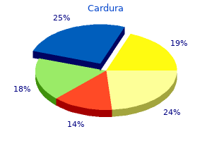
Cardura packs: 30 pills, 60 pills, 90 pills, 120 pills, 180 pills, 270 pills, 360 pills

Generic cardura 1 mg amex
Any injury to the iris arrhythmia general anesthesia generic cardura 4 mg without prescription, especially angle recession blood pressure medication kosar discount cardura 4 mg online, results in a haemorrhage within the anterior chamber (hyphaema). Topical and systemic antiglaucoma remedy is used if the intraocular stress is raised. Aspirin is to be prevented and remedy with sodium edetate could also be used to prevent rebleeding in extreme circumstances. Atropine should be instilled in iridodialysis, but avoided in ruptures of the iris or if the lens is subluxated. When the eye has settled, if the iridodialysis is gross and causes symptoms similar to diplopia, the torn peripheral edge of the iris could additionally be anchored with a 9-0 or 10-0 prolene suture right into a scleral incision just behind the limbus. It often has about the identical diameter as the contracted pupil, and is due to the impression of the iris on the lens, produced by the pressure of the blow driving the cornea and iris backwards. Minute, discrete subcapsular opacities could also be seen after resorption of the pigment. There is a tear between the circular and longitudinal muscle tissue of the ciliary body to the proper of the picture resulting in a widening of the grey ciliary physique band. A Vossius ring and inferior subluxation of the lens seen as a deepening of the anterior chamber. They frequently occur on the thinnest portion of the capsule overlaying the posterior pole of the lens. In these circumstances the entrance of aqueous is stopped and the opacity within the lens may remain stationary or even regress. Alternatively, the tear could remain open and opacification may progress to contain the entire lens. In this condition an accumulation of fluid marks out the architectural arrangement of the lens (see Chapter 1, Embryology and Anatomy). The star-shaped cortical sutures are therefore delineated and feathery traces of opacities outlining the lens fibres radiate from them. The rosette may often disappear, remain stationary or progress to total opacification of the lens-a complication which may appear quickly within a few hours after the injury, or may be delayed for a lot of months. A late rosette-shaped cataract could develop within the posterior cortex 1 or 2 years after a concussion. It is smaller and extra compact than the early sort and its sutural extensions are brief. Any surgical interference ought to be delayed for some months until the final outcome is apparent. If potential, the eye should be left till all signs of irritation have subsided, following which it must be treated as indicated for unilateral cataract. Posterior capsular integrity must be assessed by ultrasonography in order that the sort of cataract surgery employed and the intraocular lens to be inserted may be tailor-made to the attention. Dislocation of the Lens this will likely occur when the relatively fragile suspensory ligament or zonules are torn by the to-and-fro wave of strain set up by the contusion. This leads to a variation within the depth of the anterior chamber, which is deeper in the half unsupported by the lens. With the pupil dilated, the sting of the lens may be seen as a grey convex line by oblique illumination, but is extra readily and unmistakably identified as a black line with the ophthalmoscope. If the rupture to the suspensory ligament is full the lens is dislocated, often into the vitreous. Sometimes it stays clear and may be seen solely with problem, at different instances it turns opaque and appears as a yellow mass. It is extra globular than regular owing to its freedom from the restraint of the suspensory ligament, and when still clear, looks like a globule of oil in the anterior chamber. With indirect illumination it has a golden rim, due to total reflection of the sunshine. An iridocyclitis or an intractable secondary glaucoma is then set up in order that imaginative and prescient is usually fully misplaced if the anteriorly dislocated lens is allowed to remain in that place. The slackening of the suspensory ligament causes elevated curvature and lenticular myopia which, nonetheless, could additionally be greater than compensated by its backward displacement. If the lens is displaced a lot laterally that the edge crosses the pupil, uniocular diplopia is current. Through the aphakic space of the pupil the attention is highly hypermetropic, via the phakic portion it may be myopic, in addition to which the periphery of the lens acts as a prism. Ophthalmoscopic examination by the indirect methodology in these situations exhibits two photographs of the disc differing significantly in measurement, and by the direct technique the fundus may be noticed by way of the phakic or aphakic portion of the pupil. Treatment In forward dislocation the lens ought to be extracted by a cryoprobe or vectis combined with anterior vitrectomy as early as potential. Vision may be improved by appropriate glasses in cases of whole luxation into the vitreous and subluxation. A lens dislocated into the vitreous should be left there, but when uveitis or glaucoma supervene, extraction of the lens is necessary, along with a vitrectomy. Vitreous the vitreous is often disorganized to some extent by both anterior or posterior detachment or by a combination of both. The most typical occurrence is the appearance of clouds of fantastic pigmentary opacities. The vitreous framework, when examined with the slit-lamp, is bespangled with innumerable golden-brown dots derived from the uvea. The complete vitreous chamber could additionally be filled with blood so that no reflex is obtained with the ophthalmoscope, however with indirect illumination a boring purple hue may be seen, particularly if the pupil is dilated. Traumatic Macular Degeneration this will likely often seem slight and the fine pigmentary modifications are easily ignored instantly after the accident. The pigmentation, however, which mainly aggregates at the fovea, has a tendency to improve progressively and has a critical and permanent impact on central vision. Alternatively, oedema could result in cystic changes at the macula and, on the rupture of a cyst, a macular hole may be formed. This seems clinically as a round or oval, deeply red patch, as if a gap has been punched out. In the stage of cyst formation some central imaginative and prescient may stay, but when a hole is formed centrally, imaginative and prescient is misplaced. Retinal breaks may be induced and are generally superonasal dialyses caused by differential traction at the vitreous base. Retinal breaks may be precipitated in eyes already suffering from myopia or different peripheral retinal degenerations. Occasionally, notably in concussion accidents related to gunshot wounds, a rupture of the retina is related to an analogous rupture of the choroid. Such instances current a characteristic image of traumatic proliferative chorioretinopathy secondary to haemorrhage into the vitreous, leading to traction bands. Rupture of the Choroids this follows a severe contusion by a blunt body striking the front of the attention. These are treated conservatively with steroids to decrease inflammatory modifications and the extent of later chorioretinal scarring. Retina the retina may undergo oedematous or degenerative changes, be torn, or haemorrhages might happen in it.
2 mg cardura generic free shipping
Wounds involving the ciliary physique and leading to arrhythmia junctional cardura 1 mg with amex its incarceration within the scar pulse pressure 14 cardura 1 mg generic without a prescription, have at all times been considered significantly harmful. Incarceration of the iris or lens capsule are additionally extra prone to set up sympathetic ophthalmitis than others. Sympathetic ophthalmitis very hardly ever happens if precise suppuration has taken place within the exciting eye. It usually begins 4�8 weeks after the injury to the primary eye (the exciting eye) has taken place, not often earlier. The onset has been reported to occur as early as 9 days after the accident and may be delayed for many months or even years, with 80% occurring inside three months of the harm. Aetiology the aetiology of the condition is unknown but is considered to be an autoimmune, T cell-mediated illness. Uveal pigment can act as an allergen and individuals who endure from sympathetic diseases show a pores and skin sensitivity to it. Pathology Pathologically, the microscopic options in both the exciting and the sympathizing eyes are the same. In the earliest stages, examination exhibits nodular aggregations of lymphocytes and plasma cells scattered throughout the uveal tract. The pigment epithelium of the iris and ciliary body proliferates to kind nodular aggregations (Dalen�Fuchs nodules) and the tissues turn into invaded by lymphocytes and epithelioid cells. The retina can be heavily infiltrated, particularly in the neighbourhood of the vessels. In sympathetic ophthalmitis, the plastic iridocyclitis differs clinically in no respect from this form of irido-cyclitis as a result of other causes. Prodromal signs are sensitivity to light and transient indistinctness of close to objects as a end result of weak spot of lodging. The prodromal symptoms might occur in intermittent assaults, unfold over a considerable period of time. In different circumstances, the affected person first seeks advice for photophobia and lacrimation, or faulty vision in the unhurt eye (sympathetic irritation). When totally developed, all the indicators and symptoms of granulomatous uveitis are present, varying in degree in accordance with the severity of the case. Cases showing little exudation however a deep anterior chamber and keratic precipitates have a more favourable prognosis, but they might at any moment develop into the extreme plastic kind. In every case of penetrating harm, with or without the retention of a overseas physique, prophylactic and long-term therapy, including the topical and systemic administration of steroids, could also be adopted for a time. The chief causes which extend irritation are entanglement of the iris or ciliary body or lens capsule within the wound. It must be remembered that kids are extra vulnerable to the disease than adults. Sympathetic ophthalmitis not often happens after the excision of an injured eye unless it has already commenced on the time of operation. Special consideration should be directed to the presence or absence of keratic precipitates on the again of the cornea. In the rarer circumstances of delayed sympathetic ophthalmitis, the exciting eye could have passed right into a quiescent state. The thrilling eye, while showing evident traces of old iridocyclitis, should still possess helpful imaginative and prescient. The therapy of sympathetic iridocyclitis is that of iridocyclitis generally with the proviso that steroid preparations have a more dramatic effect than in most different ocular inflammations. At the earliest suggestion of inflammation, steroids must be given systemically in large doses-intravenous methylprednisolone 1 g adopted by one hundred mg of prednisone orally tapered off slowly. In all cases, 15�20 mg of prednisone ought to be continued for so much of months lest relapses observe its cessation, and the eye ought to be watched over a interval of years. Daily doses of oral steroids are employed initially however later it must be possible to change to alternate day steroid therapy. The use of steroids has fully altered the prognosis of this disease if such treatment is commenced early. If, nevertheless, the irritation has taken a agency maintain and the uvea is closely infiltrated, the outlook for imaginative and prescient is way less hopeful. Steroid-resistant instances or these with extreme corticosteroid-related side-effects require immunosuppressive remedy. Oral cyclosporin A specifically impacts T cell-mediated immuno-inflammation and is beneficial in severe instances of sympathetic ophthalmitis as an adjunct to corticosteroids. The eyes being small and delicate structures, typically even apparently minor trauma can have devastating effects. The eyes have to be totally irrigated with water or physiologic options like saline or ringer lactate, if out there. After instillation of an antibiotic ointment, the eye should be patched and the patient referred to an ophthalmologist. In case of lacerating injuries, a booster dose of tetanus toxoid and an intravenous dose of broad spectrum antibiotic must be administered, no topical drugs are to be utilized, the attention patched and patient referred urgently to the nearest eye specialist. Such instances should be asked to stay fasting to allow prompt repair under common anaesthesia as quickly as potential. Prompt recognition with early establishment of steroid therapy helps to salvage imaginative and prescient. When he was 39 years old, his left eye was removed due to phthisis bulbi, and ocular inflammation occurred with visible decline to 20/40 in the best eye. Despite 1 yr of intensive corticosteroid and cyclophosphamide (Cytoxan) systemic remedy, coupled with three plasmaphereses, the sympathetic ophthalmia in his proper eye was not introduced under control. At the time of referral, there have been many yellowish confluent and nonconfluent choroidal infiltrates in the right eye, most pronounced, as depicted right here, nasal to the optic disc. There is more swelling of the optic nerve head, enlargement of the choroidal infiltrates, and extension of the method in a circumpapillary trend toward the temporal papillomacular area. At the equatorial region, there are myriad small yellowish infiltrates at the degree of the retinal pigment epithelium, similar to Dalen�Fuchs nodules. Cyclosporine (200 mg/day) was launched along with prednisone, and a outstanding enchancment in the condition was achieved. We shall first study the extraocular muscular tissues and then their central nervous control. Position of Eyes in Orbit and in Relation to Each Other the optic axis upon which the cornea and lens are centred passes by way of the centre of rotation of the eye and roughly through the centre of the pupil. The visible axis passes by way of the nodal point and the fovea centralis, thus crossing the optic axis and making a small angle with it. Clinically this angle is assessed on the pupillary plane and is referred to as the angle kappa. In the emmetropic eye, the angle kappa is claimed to be constructive, for the reason that optic axis often cuts the retina inner to the fovea centralis. In hypermetropic eyes the angle kappa can be constructive but higher than in emmetropia and gives the appearance of pseudoexotropia or pseudodivergent squint. In myopia the angle kappa is absent or unfavorable, for the visual axis and the optic axis coincide or the latter cuts the retina exterior to the fovea centralis giving rise to a pseudoesotropia.
Cardura 2 mg buy generic line
It is greatest to study every case periodically blood pressure ranges for dogs 2 mg cardura generic with mastercard, a cautious drawing or medical photograph of the opacities being recorded at each go to hypertension jnc 7 guidelines 2 mg cardura order visa. Another widespread type of cortical senile cataract is a cupuliform cataract, consisting of a dense aggregation of opacities, usually forming a plaque, simply beneath the capsule, normally within the posterior cortex. It is tough to see with the ophthalmoscope but could be detected as a darkish shadow on distant direct ophthalmoscopy. It appears within the beam of the slit-lamp as a yellow layer and is finest seen in retroillumination towards a pink fundus reflex. Examination with this instrument is important since, being close to the nodal level of the eye, the opacity might diminish the imaginative and prescient considerably in older people and the lens may seem comparatively regular on diffuse examination. In senile nuclear sclerosis of the lens or nuclear or sclerotic cataract the other process happens; the traditional tendency of the central nuclear fibres to turn out to be sclerosed is intensified whereas the cortical fibres stay clear. This type of cataract tends to happen sooner than the cortical selection, usually soon after 40 years of age. As time progresses the nucleus becomes diffusely cloudy, the cloudiness spreading gradually towards the cortex, and sometimes it turns into tinted dark brown, dusky red or even black, owing to the deposition within the lens of yellow pigmented proteins derived from the amino acid tryptophan, altered by the motion of daylight (brown cataract: cataracta brunescens; black cataract: cataracta nigra). In maturity the sclerosis might lengthen almost to the capsule in order that the entire lens functions as a nucleus. After inflammations of the anterior phase, a non-descript opacification appears all through the cortex which normally progresses and matures quickly such as is seen in Fuchs heterochromic cyclitis (see Chapter 17). In inflammations or degenerations affecting the posterior segment a characteristic opacification often commences in the posterior a part of the cortex within the axial region (posterior cortical cataract or posterior subcapsular cataract). Ophthalmoscopically, it appears as a vaguely outlined, dark space, and with the slit-lamp the opacity is seen to have irregular borders extending diffusely in the direction of the equator and often axially forwards in course of the nucleus. In the beam of the slit-lamp the opacities have an appearance like breadcrumbs and a attribute rainbow display of colors typically replaces the traditional achromatic sheen (polychromatic lustre). Such a cataract might stay stationary within the posterior cortex for an extended time or even indefinitely; in other circumstances, the opacification spreads peripherally until all of the posterior cortex is affected, and progresses axially till the whole lens is concerned. The complete cataract formed in this manner is normally soft and uniform in look. Even in the early phases, vision is often impaired owing to the place of the opacity close to the nodal point of the eye. The operative prognosis is decided by the causal condition; however the presence of such a cataract, without obvious trigger, ought to at all times call for a cautious examination of the eye for keratic precipitates, pars planitis or different indicators of disease. In diabetic adults, compared to non-diabetics, cataracts are more prevalent, are dependent on the length of diabetes and progress more quickly. The mechanisms are believed to be glycation, carbamylation of crystallins and elevated oxidative harm. True diabetic cataract is a uncommon situation occurring usually in young folks in whom the diabetes is so acute as to disturb grossly the water steadiness of the body. With acceptable remedy to management hyperglycaemia, the speedy development to mature cataract may be arrested at this stage. Parathyroid Tetany Cataractous changes could occur due to hypo-calcaemia when the parathyroid glands turn out to be atrophic or have been inadvertently removed in the course of a thyroidectomy. Development of a cataract could also be prevented by the administration of parathyroid hormone and calcium. Clouds of small discrete opacities seem in the cortex separated from the capsule by a clear zone. These coalesce to kind large, glistening, crystalline flakes and inside 6 months the lens is usually opaque. Myotonic Dystrophy Characteristic cataracts may develop with myotonic dystrophy and could additionally be an early and outstanding feature in 90% of sufferers. In a sharply restricted zone of the cortex underneath the capsule each anteriorly and posteriorly, fantastic dust-like opacities seem interspersed with tiny iridescent spots. As the opacities mature a attribute stellate opacity appears on the posterior pole. Galactokinase deficiency is a milder disorder associated with galactosaemia and cataract, but without the opposite systemic manifestations. Galactosaemia is frequently associated with the development of bilateral cataract in early life. The cataract is usually an anterior and posterior subcapsular lamellar opacity at first, which later becomes nuclear earlier than it extends to ultimately turn out to be whole. Progression of cataract can be prevented and typically regression could occur if milk and milk merchandise are eliminated from the food regimen in the early phases; otherwise, if the affected person survives, surgical treatment should be adopted. Down Syndrome Children with Down syndrome may have punctate subcapsular cataracts. Galactosaemia this is an autosomal recessive, inherited congenital illness characterized by an inborn lack of ability of the infant to metabolize galactose. An absence of one of the three enzymes concerned within the conversion of galactose into glucose leads to a rise in galactose ranges in the blood and an accumulation of galactitol (sugar alcohol of galactose) within the lens, leading to an osmotic swelling of the lens fibres. The scientific features manifest in infancy with failure to Atopic Cataract Cataract appears regularly in these affected by extreme and widespread skin diseases-atopic eczema, poikiloderma vasculare atrophicans, scleroderma, keratosis follicularis, and others. Ultraviolet mild has been implicated as an element in the aetiology of senile cataract, a suggestion due largely to the frequent occurrence of this situation in tropical nations similar to India and Northern Australia. The average age of onset of age-related cataract in these international locations is 10 years youthful than in Europe and North America. Heat (Infrared) Cataract this may be a attribute situation which can be induced experimentally in animals and happens clinically in industry. The warmth acts not directly on the lens but is absorbed by the pigment of the iris and ciliary body and thus influences the fibres of the lens indirectly; it has thus been discovered inconceivable to produce such cataracts experimentally in flippantly pigmented or albino animals. In addition, the zonular lamella of the capsule could additionally be exfoliated, typically in giant sheets which curl up in the pupillary area. It additionally occurs in sure ironworkers, particularly tin-plate millmen and chainmakers. The attribute adjustments seem to be as a outcome of the direct action of the rays on the dividing cells and developing fibres of the lens itself. The initial modifications are found close to the equator shortly after radiation, and the first medical evidences are apparent in the cortex near the posterior pole solely after a interval of 1 or 2 years when the equatorial cells have migrated posteriorly. Such cataracts have also developed in staff in atomic vitality vegetation and occurred among the many survivors of the atomic bombs released over Japan within the Second World War. Developmental cataract thus tends to observe the architectural pattern of the lens and from its location an estimate could be made from the stage of growth at which the anomaly occurred. The deleterious influences which can cause such developmental anomalies are but largely unknown. Maternal (and infantile) malnutrition is probably one, as in zonular cataract; maternal infections by viruses one other, as in rubella; poor oxygenation owing to placental haemorrhages probably a third. Such cataracts are likely to be stationary, although progressive opacification of a senile sort can be well-known. Punctate Cataract this is the most typical manifestation and, in minute degrees, is nearly common in occurrence. Electric Cataract this will develop rapidly after the passage through the body of a powerful electrical present as from a flash of lightning or the short-circuiting of a high-voltage present.
Cheap cardura 4 mg with mastercard
Patients exhibit encephalitic and paralytic syndrome blood pressure medication blue pill cheap cardura 1 mg free shipping, which follows a prodrome stage with fever heart attack 49ers cardura 1 mg order without prescription, headache, and paresthesia on the site of the bite. Brain abscess is a uncommon however life-threatening situation, which can be indolent or fulminant, and occurs as a end result of otitis media, paranasal sinus infection, and epidural abscess. It is characterised by a focal cerebral lesion with a hypodense middle and a peripheral ring enhancement. Recent information point out that the unfold of bacterial an infection and development of mind abscess happen as a consequence of brain barrier dysfunction. As a remedy, the abscess could be aspirated, adopted by cephalosporin or penicillin with metronidazole. It affects the white matter of the parietal and occipital lobes and destroys the oligodendrocytes, producing intranuclear inclusions. Trypanosomiasis is a critical parasitic condition by which trypanosoma protozoa are found in brain tissue within the late stage of the disease. The subfornical organ is located close to the anterior pole of thalamus and the interventricular foramen of Monro, maintaining large projections to the supraoptic and paraventricular nuclei and the lateral hypothalamic space. Through these connections, it plays an necessary role in homeostasis, osmoregulation, and the circulation of blood in the choroid plexus. It is delicate to apomorphine and digitalis glycosides, regulating meals and water intake and cardiovascular functions. The neurohypophysis receives terminals of the hypothalamic neurons that convey oxytocin, neurophysin, and vasopressin via the hypothalamohypophyseal tract. The medial eminence serves as a hyperlink and transducer between the hypothalamic neurons that secrete the hormoneregulating components and the portal�hypophyseal system. The subcommissural organ is situated ventral to the posterior commissure and at the website of junction of the third ventricle and cerebral aqueduct. The cerebral hemispheres, corpus callosum, and part of the cerebellum are removed. It accommodates trace quantities of protein, primarily immunoglobulin and, to a lesser, extent albumin. It additionally accommodates a few leukocytes and a small quantity of glucose (80�120 mg/dL) and potassium (2. This is in distinction to serum, which accommodates a decrease sodium focus and better potassium and calcium. A low pH worth could occur in circumstances associated with acidosis and hypercapnia and in certain pulmonary illnesses. Increased protein focus is observed in spinal shock (complete transection of spinal cord) and in circumstances of extramedullary and intramedullary spinal cord tumors. Note the cuboidal cells of the choroid epithelium and related capillary community. This plexus is innervated by the postsynaptic sympathetic fibers that emanate from the superior cervical ganglion of the sympathetic trunk. Selective prevention of the free entrance of protein and electrolytes from the blood to brain tissue is maintained by the ependymal cells that type tight junctions. Hardened bodies known as psammoma (sand-like), that are composed of concentric rings of calcium carbonate, calcium, and magnesium phosphate, happen usually within the grownup choroid plexus. A vast variety of 5-hydroxytryptamine receptors could affect the blood circulate within the choroid plexus. This allows the creation of an environment friendly milieu for the conduction of nerve impulses. It also acts as a buffer to reduce the influence of head trauma (buoyancy effect), offers vitamins to the leptomeninges, and dramatically reduces the burden of the mind. This stress remains fixed at 50�200 mm of water unless a rise or decrease in mind size or blood volume occurs. It occurs extra often in women than males, particularly in obese ladies, but youngsters can be affected. Patients with hyperparathyroidism, Cushing and Addison disease, and iron-deficiency anemia and pregnant women are notably prone to the development of this illness. Vomiting, diplopia because of abducens nerve palsy, generalized weak spot, pulsatile tinnitus anosmia, and ataxia are additionally seen in sufferers with this disease. Lateral ventricle Hydrocephalus may be congenital or acquired: Congenital hydrocephalus (overt-infantile hydrocephalus) happens normally within the first few months of life or contained in the uterus. It can additionally be related to enlargement of the head, thinning of the scalp, visible superficial vessels, and downward (setting solar sign) place of the eyes. Parinaud syndrome, which exhibits vertical gaze palsy, may also be seen on account of the stress exerted on the pretectal area. It is attention-grabbing to observe that the cerebral cortex is less affected than the white matter, and plenty of sufferers survive this condition due to the capability of the skull to expand. Acquired (occult) hydrocephalus could additionally be caused by subarachnoid hemorrhage, postmeningitis, cysticercoids, vascular malformation, as well as tumors of the third ventricle, thalamus, and cerebral hemispheres. It is characterized by visible papilledema and fewer distinguished frontal and occipital headache. It reveals signs of frontal lobe dysfunctions such as inattentiveness, marked slowness in response, and easy distractibility. In older kids and adults with inflexible calvaria, gait apraxia and amnesia adopted by dementia and slowing of thought course of and, later, urinary incontinence may also happen. Imaging of the brain could reveal herniation of the third ventricle, erosion of the sella turcica, and atrophy of the corpus callosum. The onset of this condition is subacute and might cause mental deterioration adopted by a restriction of motion. Surgical therapy involves ventriculoperitoneal shunting, which is essentially the most commonly used method in adults. While enchancment of gait is less probably, urinary incontinence is the most likely symptom to improve with shunting, while dementia is the least prone to endure any adjustments. It might occur as a end result of stenosis of the cerebral aqueduct, atresia of foramina of Magendie and Luschka, obstruction of the fourth ventricle (as in Arnold�Chiari syndrome), or obstruction of the interventricular foramen of Monro. Obstructive hydrocephalus may be divided into speaking and noncommunicating sorts. Action monitoring and medial frontal cortex: Leading function of supplementary motor area. The circumventricular organs of the mammalian mind with special reference to monoaminergic innervation. Callosal disconnection syndrome in a patient with corpus callosum hemorrhage: A diffusion tensor tractography research. Does perinatal exposure to endocrine disruptors induce autism spectrum and a spotlight deficit hyperactivity issues Decorticate, decerebrate and opisthotonic posturing and seizures in Kenyan kids with cerebral malaria. Marchi N, Angelov L, Masaryk T, Fazio V, Granata T, Hernandez N, Hallene K, Diglaw T, Franic L, Najm I, Janigro D. Executive operate and magnetic resonance imaging subcortical hyperintensities in Vascular dementia.
Cardura 2 mg discount overnight delivery
The outcomes of operations undertaken for the management of glaucoma can only management the issue of intraocular strain arrhythmia dance company 4 mg cardura with visa. If the deterioration in vision is due primarily to a raised intraocular stress arrhythmia uk cardura 2 mg fast delivery, its surgical relief will often prevent further loss; if the intraocular strain is low and the deterioration is actually as a result of different vascular or neurogenic elements, the vision will most likely proceed to deteriorate in spite of the operation. The prognosis thus depends largely on early analysis and the institution of early and sufficient therapy to forestall cupping of the optic disc and loss of the visual field. Continuous monitoring of intraocular stress, optic nerve head and perimetry will enable the detection of tolerance to medications or progression of the glaucoma. To determine the progression of visual area defects in glaucoma, one should establish a baseline by doing no less than three chartings of the visible field in a newly recognized patient of glaucoma. Disease progression is finest assessed if the follow-up programmes and all parameters are the same as those used for the baseline. If a change in the visual field is detected at any time throughout follow-up, it should be confirmed by another check and correlated with any related clinical finding. Typically, six or more examinations at 3�6-month intervals are required to confirm progressive deterioration. The first step in evaluating the progression of a visible area defect is to research a collection of chronologically organized visible field recordings of a particular patient. Overview printouts are helpful for this purpose, as they place the grey-scale threshold worth table and likelihood plots of total and sample deviation of every examination in a row, with the rows arranged in chronological order, making it easier to scan a series of recordings. Statistical packages are available within the form of field plots which analyse changes within the global indices. The onset of retinal vascular issues also can typically mislead a glaucomatologist. Summary Glaucoma is a continual, progressive optic neuropathy with raised intraocular strain as the primary danger issue. There is a mismatch between the stress in the eye and that which the axons of the ganglion cells or optic nerve can stand up to. Open angle glaucomas could be managed medically, but surgery may be essential if not adequately controlled. Angle closure glaucomas need an initial laser iridotomy followed by medical or surgical remedy. Tonometry, optic nerve head imaging and serial perimetry are parameters used to monitor the effect of remedy which is usually lifelong. It is often a silent disease, in order that correct screening and early analysis are important. The retina is divided into a variety of zones for convenience of recording clinical findings and to allow a exact localization of retinal disorders. The retinal equator is taken into account to lie consistent with the exit of the four vortex veins and the retina posterior to that is called the posterior retina. Examination of the posterior part of the retina is undertaken with using a direct ophthalmoscope and by slit-lamp indirect biomicroscopy if additional magnification or a binocular view is required. The macula lutea (clinical posterior pole) is an space of the retina approximately 5. Within the macular area is a small, central depression known as foveola, measuring approximately zero. The imaginative and prescient is most acute on the foveola, where solely cones are discovered as every cone instantly relays to a single ganglion cell. The macular area is equipped by twigs from the superior and inferior temporal arteries, and by small branches coming straight from the disc. Occasionally, small arteries (cilioretinal) originating from the brief posterior ciliary arteries run inwards to enter the attention (near the edge of the disc) and then bend sharply outwards in course of the macula. Each trunk often divides into two, one of which sweeps up (or down) in path of the temporal facet, the opposite up (or down) in direction of the nasal side-the superior and inferior temporal and nasal arteries and veins. These divide dichotomously into innumerable branches, the mode of division being subject to nice variations, but the nasal branches run more radially than the temporal, which make a decided sweep to keep away from the macula. All the retinal vessels could have a shiny silvery streak operating longitudinally down the centre, which is extra prominent with the arteries and is due to reflection of light from the convex cylindrical surface. In some 80�90% of individuals, however, retinal venous pulsation may be seen at or close to the sting of the disc or, indeed, wherever the veins take a really sharp bend; as a result of the effect of the intraocular pressure. With each arterial pulsation, the intraocular strain raises slightly, and this increased stress on the skin of the walls of the veins tends to make them collapse. This causes a momentary impedance to the outflow of blood during systole, but the venous circulation recovers itself in the course of the arterial diastole. This strain occurs during the diastolic phase and due to this fact has been called the adverse venous pulse. If absent, the venous pulsation can be elevated or made manifest by increasing the intraocular strain by slight pressure with the finger on the globe. Ora serrata: the peripheral retina is the world bounded by the equator and the ora serrata. The ora serrata marks the tip of the choroid and retina and is grey to brownish-black in color. Retinal affections generally give rise to the next signs, only a few of which want be current in individual circumstances. There could additionally be a concentric constriction of the field of vision or scotomata could additionally be current corresponding with the areas affected. The retinal adjustments most likely originate from a state of anoxia which ends up in an increased permeability of the capillaries, the formation of a number of microaneurysms and local degenerative modifications. They are commonly small, but could type accumulations larger than the disc, and, since they disappear rapidly, they incessantly change their shape. They are formed by the arrest of axoplasmic circulate on the fringe of an ischaemic area. These hyaline or lipid deposits are generally seen as a cluster around a gaggle of leaking microaneurysms. In the macular region they have a tendency to accumulate in a radial manner around the foveal centre, the arrangement mirroring the orientation of the fibre layer of Henle to type a fan- or star-shaped determine (macular fan/star). In hypertension with involutionary sclerosis, occurring in older sufferers, the image of arteriosclerotic retinopathy appears. Changes at the arteriovenous crossings are diagnostic- nipping and a perpendicular placement of the veins-Gunn signal. In arteriolar (diffuse hyperplastic) sclerosis occurring in younger patients, the relatively youthful arterioles reply to the hypertension by proliferative and fibrous adjustments mainly affecting the media. They are slender and tortuous with nicking on the arteriovenous crossings; multiple haemorrhages are present with, within the early levels, oedema and cotton-wool patches and, within the later, exhausting exudates scattered diffusely but often forming a macular star. Malignant hypertension is an expression of accelerated development of the hypertensive state in a affected person with relatively younger arteries, undefended by sclerosis.
Cardura 2 mg order mastercard
The aponeurosis should be rigorously identified heart attack exo lyrics cardura 4 mg order on-line, the lid everted and the conjunctiva above the tarsal border ballooned with saline blood pressure lying down 1 mg cardura fast delivery. A small buttonhole incision is made through the conjunctiva on the temporal side and blunt scissors passed throughout, watching the blades via the thin conjunctiva. A ptosis clamp is passed because the scissors are withdrawn with one blade beneath the conjunctiva and the opposite on prime of the aponeurosis. With the aponeurosis in the ptosis clamp and all tissues freed from each surfaces, the horns should be rigorously incised in order to not harm the superior indirect tendon or the lacrimal gland. Three double-armed 6-0 chromic intestine sutures are passed by way of the aponeurosis from under upwards and tied securely with three knots. One needle from each of the double-armed sutures is handed by way of the outer layer of the tarsus parallel to the lid border and approximately four mm from the lid margin. Three or more additional interrupted 6-0 chromic gut sutures are added to guarantee agency fixation of the entire aponeurosis. A good fold is produced with a 6-0 silk suture for adults and vicryl or chromic catgut for youngsters. To produce a agency scar every suture ought to move through the depth of the wound about 1. When the suture is drawn up and tied, the pores and skin is firmly anchored to the aponeurosis producing a everlasting fold. A 4-0 silk Frost-type suture is inserted under the lash line for no much less than 10 mm and introduced as much as be handed by way of the pores and skin slightly below the forehead. Levator plication: In cases of delicate to average ptosis, the levator as a substitute of being resected may be merely doublebreasted over itself to produce a good outcome. Advantages embrace much less time, simpler method and no in depth dissection to isolate the levator before resection. It should be remembered that levator plication can provide good results provided that the levator perform is good/fair. The process includes isolating the levator muscle and placing three mattress sutures through the higher border of the tarsus and at acceptable vertical top of the levator. Once the correct lift is obtained then one suture is handed medially and one laterally to give a proper lid contour. This may be carried out by the use of sutures, fascia lata sling or silicon bands. If a levator resection has produced a light undercorrection a Supramid suture could additionally be used. A 4-0 Supramid additional suture with swedged-on needles facilitates easy passage by way of the outer layers of the tarsus. Infection of the suture mattress is an annoying complication and the suture should be guarded fastidiously, avoided the pores and skin and lashes during surgery and the wound in the brow must be carefully closed. To cut back the scar on the brow, a deeply placed mattress suture of 6-0 silk or Mersilene brings the muscle, fascia and skin together. A brief deep incision is made 5 mm above the medial and lateral portions of the brow. The fascial strips are drawn via the openings in the lid and then upward rising with two ends in each of the openings above the brow. The fascial strips are secured with 5-0 chromic catgut or vicryl at every brow incision. The forehead wounds are closed with deep mattress sutures adopted by a Frost-type 4-0 silk suture within the decrease lid. The sutures which be a part of the perimeters of the pores and skin incision are carried by way of the deep tissues in such a way as to stretch the skin over the tarsus and to produce a fold within the skin of the eyelid within the normal position. Acquired Ptosis Acquired ptosis is usually unilateral and its trigger must be recognized so that acceptable remedy may be instituted. Neurogenic ptosis: It may be part of the symptomcomplex involving the complete third nerve at any point in its path, or hardly ever it might be due to affection of the department supplying the levator. Isolated ptosis with out other signs of oculomotor paralysis may outcome from illness of the supranuclear pathways (see Chapter 31, Diseases of the Nervous System with Ocular Manifestations). Horner syndrome is a standard reason for neurogenic ptosis and is accompanied by miosis and anhidrosis, as sympathetic innervation is lowered. It may also be due to direct harm of the muscle or its nerve supply, as by wounds or fractures. In full paralysis of the third nerve, surgical procedure is normally contraindicated until strabismus has been corrected, since if the lid is raised in these cases diplopia turns into manifest. Bilateral, symmetrical ptosis might happen in myotonic dystrophy and persistent progressive exophthalmoplegia asymmetry is more often seen in myasthenia gravis. Treatment of the first disorder should be undertaken first, adopted by crutch spectacles to help raise the lid. Myasthenia gravis is a illness characterized by generalized muscular weakness and rapidly creating fatigue of the muscles due to an auto-immune dysfunction during which harm of the acetylcholine receptors takes place at the post-synaptic membrane. Anti-acetylcholine receptor antibodies are found in roughly 80% of sufferers with myasthenia, the degrees correlating with the severity of the disease. Attempts can be made to modify the defective immune mechanism with corticosteroids, immunosuppressives, plasmapheresis and thymectomy. The signs fluctuate and, after a short relaxation, restoration follows rapidly within the early levels. The ptosis is nearly always bilateral and is elevated by extended fixation or makes an attempt to look upwards; efficient compensation by overaction of the frontales is unimaginable. Thus, acetylcholine briefly accumulates in greater than regular amounts in the ganglia, post-ganglionic sympathetic nerve endings and in neuromuscular junctions in all types of muscle. The resultant improve in acetylcholine available at the receptor websites results in an improvement in the muscular operate, confirming the diagnosis. Aponeurotic ptosis is involutional, and is because of a weak point or disinsertion of the levator palpebrae superioris aponeurosis from the anterior floor of the tarsus. The diagnostic characteristic of this form of ptosis is the presence of a excessive lid fold with good levator motion. Surgery consists of re-insertion of the levator aponeurosis to the anterior floor of the tarsus and applicable resection of the levator. Small clear cysts frequently occur among the lashes in old folks, because of the retention of secretion of Moll glands. Xanthelasma or Xanthoma Xanthelasma or xanthoma are slightly raised yellow plaques, mostly found within the upper and lower lids near the internal canthus, and infrequently symmetrical in the two lids and on both sides. They are often seen in elderly girls, and are typically associated with diabetes and hypercholesterolaemia. Naevus or Mole Usually pigmented, this will occur on the lids, usually affects the margin and includes each the pores and skin and conjunctiva. Two may be symmetrically situated on the lids of the same eye, indicating their origin at a time when the lids had been nonetheless united. The microscopic appearance is characteristic, consisting of naevus cells, usually organized in an alveolar method.

1 mg cardura order
The inferior hypophyseal arteries kind an arterial ring across the infundibular stem and supply blood provide to the lower infundibulum and the posterior lobe prehypertension 2014 2 mg cardura order amex. These arteries establish numerous anastomoses and department repeatedly heart attack 4sh discount 1 mg cardura fast delivery, terminating into capillaries and capillary sinusoids in the medial eminence and the infundibular stem. These capillary sinusoids are the beginning of the hypophyseal portal system, which conveys blood to the epithelial tissue of the anterior lobe through major and secondary capillary venous plexuses. The adenohypophysis contains the anterior lobe, intermediate part, and tuberal area. It secretes somatotropin (growth hormone), thyrotropin, prolactin or luteotropin, adrenocorticotropin, luteinizing hormone (interstitial cell� stimulating hormone), follicle-stimulating hormone, and melanocyte-stimulating hormone. The hormone regulating elements of the hypothalamic tuberal nuclei regulate the adenohypophysis. The tuberal nuclei project to the medial eminence via the tuberoinfundibular tract and regulate the glandular function of the adenohypophysis by way of the portal hypophyseal vessels. Approximately half the quantity of the neurohypophysis consists of axonal swellings; the biggest are the Herring our bodies, which may be as massive as erythrocytes. Herring bodies provide a source of secretory granules and have a longer life span (more than 2 weeks). These hormones are secreted by the paraventricular and the supraoptic nuclei of the hypothalamus. During suckling, stimulation of the sensory nerve endings within the feminine nipple and areola activates the spinoreticular fibers, which subsequently relay the generated impulses to the supraoptic and paraventricular nuclei through the dorsal longitudinal fasciculus. The infundibulum primarily consists of fibers of the hypothalamo-hypophyseal tracts that project to the neurohypophysis. Pituitary gland dysfunctions are associated with quite lots of pathological circumstances and syndromes. Some of those circumstances primarily affect the adenohypophysis, while others may be confined to the neurohypophysis. It is associated with hypopituitarism of the anterior lobe and is noticed in individuals with postpartum hemorrhage and spasm of the infundibular arteries. Adenoma of the anterior pituitary produces quite lots of signs, depending upon the kind of tumor, which can embrace acromegaly and/or gigantism (enlargement of the face, hand, and feet); impotence; amenorrhea (cessation of menses); galactorrhea; or Cushing disease. The latter is characterized by moon facies, weight problems with distinguished fats pad within the neck and shoulder, hypertension, renal calculi, and irregular menses. Pituitary tumors most often arise from the anterior lobe and are categorised, based on the degree of their endocrine activity, into endocrine-active and endocrine-inactive tumors. Endocrine-active tumors are, for the most half, microadenomas and may be detected by radiographic imaging of the sella turcica. They may enhance secretion of many hormones such as the growth hormone, producing acromegaly or gigantism. On the opposite hand, some tumors could end in undersecretion of hormones, producing, for instance, dwarfisim (abnormally quick body stature) in childhood, amongst other deficits. Endocrine-inactive tumors, by contrast, turn into clinically vital solely upon enlargement, thus compressing the adjoining nerves or brain tissue. These tumors could produce headache, visual disturbances, and extraocular motor palsies. Surgical removing of these tumors could also be achieved by transsphenoidal approach; nonetheless, in depth subfrontal or parasellar enlargement of the tumor could require a transfrontal method. Tumors that develop laterally or wedge under the optic nerve may necessitate craniotomy. Lesions of the posterior lobe might occur in cranium fractures, suprasellar and infrasellar tumors, tuberculosis, vascular disease, or aneurysms. The onset is mostly sudden, and nocturia (excessive urination at night) is a presenting symptom in most patients. It is linked to the habenula and the posterior commissure via the laminae of the pineal stalk. FunctionAl AnD clinicAl consiDerAtion this gland secretes melanocyte-stimulating hormones (lipotropic hormones), which play an analgesic role because of its endorphin content material. Atrium (lateral ventricle) Pineal gland Through these secretions, the pineal gland could exert a regulatory influence, modifying the exercise of the pituitary, adrenal, and parathyroid glands, as well as gonads. Darkness prompts the secretion of melatonin, inhibiting sexual improvement through a series of neuronal chains within the retina, hypothalamus, reticular formation, and spinal cord. Bilateral lesions of the suprachiasmatic nuclei could abolish the rhythmic actions of the N-acetyltransferase and produce low ranges of hydroxyindole-o-methyltransferase exercise, leading to disruption of the circadian rhythms associated with the sleep�wakefulness cycle and spontaneous motor activities that pertain to drinking and feeding. Pinealocytes also contain tryptophan hydroxylase and fragrant amino acid decarboxylase, which are involved within the synthesis of serotonin. Diencephalon 125 the most typical malignant tumor of this gland is germinoma (atypical teratoma), which is incessantly seen in younger males. Pinealoma (pineal gland tumor) may compress the tectum and the posterior commissure, producing signs of Parinaud syndrome, which is characterised by bilateral vertical gaze palsy, hydrocephalus, and lack of pupillary light reflex. Cysts or tumors related to the pineal gland might compress the hypothalamus, resulting in obesity and hypogonadism. Pineal (sand) concretions or corpora arenacea within the astrocytes were considered as essential radiographic sites for detection of a brain shift�associated spaceoccupying intracranial mass on the contralateral side. However, it should even be remembered that due to the comparatively giant size of the proper cerebral hemisphere, a traditional pineal gland could slightly deviate to the left side. It contains afferents to the habenular nuclei from the septal area that receives input from the amygdala, hippocampal formation, and anterior perforated substance. The lateral habenular nucleus receives afferents from the globus pallidus, substantia innominata, tectum, lateral hypothalamus, prepyriform cortex, lateral preoptic space, basal nucleus of Meynert, midbrain raphe nuclei, olfactory tubercle, and pars compacta substantia nigra. Input from the prepyriform cortex, tectum, nucleus Medial pallidal segment Habenulopeduncular tract (fasciculus retrofasciculus) Interpeduncular nucleus Anterior thalamic nucleus, prepyriform cortex, basal nucleus Tectohabenular tract of Meynert, and preoptic areas Stria medullaris thalami Tectum Midbrain reticular formation Hypothalamus Medial habenular nucleus Lateral habenular nucleus Habenular nuclear complex Ar O. However, no recognized symptoms are related to lesions of this commissure, though impairment of visual monitoring motion has been reported. It accommodates the subthalamic nucleus, zona incerta, cranial portions of the pink nucleus, and substantia nigra. It additionally accommodates the spinal and medial lemnisci and efferent fibers from the globus pallidus (ansa lenticularis, lenticular, thalamic, and subthalamic fasciculi) and cerebellum, which are destined to the thalamus. It receives input from the lateral phase of the globus pallidus, reticular formation, and the motor and prefrontal cortices. It initiatives to both segments of the globus pallidus via the subthalamic fasciculus and to the pars reticulata of the substantia nigra. It is also related to the ipsilateral red nucleus, mesencephalic reticular formation, and zona incerta. The subthalamic nucleus integrates motor activities by way of its connections with the basal nuclei, substantia nigra, and tegmentum of the midbrain. The lateral habenular nucleus tasks back to the substantia nigra and also to the midbrain reticular formation and the hypothalamus. The medial habenular nucleus, the smallest component of the habenular nuclear complex, receives fibers from the serotonergic neurons of the midbrain reticular formation and from the septofimbrial nucleus. The latter nucleus receives enter from the amygdala and the hippocampal formation.
2 mg cardura buy with visa
The median nerve can also be entrapped in the bicipital aponeurosis blood pressure essential oils buy cardura 4 mg with mastercard, producing indicators of lacertus fibrosus syndrome heart attack high blood pressure 1 mg cardura order free shipping. This sort of entrapment produces pain upon forced pronation of the flexed and supinated forearm. This kind of harm produces loss or weakened thumb opposition as in thumb pinching (thumb strikes inward throughout the palm to oppose the fifth digit), atrophy of the thenar muscles, and ache and paresthesia or anesthesia in the lateral two-thirds of the palm. Spinal Nerves 243 the palmar surfaces of the thumb, index and center fingers, as properly as the lateral one-half of the ring finger and the dorsal surfaces of the medial four digits so far as the middle phalanges. Weakened thumb abduction (inability to separate the thumb and index finger) and inadequate thumb rotation could produce "bottle signal," a deficit by which the subject is unable to maintain a grip round a bottle. Flattening and atrophy of the thenar eminence and the counter pull of the extensor and the abductor pollicis longus muscle tissue of the thumb end in pulling the thumb to the same level with the opposite digits and producing the ape hand configuration. Compression of the median nerve at the wrist may also occur inside the carpal tunnel producing signs of carpal tunnel syndrome. This syndrome, a unilateral and generally bilateral situation that often includes the dominant hand, is frequent in pregnancy and in middle-aged girls in the course of the premenstrual interval. Since repetitive movements on the wrist displace the flexor tendons towards the palmar aspect of the carpal tunnel, continuous and sustained flexion and extension of the wrist could additionally be a predisposing issue for this condition. Congenital arteriovenous fistula or surgically induced Cimino�Bresica fistula and a persistent median artery can produce compression of the median nerve in the carpal tunnel. Diabetes mellitus, a quantity of myeloma, main amyloidosis, and insect or snake bites can set off manifestation of this situation. Transducers inserted into the canal may measure the rise in intracarpal pressure. Demyelination of the median nerve, subsequent weak point or atrophy of the thenar muscles, and associated autonomic disturbances similar to swelling and alteration in the texture of the skin in areas of distribution of the median nerve are also widespread manifestations of this illness. Opposition and, to a lesser degree, abduction of the thumb might eventually be affected, although variations of manifestations that vary from numbness confined to the center finger or atrophy of the muscle that act on the index finger without thenar involvement also can happen. Anomalous innervation, as in Martin�Gruber anastomosis, have to be ruled out in order to affirm the diagnosis of this situation. In general, injury to the median nerve is commonly related to a burning and tearing ache sensation within the digits and palm of the hand, accompanied by vasomotor and sudomotor adjustments in the cutaneous areas wider than the world of distribution of the median nerve (causalgia). The affected part of the hand and digits becomes extraordinarily sensitive to contact, including contact with garments or air. Sympathectomy or blockade of the corresponding sympathetic ganglia could relieve it. Muscles of the hand that are innervated by the ulnar nerve may be affected if the motor fibers to these muscles run inside the anterior interosseous nerve before joining the ulnar nerve (Martin�Gruber anastomosis). Similarly, digital branches can develop neuropathy in cheerleaders as a consequence of clapping and turning. As a result, the hand turns into hyperextended (unopposed by the flexors), and the forearm assumes supinated place due to paralysis of the pronators. Fingers are hyperextended on the metacarpophalangeal joints due to paralysis of the interossei and lumbricals. Thumb opposition and adduction as well as atrophy of the thenar and hypothenar muscles are additional deficits of this injury. Damage to the axillary nerve might occur as a outcome of anterior inferior dislocation of the humeral head, manipulation to scale back dislocation, fracture of the surgical neck of the humerus, hereditary neuralgic amyotrophy, blunt trauma, although hardly ever, intramuscular injections, or pressure from the usage of crutches. Sleeping in a prone place or lying on a surgical table with an abducted arm above 90� and forearm placed horizontally can traumatize this nerve. Axillary nerve palsy is characterized by atrophy of the deltoid and teres minor, lack of shoulder contour, and severe weak spot of arm abduction up to 90�. Abduction is lost completely due to intactness of the supraspinatus, upper fibers of the trapezius, and serratus anterior muscular tissues. Weakened lateral rotation (due to paralysis of the teres minor) and limited lack of cutaneous sensation from the shoulder are additional deficits of this situation. In quadrangular house syndrome seen in baseball pitchers, the axillary nerve could also be affected along side the posterior humeral circumflex. Patients usually report anterior shoulder ache and paresthesia, which become intense upon abduction and exterior rotation. It derives from the posterior cord and descends posterior to the axillary artery and anterior to the latissimus dorsi and teres main. Before it reaches the posterior surface of the humerus, it maintains important relationships to the lengthy and medial heads of the triceps brachii and additional distally to medial and lateral heads of the same muscle. On the posterior surface of the humerus, it descends within the radial (spiral) sulcus accompanied by the deep brachial vessels. It innervates the triceps brachii by a branch that arises from the radial nerve inside the sulcus and above the mid-diaphysis of the humerus. The web site of origin of this department accounts for intactness of triceps brachii muscle in a fracture involving the radial nerve on the distal third of the humerus. During its course on the lateral aspect of the arm, the radial nerve passes between the brachialis and brachioradialis and then between brachioradialis and extensor carpi radialis longus. In the lower medial arm, the radial nerve offers rise to muscular, cutaneous, and articular branches. The muscular branches innervate the anconeus, brachioradialis, and extensor carpi radialis longus. Cutaneous branches distribute to the posterior arm (posterior brachial cutaneous nerve) and lower lateral arm and posterior forearm (posterior antebrachial cutaneous nerve). Elbow joint is the principle recipient of the sensory articular branches of the radial nerve. At the decrease border of the subscapular muscle, it pursues a curved course and enters the quadrangular space bounded by teres main distally, teres minor proximally, humerus laterally, and the long head of triceps medially. During its course in the quadrangular space, the axillary nerve is accompanied by the posterior humeral circumflex vessels. The anterior department of this nerve supplies the deltoid muscle and the skin that covers the muscle and the upper lateral arm, whereas the posterior branch provides the teres minor and the posterior a part of the deltoid, carrying sensations from the lower lateral a half of the arm. Function of the deltoid is checked by abduction of the arm towards resistance above 90�. To take a look at the operate of the teres minor, the patient is placed on inclined position and asked Spinal Nerves 245 Radial nerve variations in the website of origin of varied branches of the radial nerve. In some people, branches to the supinator and extensor carpi radialis brevis come up from the radial nerve proximal to the arcade of Frohse and before its division into superficial and deep branches directly from the posterior interosseous nerve. The superficial terminal branch descends posterior (deep) to the brachioradialis and lateral to the radial artery after which anterior to the flexor digitorum superficialis and flexor pollicis longus. Proximal to the wrist, it encircles the distal end of the radius and courses deep to the tendon of brachioradialis to achieve access to the dorsum of the hand.