
Ciprofloxacin
Ciprofloxacin dosages: 1000 mg, 750 mg, 500 mg, 250 mg
Ciprofloxacin packs: 30 pills, 60 pills, 90 pills, 120 pills, 180 pills, 270 pills, 360 pills
250 mg ciprofloxacin discount otc
Other intralaminar nuclei receive input from ascending ache pathways and project to the somatosensory and parietal cortex antibiotics for uti flucloxacillin discount ciprofloxacin 500 mg with amex. Each thalamic nucleus (with a few exceptions) provides rise to efferent projections (thalamocortical fibers) that target some portion of the cerebral cortex antibiotic that starts with r purchase ciprofloxacin 750 mg free shipping. That region of cortex then usually supplies a reciprocal projection (corticothalamic fibers) that returns to the unique thalamic nucleus. Some thalamic nuclei are primarily associated with a selected function and in flip with a particular gyrus (and practical area) of the cerebral cortex. The anterior nucleus projects primarily to the cingulate gyrus and functions within the broad space of habits. The nuclei of the thalamus have been classified based on their connections as either relay nuclei or association nuclei. A relay nucleus is one which receives input predominantly from a single source, similar to a sensory pathway or a cerebellar nucleus, or from the basal nuclei. The incoming neural info is processed after which sent to a localized region of sensory, motor, or limbic cortex. However, their position in a modalityspecific pathway linking one particular source to one particular destination makes the word "relay" a useful designation. In distinction, an affiliation nucleus receives input from numerous completely different structures or cortical regions and often sends its output to more than one of many association areas of the cerebral cortex. Association nuclei embrace dorsomedial, lateral dorsal, lateral posterior, and the nuclei of the pulvinar complicated. A thalamic nucleus can be designated specific or nonspecific on the idea of thalamocortical alerts generated in response to electrical stimulation delivered to a localized web site in that thalamic nucleus. Focal electrical stimulation of a specific nucleus produces a rapidly conducted, sharply localized evoked response in the ipsilateral cerebral cortex. Focal electrical stimulation of a nonspecific nucleus produces widespread activity in the cortex of each hemispheres, at a considerably longer time delay than with stimulation of a specific nucleus. It is assumed that nonspecific nuclei play a job in modulating the excitability of enormous areas of cortex. The genu is positioned immediately lateral to the anterior thalamic nucleus, at about the same degree because the interventricular foramen. The anterior limb extends rostrolateral from the genu and is insinuated between the caudate and lenticular nuclei. The posterior limb extends caudolateral from the genu and separates the thalamus from the globus pallidus. As its name implies, the retrolenticular limb is the white matter located immediately caudal to the lenticular nucleus (Latin retro-, for "behind"). Even though this construction consists principally of axons that reciprocally hyperlink the thalamus and cerebral cortex, it additionally accommodates cortical efferent fibers that project to the brainstem (corticorubral, corticoreticular, corticonuclear-corticobulbar) or spinal twine (corticospinal). The hypothalamus and related limbic structures obtain sensory input concerning the inner setting and in turn regulate the motor methods that modify the inner environment via 4 mechanisms. First, the hypothalamus is a principal modulator of autonomic nervous system operate. Third, the hypothalamus regulates the activity of the anterior pituitary through the production of releasing factors (hormone-releasing hormones). Fourth, it performs an endocrine perform by producing and releasing oxytocin and vasopressin into the overall circulation throughout the posterior pituitary. The periventricular zone consists of the neurons that border the ependymal surfaces of the third ventricle. The latter, which kind the medial forebrain bundle, are diffusely organized within the human mind. No discrete named nuclei are present in this lateral space, though the supraoptic nucleus is taken into account by some authorities to be part of it. Cells of the lateral hypothalamic space are involved in cardiovascular operate and within the regulation of meals and water intake. The medial hypothalamic zone contains discrete groups of neurons whose function and connections are established. Nuclei within the chiasmatic area are typically involved in regulating hormone launch (preoptic, supraoptic, periventricular), cardiovascular function (anterior), circadian rhythms (suprachiasmatic), and physique temperature and heat loss mechanisms (preoptic). Bilateral lesions of this hypothalamic region produce hyperphagia, a greatly elevated meals consumption with resultant weight problems. Cells of the arcuate nucleus ship peptides to the portal vessels and, by way of these channels, to the anterior pituitary. Some of those peptides are releasing components, which cause an increase within the secretion of specific hormones by the anterior pituitary, and a few are inhibiting elements, which inhibit the secretion of specific hormones by the anterior pituitary. In people, the mammillary nuclei consist of a large medial nucleus and a small lateral nucleus. Although both of those nuclei obtain enter through the fornix, only the medial nucleus projects to the anterior thalamic nucleus by way of the mammillothalamic tract. The stria medullaris thalami has disappeared at this degree because its fibers have dispersed to finish within the habenular nuclei. The neurons of the posterior nucleus are involved in actions that embrace elevation of blood stress, pupillary dilation, and shivering or physique warmth conservation. The mammillary nuclei are involved in the control of assorted reflexes related to feeding in addition to in mechanisms referring to memory formation. Afferent Fiber Systems Although many axonal techniques lengthen into the hypothalamus, only four inputs are talked about right here (see Chapter 30). As talked about earlier, the medial forebrain bundle passes bidirectionally by way of the lateral hypothalamic area. This composite fiber bundle consists of ascending axons that originate in areas throughout the neuraxis and terminate in the hypothalamus and different axons that exit the hypothalamus to reach forebrain and brainstem targets. Patients with this involuntary movement disorder exhibit rapid and forceful flailing actions, which normally involve the contralateral higher extremity. These movements could be very debilitating as a result of the affected person has no control over their initiation or length. The zona incerta accommodates output neurons that project to quite lots of places, including the cerebral cortex, the superior colliculus, the pretectal area, and the basilar pons. Afferent projections come up from the motor cortex and as collaterals from the medial lemniscus. Several nuclei give rise to descending fibers that contribute to the dorsal longitudinal fasciculus and the medial forebrain bundle and to diffuse projections that cross into the tegmentum. These fiber systems project directly to quite a few brainstem nuclei as nicely as to preganglionic sympathetic and parasympathetic neurons in the spinal twine. Other projections reach the thalamus and frontal cortex, and still others prolong to the posterior pituitary or to the tuberohypophysial portal system for supply of drugs to the anterior pituitary. As the term ventral thalamus (or subthalamus) implies, these cell teams are positioned ventral (anterior) to the large expanse of the dorsal thalamus. The cells of the subthalamic nucleus receive enter from motor areas of the cerebral cortex, project to the substantia nigra, and are reciprocally linked with the globus pallidus.
Diseases
- Stuve Wiedemann dysplasia
- Uncombable hair syndrome
- Albinism oculocutaneous, Hermansky Pudlak type
- Gingival fibrosis
- Incontinentia pigmenti achromians
- Short limb dwarf oedema iris coloboma
- Hypercholesterolemia
- Friedman Goodman syndrome
Ciprofloxacin 500 mg buy with visa
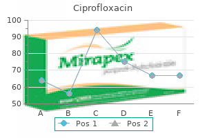
The attribute frequency is the frequency at which the fiber has the lowest threshold antibiotics for sinus infection for sale ciprofloxacin 250 mg generic line. For low-frequency fibers antibiotic gentamicin cheap ciprofloxacin 250 mg visa, the timing of every impulse is section locked with the stimulus cycle, so that the fiber output preserves the timing information of the signal. Intensity is coded both by the discharge fee of cochlear nerve fibers and by recruitment of activity in extra afferents as stimulus depth will increase. At higher stimulus intensities, further cochlear nerve fibers having sequentially greater thresholds are recruited. Sensorineural Deafness Sensorineural deafness (this may sometimes be referred to as nerve deafness) outcomes from injury to the cochlea or to the cochlear root of the vestibulocochlear nerve. The causes of sensorineural deafness are diversified and will embrace repeated publicity to loud noises, therapy with sure antibiotics, infections (such as rubella, mumps, or bacterial meningitis), and tumors at totally different ranges of the neuraxis. As is the case with the center ear, trauma in the form of cranium fracture may lead to sensorineural deafness. The deficits experienced by the affected person are deafness within the ear on the affected side, varying levels of tinnitus, a notion of ringing within the ears if the cochlea is damaged, and extra indicators and symptoms indicative of injury to the adjoining vestibular root. Immediate sensorineural listening to loss may occur after even a single publicity to a sudden loud blast. Frequency tuning curves (A), poststimulus time histogram of discharges by way of the period of a tone burst at the attribute frequency of a primary afferent fiber (B), and rate-intensity curve illustrating how a restricted vary of depth may be coded in the response of primary afferent fibers to growing depth of a tone at its characteristic frequency (C). To take a look at bone conduction, a vibrating tuning fork (256- or 512-Hz frequency) is positioned directly on the vertex of the skull or the mastoid course of. Perceiving these vibrations as sound means that the sound (or vibration) bypasses the exterior and center ear and is transmitted through bones of the skull directly to the cochlea within the inner ear. Perception of the vibration (sound) requires that the sound waves generated be transmitted across the tympanic membrane and the ossicles of the center ear to reach the oval window and cochlea; illness or harm in both the exterior or the center ear would lead to diminished or absence of hearing in this ear, referred to as a conductive listening to deficit. Ideally, each the Weber check and the Rinne test should be administered to every patient, and the observations from the Weber take a look at should correlate with those of the Rinne test. That the sound would be perceived as louder within the faulty left ear seems paradoxical but, though not fully understood, is probably due to masking of ambient room noise within the left ear. The tuning fork is then turned and positioned in entrance of the exterior acoustic meatus of the left ear. With normal listening to (positive Rinne), air conduction is louder (and longer) than bone conduction. It may also be noted in the Rinne take a look at that the length of audible sound is normally longer for air conduction than for bone conduction. The findings for normal hearing and for conductive listening to loss replicate the effectivity of conduction of sound from air to fluid by the middle ear. Sound was not perceived by air conduction when sound notion by bone conduction ceased. This indicated the presence of either a conductive deficit in the left ear or a sensorineural deficit in the proper ear. Within this tonotopic framework, projections join related frequency regions of successive nuclei. Information processing is due to this fact hierarchical with increasing complexity of feature extraction. As cochlear information ascends to the auditory cortex, info is distributed by way of a quantity of parallel pathways that finally converge in the inferior colliculus. The hierarchy of auditory nuclei involved in these parallel pathways contains the cochlear nuclei, nuclei of the superior olive and trapezoid physique, nuclei of the lateral lemniscus, and inferior colliculus. Specific fiber bundles that convey this information from one level to the next are the trapezoid physique, acoustic stria, and lateral lemniscus. From the midbrain, auditory info is conveyed from the inferior colliculus by its brachium to the medial geniculate nucleus of the thalamus after which through the sublenticular limb of the interior capsule to the auditory cortex. Although fibers conveying auditory enter decussate at several levels, this data is routed in considered one of two orderly ways: (1) monaural information (information about sounds at a single ear) is routed to the contralateral facet and (2) binaural info (information about differences between sounds at each ears) is dealt with by central pathways that obtain, evaluate, and transmit this enter. Binaural pathways carry out the neural computation wanted to localize brief sounds and to extract sign embedded in background noise. Inhibitory as properly as excitatory connections are attribute of those circuits. Unilateral injury to the cochlear nerve or cochlear nucleus leads to ipsilateral monaural deafness. Central hearing dysfunction includes lack of ability to localize sounds, inattention to stimuli on the contralateral side, or lack of ability to follow conversations in a loud room. C, For a patient with a unilateral listening to deficit, the Weber check revealed that sound lateralized to the left ear, indicating the presence of both a conductive deficit in the left ear or a sensorineural deficit in the right ear. D and E, Two potential observations to be revealed following Rinne testing of patient in C. In D, results of Rinne testing in the best ear indicate that both air and bone conduction are diminished, which suggests a sensorineural hearing loss (in the right ear). E, Results for Rinne testing in the left ear demonstrate a conductive hearing deficit (in left ear), the place bone conduction is louder than air conduction. F and G, For the precise patient in C, Rinne testing within the left ear (G) revealed regular hearing, whereas in the best ear (F), Rinne testing indicated that both air and bone conduction had been diminished, and thus a sensorimotor deficit was present in the best ear. The blood supply to the cochlea and the auditory nuclei of the pons and medulla originates from the basilar artery. This lesion may also harm the emerging fibers of the facial nerve and the pontine gaze center, resulting in monaural deafness combined with ipsilateral facial paralysis and an incapability to look toward the facet of the lesion. Vascular lesions greater in the ascending auditory system necessarily interrupt pathways conveying information from both ears. The superior olivary complex and lateral lemniscus are mainly equipped by quick circumferential branches of the basilar artery. The superior cerebellar and quadrigeminal arteries supply the inferior colliculus, and the medial geniculate bodies lie in the vascular territory of the thalamogeniculate arteries. The blood supply to the primary auditory and association cortices is via branches of the M2 segment of the middle cerebral artery. When damage happens to neural tissue from vascular insults, tumors, or demyelinating ailments corresponding to multiple sclerosis, the effect on conduction time and activity ranges within the auditory system can be used in medical neurophysiology to assist in localization of the pathologic process. A pattern of seven waves occurs in the auditory evoked response, with peaks at common latencies after the clicks which might be correlated with activity ranges of the ascending auditory system. The vertical traces indicate the approximate correlation of wave and auditory structure, although multiple structure is prone to contribute to various waves. The posterior cochlear nucleus drapes over the restiform physique simply inferior to the pontomedullary junction. As these fibers enter the brainstem on the cerebellopontine angle, they divide into ascending and descending bundles. Fibers within the ascending bundle synapse within the anterior a part of the anterior cochlear nucleus, whereas fibers within the descending bundle synapse in the posterior part of the anterior cochlear nucleus and within the posterior cochlear nucleus. In the cochlear nuclei, every afferent nerve fiber makes specialized synaptic contacts with several totally different cell types. These projections are subdivided into pathways conveying monaural info to the inferior colliculus and those offering enter to the superior olivary complex for binaural processing.
Generic ciprofloxacin 1000 mg with visa
The infarct created known as a venous infarct because of its origin from venous structures antibiotic powder for wounds ciprofloxacin 1000 mg discount visa. The deficits experienced by the affected person might replicate the overall location of the venous thrombosis virus like chicken pox order 1000 mg ciprofloxacin amex. For example, thrombosis of the center parts of the superior sagittal sinus could result in increased tone and weak point of the alternative lower extremity or both decrease extremities because of the proximity of the occluded sinus to the motor cortex. Thrombosis of the caudal portions of the superior sagittal sinus may lead to visual deficits due to venous stasis of the superior sagittal sinus. The terminal branches of the medullary arteries contribute to the formation of the arterial vasocorona. At about levels T10 to , L1, or L2, one spinal medullary (or presumably a radicular) artery, often on the left, is very large. As within the case of the cerebral and cerebellar arteries, the spinal cord has watershed zones and may be susceptible to watershed infarcts, particularly in midthoracic ranges. Due to its diameter, even a small watershed infarct in the cord may lead to critical deficits. The anterior spinal artery provides off central (or sulcal) branches, which pass alternately to the proper and to the left to serve central regions of the spinal cord. The posterior spinal arteries course on the floor of the spinal wire medial to the posterior root entry zone. The blood provide to the spinal twine is supplemented at most spinal levels by branches of segmental arteries. Because the radicular arteries provide the posterior and anterior roots, every spinal degree has these branches. However, the spinal medullary arteries serve to supplement the In general, the venous drainage of the spinal twine mirrors its arterial supply. There is intensive communication between spinal veins and the inner and exterior venous plexuses found adjacent to the dural sac and vertebral our bodies. The veins forming these plexuses apparently lack valves, and the flow in these channels is definitely reversed. For instance, along with low again pain and occasional sensory or motor issues, patients regularly expertise impaired micturition. Although this can be a physiologic barrier to the motion of many substances into or out of the mind, the blood-brain barrier has anatomic options that correlate with its operate. In contrast, capillaries of the overall circulation have fenestrations and infrequently lack tight junctions. Including the endothelium and its tight junctions, the subjacent basal lamina, and the astrocyte perivascular end-feet is a extra inclusive view of the structure of the blood-brain barrier. Under regular (healthy) situations, the blood-brain barrier prohibits the movement of high-molecular-weight substances (such as proteins, penicillin, and dopamine) and very important dyes into the mind. In addition, glucose and different lipid-soluble substances move through the blood-brain barrier, aided by selective transporter molecules. As a result, the endothelium of those tumor capillaries develops fenestrations however no tight junctions. The correlation of the blood supply to the human mind stem with clinical mind stem lesions. The arterial and venous blood supplies to the forebrain (including the internal capsule) of primates. Deep cerebral venous system in man: A microangiographic research on its areas of drainage and its anastomoses with superficial cerebral veins. Arterial sample of human mind stem: normal look and deformation in expanding supratentorial situations. I, Microsurgical Anatomy of the Basal Cisterns and Vessels of the Brain, Diagnostic Studies, General Operative Techniques and Pathological Considerations of the Intracranial Aneurysms. Yezierski Overview-138 Development-138 Neural Plate-138 Neural Tube-139 Neural Tube Defects-139 Spinal Cord Structure-140 Surface Features-140 Spinal Meninges-140 White Matter-141 Gray Matter-141 Blood Supply-142 Regional Characteristics-142 Cervical Levels-142 Thoracic Levels-142 Lumbar Levels-142 Sacral Levels-142 Spinal Nerves-144 Contemporary View of the Functional Components of Spinal Nerves-144 Sensory Components of the Spinal Nerve-144 Neurotransmitters of Primary Sensory Neurons-145 Deafferentation Pain and the Posterior (Dorsal) Root Entry Zone Procedure-145 Motor Components of the Spinal Nerve-146 Neurotransmitters of Spinal Motor Neurons and Myasthenia Gravis-146 Spinal Reflexes-146 Muscle Stretch Reflex-147 Flexor Reflex-147 Crossed Extension Reflex-148 Peripheral Nerve Lesions-148 Radiculopathy-148 Mononeuropathy-148 Polyneuropathy-148 Pathways and Tracts of the Spinal Cord-149 Functions of Ascending Tracts-149 Functions of Descending Tracts-150 Deficits Characteristic of Spinal Cord Lesions-151 Syringomyelia-151 Brown-S�quard Syndrome-151 High Cervical Cord Lesion-151 Acute Central Cervical Spinal Cord Syndrome-151 contains fibers and cells that control the motor components found in these constructions. Consequently, damage to the spinal wire, especially at cervical levels, may trigger permanent and catastrophic deficits or demise. Recall that many cranial nerves contain sensory fibers that receive info from, and motor fibers that project to , diverse body areas; these bypass the spinal wire. The spinal twine conveys sensory input from the upper limbs, trunk, lower limbs, and most of the viscera and 138 the spinal twine participates in 4 important functions. First, it receives major sensory input from receptors in skin, skeletal muscles, and tendons (somatosensory fibers) and from receptors in thoracic, abdominal, and pelvic viscera (viscerosensory fibers). This sensory input may take part in spinal reflexes, be conveyed to larger levels of the neuraxis, or each. In addition, somatosensory information from the periphery contributes to the formation of ascending tracts/systems that convey pain and thermal sense and proprioception, vibratory sense, and discriminative sense. Second, the spinal wire contains somatic motor neurons that innervate skeletal muscle tissue and visceral motor neurons that, after synapsing in peripheral ganglia, influence clean and cardiac muscle and glandular epithelium. Any illness process that damages the somatic motor neuron (as in poliomyelitis), compromises its ability to elicit a response within the skeletal muscle (as in myasthenia gravis), or affects both descending fibers and decrease motor neurons (as in amyotrophic lateral sclerosis, also known as Lou Gehrig disease) will lead to weak spot or paralysis. Third, somatosensory fibers enter the spinal twine and contribute to the formation of sensory pathyays that convey information to the thalamus and cerebral cortex for conscious recognition, whereas others concurrently influence anterior horn motor neurons both immediately or not directly by way of interneurons. These activated motor neurons, in turn, produce fast involuntary contractions of skeletal muscles. The sensory fiber, the related motor neuron, and the resultant involuntary muscle contraction represent the circuit of the spinal reflex. Reflexes are important to normal operate and are widely used as diagnostic tools to assess the practical integrity of the spinal wire. Fourth, the spinal twine accommodates descending fibers that influence the exercise of spinal neurons. These fibers originate within the cerebral cortex and brainstem, and injury to them adversely influences the exercise of spinal motor and sensory neurons. In many instances, the place of a lesion within the brainstem or spinal twine might give rise to a predictable or attribute series of deficits, corresponding to in decorticate rigidity or an alternating hemianesthesia. Fifth, harm to peripheral nerves will result in motor or sensory deficits distal to the lesion. These are most noticeable within the extremities and could additionally be manifested as motor deficits (flaccid paralysis), a significant decrease or loss of essential spinal reflexes (hyperreflexia, hyporeflexia, areflexia), a loss of sensation (anesthesia), or irregular sensations (paresthesia). Cross-sectional (A) and dorsal (B) views of the neural plate and the correlation of neural tube buildings with the grownup twine (C). The white space between the blue (posterior horn) and pink (anterior horn) of the grownup spinal wire represents the approximate position of the intermediate grey. Malformations involving defects of the nerve tissue or surrounding bone include rachischisis (D), meningocele (E, solid cord) or meningomyelocele (E, dashed cord), and anencephaly with rachischisis (F). The neural plate gives rise to the cervical, thoracic, and lumbar ranges, whereas the caudal eminence offers rise to the sacral and coccygeal levels. This closure simultaneously proceeds in rostral and caudal instructions, finally creating small openings at either finish between the cavity of the neural tube and the surrounding amniotic cavity.
Ciprofloxacin 250 mg discount with mastercard
The subplate is a slender region positioned instantly inside to the cortical plate antibiotics for uti make me feel sick 500 mg ciprofloxacin generic with visa. The histogenesis of the cerebellar cortex is a slight modification of the cerebral cortex plan as a end result of antibiotic spectrum chart ciprofloxacin 250 mg buy generic line the presence of an exterior germinal layer. This exterior germinal layer originates from the rhombic lip, an alar plate derivative, and is located throughout the marginal layer. It defines the longitudinal axis of the embryo, determines the orientation of the vertebral column, and persists because the nucleus pulposus of the intervertebral disks. Associated with this course of is the manufacturing of cell adhesion molecules within the notochord. These molecules diffuse from the notochord into the neural plate and performance to be a part of the primitive neuroepithelial cells into a tight unit. Within the neuroectoderm, some neuroepithelial cells elongate and become spindle shaped. Most of the neural tube forms from the neural plate by a strategy of infolding referred to as main neurulation. This a part of the neural tube will give rise to the brain and to the spinal twine via lumbar ranges. The most caudal portion of the neural tube, which is ready to give rise to sacral and coccygeal levels of the wire, is fashioned by a course of called secondary neurulation. This thickening A Ventricular zone Marginal zone Ventricular zone B Intermediate zone Marginal zone Subventricular zone Ventricular zone Intermediate zone elevates the edges of the neural plate to kind neural folds. At about 20 days, the neural folds first contact one another to begin the formation of the neural tube. The rostral opening, the anterior neuropore, closes at about 24 days, and the caudal opening, the posterior neuropore, closes about 2 days later. Neurulation is caused by morphologic adjustments in the neuroblasts, the immature and dividing future neurons in the ventricular zone. As talked about previously, these cells are elongated and are oriented at right angles to the dorsal floor of the neural plate, which would be the internal wall of the neural canal. Microfilaments in every cell type a circular bundle parallel to the lengthy run luminal surface, whereas microtubules lengthen along the size of the cell. The contraction of the circular bundle of microfilaments causes the microtubules to splay out like the rays of a fan. This varieties an elongated conical cell with its apex at the neural groove and its base at the fringe of the neural fold. Congenital malformations associated with faulty neurulation are called dysraphic defects. The process of induction also implies that the right improvement of a structure is dependent on the proper improvement of its neighbors. There is an intimate relationship of neural tissue to the encompassing bone, meninges, muscles, and skin. Because of this relationship, a failure of neurulation typically impairs the formation of these surrounding constructions. Several well-controlled scientific trials have confirmed that supplementation with the vitamin folic acid can scale back the incidence of neural tube defects. In the Medical Research Council Vitamin Study, carried out in Great Britain and revealed in 1991, women who had beforehand been delivered of a child with a dysraphic defect had been assigned to either a folic acid supplementation group or a management group during a subsequent pregnancy. Folic acid supplementation lowered the incidence of neural tube defects by about 70% relative to that in untreated controls. Dysraphic defects have additionally been observed in infants born to mothers who had circulating antibodies to the folate receptor. Department of Agriculture has established a Recommended Daily Allowance for folic acid supplementation of 600 g/day earlier than and through pregnancy. In addition, medicine taken for epilepsy, such as valproic acid and carbamazepine, could cause dysraphic defects. Rostral to the spinal twine, the growing neural tube differentiates in a extra complicated method (D) to accommodate more complicated buildings such because the cerebellar and cerebral cortices. Most dysraphic issues happen on the location of the anterior or posterior neuropore. The defect extends from the extent of the lamina terminalis, the positioning of anterior neuropore closure, to the region of the foramen magnum. Encephaloceles are commonest in the occipital region, however they may additionally happen in frontal and parietal locations. This defect could go unnoticed until early adulthood and is usually related to a cavitation of the spinal twine (syringomyelia) or of the medulla (syringobulbia). Defects in the closure of the posterior neuropore trigger a range of malformations recognized collectively as myeloschisis. The defect all the time includes a failure of the vertebral arches at the affected ranges to kind fully and fuse to cover the spinal cord (spina bifida). In the latter case, the neural tissue could be the decrease a half of the spinal wire or, more generally, a portion of the cauda equina. Infants with meningomyelocele may be unable to transfer their lower limbs or may not perceive ache sensations from skin innervated by nerves passing by way of the lesioned area. A cell mass, the caudal eminence, appears just caudal to the neural tube after which enlarges and cavitates. The caudal eminence joins the neural tube, and its cavity becomes steady with the neural canal. Magnetic resonance image of meningohydroencephalocele (A) and drawings of meningocele (B), meningoencephalocele (C), and meningohydroencephalocele (D). The malformation is roofed with skin generally, but the website may be marked by unusual pigmentation, hair progress, telangiectases (large superficial capillaries), or a prominent dimple. A common abnormality is tethered cord syndrome, in which the conus medullaris and filum Pons Syringobulbia terminale are abnormally fastened to the faulty vertebral column. The sustained traction damages the spinal twine and causes variable weakness, sensory loss and asymmetric progress of the legs and ft, and issues with bowel and bladder management. Infants born to moms with diabetes mellitus can have caudal regression syndrome, which impacts the development of the embryonic structures in the caudal area including the spinal wire. A second bend in the neural tube at the degree of the mesencephalon is the mesencephalic (or cephalic) flexure. The pontine flexure divides the hindbrain into the myelencephalon caudally and the metencephalon rostrally. The telencephalon (meaning "end-brain") varieties as an outpocketing of the forebrain and expands enormously, with its complex lobes, gyri, and sulci, to turn out to be the biggest a part of the brain. The major constructions of the forebrain develop in the course of the second month of gestation. Magnetic resonance picture (A) and corresponding views displaying spina bifida occulta (A, B) and spina bifida cystica (C, meningocele; D, meningomyelocele). At about the end of the fifth week, the telencephalon provides rise to two lateral expansions referred to as the telencephalic (cerebral) vesicles. Their adult derivatives embrace the cerebral cortex and the subcortical white matter (including the internal capsule), the olfactory bulb Rhombencephalon and tract, portions of the basal nuclei, the amygdala, and the hippocampus.
Acacia. Ciprofloxacin.
- Lowering cholesterol levels.
- Dosing considerations for Acacia.
- How does Acacia work?
- Are there any interactions with medications?
- What is Acacia?
- Are there safety concerns?
Source: http://www.rxlist.com/script/main/art.asp?articlekey=96291
Order ciprofloxacin 750 mg on line
Bilateral anosmia is often sustained in response to head damage or the common chilly antibiotics cvs order ciprofloxacin 1000 mg with mastercard. However antibiotic cream for dogs discount 1000 mg ciprofloxacin otc, the absence of scent from either nostril has been noted in superior cases of olfactory groove meningioma. Typically, the loss happens progressively, and the patient typically fails to discover these modifications. Olfactory dysfunction can additionally be encountered in neurodegenerative diseases, such as Alzheimer and Parkinson illnesses, or in Huntington chorea. Most notably, these olfactory deficits appear very early in the midst of the illness and could additionally be amongst its first manifestations. Interestingly, recent evidence means that individuals in the early stages of Alzheimer illness may experience difficulties in perceiving the odor of peanut butter. Disorders of olfaction are also associated with epilepsy and varied depressive and psychiatric disorders, similar to schizophrenia and Korsakoff psychosis. The sufferers incessantly experience parosmia (dysosmia), a distortion in a scent expertise or the notion of a smell when no odor is current (olfactory hallucination or phantosmia). Such episodes, resulting presumably from abnormal sequences of neuronal exercise, may be elicited by an irritative lesion of the anterior medial temporal lobe, hippocampus, amygdala, or medial dorsal thalamic nucleus. In the case of epilepsy, focal (partial) seizure activity in the uncal area of the temporal lobe has been discovered to correlate with the formation of repugnant or disagreeable olfactory auras (cacosmia). This sort of assault, often known as an uncinate match, could happen in people who are genetically predisposed to seizure-like discharge or in individuals who show tumor growth, a vascular disorder, head trauma, an infection, drug use, or substance withdrawal. It is now broadly accepted that there are functionally distinct populations of cells within the style bud. The apical ends of those cells are coated with microvilli of variable lengths that stretch right into a style pore. Numerous junctional complexes situated between the apices of style cells limit access of stimuli to the microvilli, the place taste transduction occurs. The style pore is crammed with a protein-rich substance by way of which substances should pass to reach the taste cell microvilli. Taste pore Epithelium Taste buds are discovered, in variable numbers, on the human tongue, palate, pharynx, and larynx. These buildings are scattered among the many more quite a few nongustatory filiform papillae distributed over the surface of the tongue. The dimension, shape, and variety of fungiform papillae vary extensively, and normally two to four style buds are discovered within the dorsal epithelium of every. Associated with both the vallate and foliate papillae are the von Ebner lingual salivary glands. These glands drain into the bottom of the clefts and influence their microenvironment. Taste stimulation of vallate and foliate papillae influences the secretions of the von Ebner glands by way of circuits positioned in the brainstem. It was as soon as believed that completely different areas of the tongue were specialised for the detection of explicit style qualities. It is now recognized that all taste qualities are detected in all regions of the tongue, although sensitivity to the completely different taste qualities and style transduction mechanisms might range by tongue region. Microvilli Extralingual Taste Buds Additional style buds are positioned on the human taste bud, oral and laryngeal pharynx, larynx, and higher esophagus. Palatal style buds are situated on the juncture of the onerous and soft palates and on the soft palate. Laryngeal style buds are found on the laryngeal surface of the epiglottis and adjacent aryepiglottic folds. The number of extralingual style buds is substantial, and so they could contribute to the style experience. Stimulation of some extralingual taste buds, significantly these close to the larynx, elicits brainstem-mediated reflexes that prevent unintentional aspiration of ingested supplies. As research has begun to clarify the mechanisms underlying taste transduction, it has turn into clear that particular person taste qualities or even individual style compounds use a quantity of transduction mechanisms. In basic, style transduction is initiated when soluble chemical substances diffuse via the contents of the style pore and work together with receptors located on the exposed apical microvilli of the taste cells. Several different receptor varieties have just lately been cloned, including a novel variant of the metabotropic glutamate receptor that functions as an umami style receptor and G protein�coupled receptors that function as bitter receptors. The interaction of the chemical stimulus with the style cell receptor leads to either depolarization or hyperpolarization of the taste cell microvilli. This calcium launch results in a launch of chemical transmitters at the afferent synapse, which in flip leads to an action potential within the afferent fiber. New style cells are thought to arise from polygonal basal cells situated in basolateral areas of the style bud. Taste afferent fibers kind the postsynaptic factor of a chemical synapse near the base of the taste cell. Each taste bud is typically innervated by a couple of afferent fiber, and a person fiber may innervate a quantity of taste buds. Light micrographs of transverse sections via human fungiform (B), vallate (C), and foliate (D) papillae. Transduction of stimuli resulting in salty and maybe some sour and bitter tastes seems to be the results of a direct interplay of these tastants with specific ion channels located within the apical membrane of style cells. One mechanism for the transduction of sodium salts, such as sodium chloride, involves motion of sodium into the taste cell by way of apically located amiloride-sensitive cation channels. One pathway put forward for transduction of some sour and bitter stimuli entails blockage of apical voltage-sensitive potassium channels. Protons provided by sour stimuli, corresponding to hydrochloric acid, are thought to block this outward current, inflicting the cell to depolarize. Such findings emphasize the position of intracellular acidification in bitter transduction. One pathway entails binding of amino acids by receptors which are instantly coupled to cation channels having properties similar to those of the nicotinic acetylcholine receptor. Some bitter and bitter substances are transduced by the closing of apical voltage-sensitive potassium channels. The transduction of salts, corresponding to sodium chloride, involves the movement of ions (such as sodium and potassium) through amiloride-sensitive cation channels in the apical membrane. Special visceral afferent fibers for taste (in red) terminate in the rostral (gustatory) areas of the solitary nucleus, whereas basic visceral afferent fibers (in blue) terminate within the caudal portion of the nucleus. The chorda tympani department of the facial nerve innervates style buds within the fungiform papillae on the anterior two thirds of the tongue and in probably the most anterior clefts of the foliate papillae. The higher superficial petrosal nerve, also a branch of the facial nerve, innervates taste buds on the soft palate. The cell our bodies of facial nerve fibers subserving taste are positioned within the geniculate ganglion, and their central processes enter the brainstem on the pontomedullary junction within the intermediate nerve, which is definitely a part of the facial nerve. Color coding demonstrates the projections of first-order (blue), second-order (red), and third-order (green) neurons conveying gustatory information. These visceral fibers are involved in the central control of respiration, cardiac perform, and certain features of swallowing.
500 mg ciprofloxacin purchase fast delivery
Water permeation across organic membranes: mechanism and dynamics of aquaporin-1 and GlpF antibiotics chart 1000 mg ciprofloxacin discount visa. Epithelial sodium channel/degenerin family of ion channels: a selection of functions for a shared construction antibiotic resistance rates buy 500 mg ciprofloxacin free shipping. Aquaporin water channels: atomic construction molecular dynamics meet medical drugs. The voltage-sensitive sodium channel is a bell-shaped molecule with several cavities. The construction and function of particular person synapses are customized in accordance with the requirements of a specific task. The synapse must regulate the synthesis, storage, and launch of one or more transmitters, and sign transduction should embrace a number of receptors at the target cell and a mechanism to terminate the sign. This article will focus on the individual classes of transmitters in some element, using every to illustrate a number of of those six principal options of chemical transmission. While the postsynaptic element is mostly a dendrite and an axon terminal is usually the presynaptic factor, neuron cell our bodies, axons, and dendrites can all be presynaptic or postsynaptic in nature. Regardless, in every case the elements are tailored to a particular function: to optimize transmitter storage, its launch, its results, and its disposition. Transmitters and Modulators the overwhelming majority of vesicles within the nervous system comprise one or the opposite of the widespread transmitters (Table four. Most synapses additionally use modulators of synaptic exercise whose release is tightly regulated, such because the biogenic amines (dopamine, norepinephrine, or serotonin) and the neuropeptides. Solute and first active transporters are tailored to specific storage and uptake needs (Table four. Synapses are the chemically and anatomically specialized websites where neurons ship chemical alerts to goal cells (left). In the standard neuron to neuron synapse, anterograde axon transport delivers synaptic vesicles to the nerve terminals, the place they advance to the energetic zones to be primed for launch. When an motion potential depolarizes the terminal, voltagedependent calcium channels open, permitting calcium to enter. It is that this calcium that triggers docking proteins to fuse the vesicle membrane to the plasma membrane, releasing the neurotransmitter into the synaptic cleft, where it binds to receptors on the target cell. The chemical sign is terminated when the neurotransmitter is transported again up into the nerve terminal for reuse or changed into an inactive metabolite by an enzyme on the postsynaptic membrane or within the synaptic cleft. The particular particulars of synaptic function differ significantly from one site to the subsequent, and the glutamatergic synapse (right) diverges from the general schema in a selection of ways. Release Release of the vesicle-stored transmitters is tightly regulated, with synapses in a place to launch variable numbers of vesicles according to the strength of the signal to the nerve terminal. Vesicles themselves may open transiently, releasing only part of their contents, or they may fuse utterly with the plasma membrane of the nerve. Termination Transduction All transmitters act by binding to some molecule, and that molecule is thus called its receptor. Occasionally, synapses consist of a single kind of transmitter performing on a single kind of receptor, leading to a single kind of response, however most synapses contain multiple receptor varieties and use more than one transmitter. Receptors themselves can be easy, with a single binding web site for the neurotransmitter and a single function, but most exhibit exuberant extracellular and intracellular structures which are the results of all kinds of translational and posttranslational modifications and which may be topic to allosteric modulation by metabolites, medication, and chemical modifications. Transmitter that escapes the cleft is sequestered by solute transporters on the astrocytes or the postsynaptic cell or is removed by the cerebrospinal fluid or the vasculature (Table four. In the particular case of neurohormones, launch is designed to escape degradation as a end result of the target is distant. Common examples are listed along with the extra common naturally occurring therapeutic and poisonous agonists or antagonists. Phylogenetic studies of gene homologies have grouped these gene families into 23 superfamilies, four of that are essential to the mechanisms discussed on this chapter. If the GluA2 subunit is current, which is the identical old case, the channel is permeable to sodium and potassium ions solely. This arginine adds an extra positive charge to the selectivity filter of the ion pore, thus excluding divalent cations corresponding to calcium. When a third glutamate binds, the channel opens for longer, giving extra current, and with a fourth glutamate, the channel is open most of the time with still more present flowing. This schema preserves microscopic electoneutrality, ultimately allowing the hydrogen ions to turn out to be 30-fold concentrated relative to the cytoplasm, a pH of 1. Uniquely among the small neurotransmitters, the ultimate step of norepinephrine synthesis occurs within the synaptic vesicle, the place dopamine -hydroxylase adds a hydroxyl group to dopamine. Thus, on this example, the synaptic vesicle will include two neurotransmitters, the cation norepinephrine and the anion glutamate. Extracellular magnesium binds to the electronegative oxygens of these twin asparagines and is released solely slowly when the membrane is depolarized from rest, a change in voltage that gives the electrical drive to drive the divalent ion out of the channel. For instance, stargazin will increase the rate of channel opening and slows desensitization. This calcium modulates many and various cell processes by activating Ca2+/calmodulin-dependent kinases, protein kinase C, phospholipase C and A2, calcineurin, and nitric oxide synthase. It has been tentatively implicated in Alzheimer and Parkinson ailments, depression, schizophrenia, and ischemic neuron dying after a stroke. All monomers bind one glutamate, but only after the two GluN1s have sure a different ligand: d-serine. Glia synthesize d-serine (one of the one two d-amino acids to be used by vertebrates) from l-serine via serine racemase and launch it into the synaptic area. Glycine, the inhibitory neurotransmitter of the brainstem and spinal wire, can substitute for d-serine. Eating tainted shellfish can end result in neurotoxic ranges of calcium influx by way of kainate receptors within the hippocampus and amygdala. This part will current particulars concerning the structure of the synapse, about how the transmitter is stored and released, how failures in synaptic function happen, and, finally, how other transmitters use this receptor type. Release occurs when an motion potential depolarizes the membrane and opens voltage-dependent calcium channels that deliver the calcium sign to launch the vesicles (B). The folds of the postsynaptic membrane closest to the nerve terminal comprise receptors that recognize the newly released neurotransmitter, opening nonspecific cation-permeant channels that provoke an action potential in the skeletal muscle. The stimulus is rapidly terminated when the neurotransmitter diffuses into the synaptic folds away from the receptors and toward acetylcholinesterase molecules that eliminate the transmitter by hydrolysis. Chemical Signaling within the Nervous System sixty one transmitter-filled vesicles fill the nerve terminal, each large and small. First, transient -shaped encounters end result in a flickering fusion pore that extends from the vesicle inside to the extracellular space through a brief stalk. At this level, the fusion pore is 1 nm in diameter with a conductance of eighty ps, placing it within the realm of the large nonspecific cation channels. Although this stage may quickly proceed to a full fusion and full release of vesicle contents, the encounter may break off prematurely if the fusion pore closes earlier than full fusion (yclept "kiss-and-run"). As with mitochondria and tons of other organelles descended from gram-negative bacteria, synaptic vesicles processes are pushed by a proton gradient, on this case with the vesicle inside being acidic and electrically positive.
Purchase ciprofloxacin 500 mg with visa
Important components of these buildings in the keratinocyte include the transmembrane link antibiotics for uti with least side effects ciprofloxacin 250 mg order with mastercard, E-cadherin infection resistant legguards cheap ciprofloxacin 500 mg fast delivery, and the intracellular actin-binding -catenin. Gap junctions are formed from aggregation of connexins into connexons that join adjacent cells to allow intercellular transport of ions and other small molecules. The keratinocyte life cycle as described earlier ranges from 5 to 30 days, relying on the anatomic website and state of well being of the skin. A few specialized cell types present in the skin deserve particular point out with respect to perform of the pores and skin as an organ. Melanocytes the melanocyte provides pigmentation to the skin or fur essential for behavioral features of survival corresponding to camouflage in certain species. In addition to their presence in the epidermis, melanocytes are also found within the eye, cochlea of the ear, the brain/meninges, and the center. Melanin also can bind certain probably dangerous compounds such as cations and metals. Each melanocyte "serves" about 30�40 keratinocytes within the basal layers of the epidermis. Melanosomes are organelles derived from the endoplasmic reticulum of melanocytes and function packages for switch of melanin. Although the number of melanocytes stays constant, the manufacturing and transfer of melanosomes to keratinocytes can be up- or downregulated. In healthy skin, these cells are comparatively inactive and even act to attenuate inflammatory response. They are in a position to preferentially reply to particular or extreme international antigens, thereby preventing continual upregulation of inflammatory mediators every time international antigens are sensed, which is type of constant in the skin. The dermis gives the pores and skin tensile power via collagen, contributes to movement by allowing stretch via elastic fibers, has immune regulatory capabilities, incorporates vascular and neurologic parts necessary for communication with the dermis and setting, and forms the matrix for adnexa. In species lacking epidermal rete ridges, these subregions are normally merely described as superficial and deep, respectively. The major parts of the dermis are collagens and elastins, conferring tensile energy, and proteoglycans, glycosaminoglycans, and hyaluronans, which assist dissipate stress forces. They represent a cell kind distinctive to the pores and skin and oral mucosa, able to appearing as mechanoreceptors with dense granules containing neurotransmitter-like mediators. The dendrites of the Merkel cells contact unmyelinated axons within the dermis, the place they perform collectively as a unit (tylotrich pads) to sign adnexal secretions (sweat), changes in blood flow, tactile sensation, and presumably serve in a paracrine perform together with other cells of the skin. Motor innervation is from sympathetic autonomic nerves and responds to both cholinergic or adrenergic stimuli. Adrenergic responses embrace vasoconstriction, apocrine gland secretion, and hair follicle positioning. Eccrine sweat glands are underneath cholinergic control; however, these glands are largely absent in most areas of nonhuman mammalian skin. Superficial, middle, and deep vasculature plexuses supply corresponding layers of the skin. Arteriovenous anastomoses are common within the pores and skin of the extremities specifically, necessary for adaptation to frequent modifications in temperature and blood strain. These junctions respond to vasoconstrictors and vasodilators such as epinephrine and histamine. Many vessels of the skin have comparatively thick walls compared to those of comparable dimension in internal organs, a needed adaptation for defense in opposition to the shear and stress forces to which the skin is often subjected. Also present in the dermis are the fibroblast-like veil cells that outline spaces for vessels throughout the dermis, surrounding microvasculature, and creating a perivascular space. Cutaneous lymphatics arise as capillaries within the superficial dermis, but under the extent of superficial blood capillaries. Gaps throughout the lymphatic vessels type channels by linking with the dermal matrix, directing extra interstitial fluids from the dermis into the lymphatic system. Lacking clean muscle and pericytes, lymphatics of the dermis rely on subcutaneous muscle contraction, strain of the encircling matrix, and associated blood vessel actions to initiate flow of immunologic, waste, and even degraded pathogenic and xenobiotic supplies away from the dermal interstitium. In the deeper subcutis, lymphatics have clean muscle partitions and are actively contractile immediately directing circulate of lymph to and from the skin. The Adnexa the cutaneous adnexal unit refers to a hair follicle, the associated arrector pili muscle, and associated glands. The hair shaft consists of an inside medulla, a surrounding cortex, and outer cuticle. The hair follicle has subanatomic areas generally known as the infundibulum (from the surface epidermis to the sebaceous gland duct entrance to the hair follicle), the isthmus (just beneath the infundibulum, from sebaceous duct to the arrector pili muscle insertion), and the inferior segment from the isthmus deep to the dermal papilla. The outer root sheath that contains the hair is contiguous with the floor epidermis. The internal root sheath is attached to the cuticle of the hair and is distinguishable in tissue sections by the presence of eosinophilic cytoplasmic trichohyalin granules. In humans and mice, a construction often identified as "the bulge" is present and attached to the outer root sheath, near the insertion of the arrector pili muscle. The bulge provides the adnexal unit and, in some instances the floor dermis. The bulge area could also be absent in different species, with stem cells being present in infundibular and isthmus areas of the follicle itself, though some analysis does help bulge cell presence within the dog. Hair growth is split into the following levels: anagen (active growth), catagen (transition phase), telogen (resting stage), and exogen (shedding). During hair growth, an ordered array of keratinized cells is progressively pushed upward within the form of hair shafts. These cells give rise to hair by a means of terminal differentiation, analogous to , however more difficult than, the method described for epidermal cornification. Keratinization of hair follicle cells is of four morphologic subtypes: infundibular (like the floor epidermis, that includes keratohyalin), trichilemmal (important in identification of catagen hairs, with dense eosinophilic keratin "flames"), trichogenic/matrical ("ghost cell" keratinization), and medullary/inner root sheath (with deeply eosinophilic trichohyalin granules). The Subcutis the subcutis is the adipose-rich tissue beneath the dermis responsible for attachment to underlying muscle, fascia, or periosteum. Connective tissue septa present throughout the subcutis facilitate motion and help dense vessel and nerve networks in the tissue. This layer additionally serves to take up shock to underlying structures, form the exterior features of the organism, and regulate temperature. The wealthy triglyceride shops of the subcutis could be utilized as an power retailer and also serve to protect underlying tissues from temperature extremes. Visceral and subcutaneous adipose stores are additionally necessary in the secretion and concentrating on of various hormones and cytokines. Zone of dividing cells of the hair matrix, comparable with the stratum basale of the epidermis. This zone accommodates melanocytes that give color to the hair by passing melanin to the matrix cells. Eccrine glands participate in thermoregulation by secreting water and salts on to the epidermal floor. Abundant throughout the pores and skin in great apes and humans, in home and laboratory animals, the eccrine sweat glands are largely restricted to the foot pads in canine, and the nasal planum and carpus of pigs.
Ciprofloxacin 1000 mg fast delivery
For example antibiotic resistance over prescribing cheap ciprofloxacin 250 mg free shipping, a spinal wire hemisection at T8 would have an effect on the body below that level however would spare the higher trunk and higher extremity virus b discount ciprofloxacin 250 mg amex. A lesion involving the posterior columns bilaterally would result in proprioceptive and discriminative contact losses beneath the extent of the lesion however would spare pain and thermal sensations. In our examine of systems neurobiology, we will discover these and other examples of dysfunction ensuing from spinal cord lesions. Organization within the Spinal Cord: the Anatomy and Physiology of Identified Neurons. Spinothalamic and spinohypothalamic tract neurons within the cervical enlargement of rats: I. Spinal wire: cytoarchitectural, dendroarchitectural, and myeloarchitectural group. The ache system, the neural foundation of nociceptive transmission in the mammalian nervous system. Spinomesencephalic tract: projections from the lumbosacral spinal twine of the rat, cat, and monkey. For example, the central regions of the medulla comprise the cranial nerve nuclei affiliated with the medulla. This medullary space is rostrally continuous with the pontine tegmentum, which incorporates the cranial nerve nuclei related to the pons. The basilar pons is bulbous and fairly characteristic of the anterior aspect of the pons. The pontine tegmentum contains parts of the trigeminal nuclei and the vestibular nuclei and, just rostral to the pons-medulla junction, the facial motor nucleus, superior salivatory nucleus, and abducens nucleus. These are the inferior cerebellar peduncle, the middle cerebellar peduncle (or brachium pontis), and the superior cerebellar peduncle (or brachium conjunctivum), connecting the cerebellum to the medulla oblongata, basilar pons, and midbrain, respectively. Pons the time period brainstem (sometimes written brain stem) can mean both the portion of the brain that consists of the medulla oblongata, pons, and midbrain or the portion that consists of those structures plus the diencephalon. For our functions, subsequently, the brainstem consists of the rhombencephalon, comprising the myelencephalon and metencephalon (but excluding the cerebellum), and the mesencephalon. These areas of the brainstem share a basic group, which is the subject of this chapter. The medulla is slightly cone formed and enlarges in diameter because it extends rostrally from the medulla�spinal twine junction towards the pons-medulla junction. The nuclei of the hypoglossal, vagal, and glossopharyngeal nerves as well as portions of the nuclei of the trigeminal nerve are located in the medulla. The exit of the trochlear nerve is considered the pontomesencephalic junction on the posterior side of the brainstem; together with its decussating fibers, it composes the isthmus rhombencephali (the transitional zone from pons to midbrain). In C, the cerebellum is removed to expose the posterior surface of the brainstem and the fourth ventricle. The posterior side of the midbrain is characterised by the superior and inferior colliculi, and their respective nuclei, and the anterior side by the crus cerebri and interpeduncular fossa. The tegmental and basilar areas and contiguous areas of the medulla are shown in light and dark gray, respectively. Medulla Cisterna magna Tegmental and Basilar Areas the central core of the midbrain and the pons is known as the tegmentum, and their anterior (ventral) components are the basilar areas. Basilar buildings of the brainstem embrace the descending fibers of the crus cerebri (midbrain), basilar pons, and pyramid (medulla) and particular populations of neurons in the midbrain and pons that originate from the alar plate of the embryonic mind. The cerebral aqueduct is a slender channel, 1 to three mm in diameter, that connects the third ventricle (the cavity of the diencephalon) with the fourth ventricle (the rhombencephalic cavity). The cerebral aqueduct incorporates no choroid plexus; its walls are formed by a steady mantle of cells collectively known as the periaqueductal grey. The fourth ventricle is steady rostrally with the cerebral aqueduct, caudally with the central canal of the caudal medulla and cervical spinal cord, and laterally with the subarachnoid house through the midline foramen of Magendie and the two lateral foramina of Luschka. The medial portions on 154 Regional Neurobiology this cistern on the interface of the medulla, pons, and cerebellum comprise the cerebellopontine angle. The tela arises from the inferior surface of the cerebellum and sweeps caudally to attach to the V-shaped edges of the medullary portion of the ventricular space. There are two slight depressions along the course of the sulcus limitans, somewhat like deep spots within this sulcus. In some surgical procedures involving the fourth ventricle or medulla, the sulcus limitans and the superior and inferior foveae symbolize important landmarks. The rostral edge of these fibers is usually regarded as the pons-medulla junction within the ground of the fourth ventricle. Medial to the sulcus limitans, the hypoglossal and vagal trigones characterize the underlying hypoglossal and dorsal motor vagal nuclei. In the caudal pontine area, the facial colliculus, situated medial to the sulcus limitans, marks the location of the underlying abducens motor nucleus and the interior genu of the facial nerve. Lateral to the sulcus limitans within the medulla and caudal pons is a flattened area known as the vestibular area, which marks the situation of the vestibular nuclei. The cerebellum is removed to expose the posterior aspect of the medulla and midbrain and the rhomboid fossa. Cranial nerves, like spinal nerves, comprise sensory or motor fibers or a mix of those fiber types. These numerous fibers are classified on the basis of their embryologic origin or widespread structural and practical traits. Primary sensory fibers, somatic motor neurons, and preganglionic and postganglionic visceromotor neurons that exhibit "like anatomical and physiological characters in order that they. For example, fibers conveying sharp ache, a selected sort of input, from broadly separated body parts (the foot, hand, and face) have the identical practical component. An Overview of the Brainstem one hundred fifty five introduced in relation to spinal nerves (see Chapter 9), can additionally be directly applicable to cranial nerves. Early in growth, the rostrocaudally oriented cell columns forming the alar and basal plates of the spinal cord primarily prolong all through the brainstem. As improvement progresses, maturing neurons in alar and basal plates (these can also be called alar and basal cell columns) begin to migrate to kind their adult structures, and the caudocephalic continuity of the cell columns could also be disrupted. Motor nuclei of cranial nerves arise from basal plate neurons, whereas the nuclei that receive primary sensory input through cranial nerves originate from the alar plate. The alar plates shift laterally (A), where the fourth ventricle flares open on the obex, and then shift back to a posterior position, where the ventricle funnels into the cerebral aqueduct. The basic position of structures derived from the alar (blue) and basal (red) plates in relation to the sulcus limitans and the ventricular space is shown for the medulla (B, C), the pons (D), and the midbrain (E). The sulcus limitans, an embryologic landmark that persists within the medulla and pons of the adult, separates structures derived from the basal plate from these derived from the alar plate. This is very the case for cell columns within the spinal twine which are steady with functionally similar cell columns of the brainstem.
Order 750 mg ciprofloxacin with amex
This area is called the vasopressor middle as a end result of stimulation ends in elevated peripheral vascular resistance and increased cardiac output antibiotic amoxicillin discount 250 mg ciprofloxacin fast delivery. Areas such as this have been designated centers infection years after root canal generic ciprofloxacin 750 mg free shipping, for example, the respiration heart, micturition middle, and vomiting heart. The significance of the supraspinal control of autonomic perform is illustrated by some of the deficits related to spinal wire injuries at larger ranges (T6 or above). Initially, the interruption of descending reticulospinal and hypothalamospinal fibers that regulate sympathetic preganglionic neurons in the intermediolateral cell column is manifested as an overall discount in sympathetic activity. Thus medical signs in the acutely injured patient embrace lowered blood strain, orthostatic hypotension, and lowered heart rate (bradycardia). With time, nevertheless, hyperactivity of sympathetic reflexes (termed autonomic dysreflexia) develops, most likely as a end result of denervation hypersensitivity of sympathetic neurons and goal tissues. Signs and symptoms include hypertension, urinary retention, piloerection, profuse sweating, and reduction of blood move to peripheral tissues in response to any of all kinds of noxious stimuli below the extent of the spinal wire damage. A sudden movement to an upright position causes a drop in pressure within the aortic and carotid sinuses. This ends in modification of neuronal exercise (red) in these pathways to produce the compensatory adjustments within the cardiovascular system. Failure of this reflex results in orthostatic hypotension, a severe drop in blood strain when the affected person assumes an upright place. Projections of solitary neurons affect the tonic activity of parasympathetic (vagal) output to the center and sympathetic output to the guts and peripheral blood vessels. The first of those targets, the vagal preganglionic parasympathetic neurons that suppress heart fee and cardiac output, receive an excitatory drive from neurons of the solitary nucleus. Thus a reduction of baroreceptor discharge results in a launch of the heart from this inhibitory parasympathetic drive. The neurons on this region have intrinsic pacemaker features and obtain inhibitory inputs from the solitary nucleus. Reduced inhibition of this excitatory projection leads to elevated cardiac output and elevated resistance in vascular beds of skeletal muscle and stomach visceral organs however not of the skin, coronary heart, and brain. Some of this solitary input to vasopressor neurons could also be relayed by way of vasodepressor cell teams within the caudal ventrolateral medulla. Thus when an individual strikes from a reclining to a standing posture, the resultant pooling of blood in the lower half of the physique is rapidly countered by elevated vascular tone and elevated cardiac Visceral Motor Pathways 441 output. Without this reflex, movement to a standing position results in dizziness or fainting (reflex syncope) due to decreased blood flow to the mind. The chemoreceptor reflex maintains homeostasis of blood fuel composition by adjusting respiration, cardiac output, and peripheral blood flow. Decreased partial pressure of oxygen (Po2) and increased partial strain of carbon dioxide (Pco2), detected by receptors in the carotid and aortic bodies, are signaled by glossopharyngeal and vagal afferents that terminate within the solitary nucleus. Within the medulla, the reflex pathway for cardiovascular results parallels that for the baroreceptor reflex. A lower in blood Po2 prompts this reflex and promotes elevated heart fee and vascular tone. These adjustments lead to a decreased blood move to skeletal muscular tissues and viscera, whereas blood circulate to the mind is maintained. Thus proportionately extra oxygenated blood is available to the mind than to skeletal muscle and viscera. The cardiovascular component of the chemoreceptor reflex is carefully coordinated with respiration, a somatic motor operate coordinated by other neurons of the brainstem reticular formation. For example, if breathing is suspended (as in diving), heart rate is slowed (bradycardia) somewhat than accelerated (tachycardia). Contraction of the detrusor and inhibition of the internal sphincter are mediated by parasympathetic outflow. The exterior urethral sphincter, which is subject to each reflex and voluntary management, is provided by alpha motor neurons in spinal twine segments S3 and S4. The low exercise of the sensory neurons leads to (1) low activity of parasympathetic excitatory innervation to the detrusor and inhibitory innervation of the interior sphincter, (2) tonic exercise of sympathetic neurons that inhibit each the parasympathetic ganglion cells in the bladder wall and the detrusor muscle instantly, and (3) tonic exercise of sacral somatic motor neurons mediating constriction of the exterior sphincter. As urine accumulates, stress on the bladder wall prompts rigidity receptors until bladder afferent exercise rises to a threshold level. This elevated exercise of bladder afferents induces micturition by the use of each spinal and brainstem reflexes that result in inhibition of sympathetic outflow, activation of parasympathetic outflow, and inhibition of somatic motor neurons supplying the exterior sphincter muscle. Dysfunction of the bladder is a common consequence of dysautonomias and should manifest variably as urgency, incontinence, or retention. Autonomic problems that promote urinary retention are associated with increased occurrence of urinary tract infections, which can cause irritation of the afferent innervation of the bladder lining and consequent activation of the micturition reflex. Thus the contraction of the detrusor muscle can induce a powerful urge to void the bladder even when urine quantity is low. Causalgia and reflex sympathetic dystrophy: does the sympathetic nervous system contribute to the generation of pain Neuropeptides in the sympathetic system: presence, plasticity, modulation, and implications. Cardiovascular penalties of loss of supraspinal management of the sympathetic nervous system after spinal cord damage. Axon transport and neuropathy: related perspectives on the etiopathogenesis of familial dysautonomia. Physiology and pathophysiology of the interstitial cell of Cajal: from bench to bedside: I. Perkins mind volume) is dwarfed in dimension by the relaxation of the mind (weighing roughly 1300 to 1400 g). The lamina terminalis separates the hypothalamus from the extra rostrally situated septal nuclei. Medially, the hypothalamus is bordered by the inferior portion of the third ventricle. Externally, the boundary between the hypothalamus and the midbrain is represented by the caudal edge of the mammillary physique. This is an especially good landmark to use when viewing a sagittal magnetic resonance picture in the prognosis of hypothalamic lesions. In addition to its role in regulating visceromotor functions, the hypothalamus, via a variety of circuits, influences circadian rhythms, neurohormones, reproductive capabilities, common homeostasis, and behavior. Although its major role is in the upkeep of homeostasis, the hypothalamus partially regulates quite a few functions, including water and electrolyte balance, meals consumption, temperature, blood stress, possibly the sleep-waking mechanism, circadian rhythmicity, and common body metabolism. The infundibulum is positioned immediately caudal to the optic chiasm, is somewhat funnel shaped (hence its name), and accommodates a small portion of the third ventricle, the infundibular recess. The smaller parts of the pituitary, the tuberal part (pars tuberalis) and the intermediate half (pars intermedia), additionally originate in affiliation with the anterior lobe. In addition, the extension of the hypophysial stalk and infundibulum through the diaphragma sellae is a vulnerable relationship.