
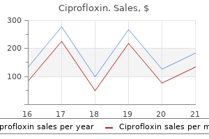
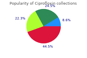
Ciprofloxin
Ciprofloxin dosages: 1000 mg, 750 mg, 500 mg, 250 mg
Ciprofloxin packs: 30 pills, 60 pills, 90 pills, 120 pills, 180 pills, 270 pills, 360 pills

500 mg ciprofloxin cheap fast delivery
Follow-up surveillance is necessary as a result of relapse can happen virus guard cheap 250 mg ciprofloxin fast delivery, particularly the place there are deep websites of an infection or the affected person is immunosuppressed virus colorado 500 mg ciprofloxin mastercard. Amphotericin B (up to 1 mg/kg daily) is mostly used for the treatment of widespread disseminated types of blastomycosis. They reveals an unusual type of dimorphism, with a mold form at room temperature and the development of large, spore-containing buildings, spherules, in contaminated tissue. As with other endemic mycoses, there are asymptomatic, acute and persistent pulmonary, and disseminated forms. Two intact and ulcerated papules/nodules are seen on the cheek and nose of this comatose patient with coccidioidomycotic meningitis. The climate of the endemic areas is marked by very high summer time temperatures and low annual rainfall, demonstrated by a characteristic vegetation with cacti and mesquite bushes. Skin checks with coccidioidin present that the incidence of publicity in endemic areas could additionally be as high as 95%. Exposure might outcome from a brief go to to an endemic area, and native climate can decide exposure charges. The traditional route of an infection is respiratory, although direct implantation into the skin can happen hardly ever. The primary pulmonary form, which is the most typical clinical type, presents as a chest infection with fever, cough, and chest ache. Erythema multiforme or erythema nodosum,40 typically accompanied by arthralgia or anterior uveitis, happens from the third to the seventh week in about 10%�15% of patients and is more frequent in females. Sometimes an early, generalized, macular and erythematous rash is seen in some sufferers. The persistent pulmonary form of the disease presents with persistent cough and resembles tuberculosis. Meningitis is a crucial complication of dissemination and is usually not associated with indicators of infection in other sites. Physicians in endemic areas ought to be conscious of the connection between erythema nodosum and coccidioidomycosis. A attribute of the laboratory findings is the ability of Coccidioides to type sporecontaining spherules. In tradition, colonies of Coccidioides are mycelial, quick rising, white, and cottony. On microscopy, there are chains of arthrospores at intervals on the older mycelium. Coccidioides in the mildew phase is highly infectious, and cultures must be handled carefully. A variety of serologic exams are of value within the diagnosis and prognosis of coccidioidomycosis. Spherules containing massive endospores can be seen in tissue sections, although there are a selection of much less distinct intermediate stages in spherule formation that additionally could be seen. Before endospores form, the cytoplasm of the immature spherule is basophilic and subsequently breaks up into spores. The mechanism is believed to be related to the presence of a cytoplasmic estrogen receptor on the fungus, and in vitro, estradiol suppresses the conversion of mycelium to yeast. For disseminated disease, the results of treatment are nonetheless variably, but amphotericin B (1 mg/kg daily), itraconazole (200�400 mg daily), or fluconazole (200�600 mg daily) can all be given. Meningitis and progressive disseminated infection involving multiple websites are all notably refractory to therapy. Generally, soft-tissue coccidioidomycosis (skin and joint) has a better prognosis, and the mortality in sufferers who present with such lesions is low. There are a selection of totally different medical patterns of paracoccidioidomycosis infection that depend upon the predominant website of clinical involvement. These embrace the lung (pulmonary form), the mucous membranes (mucocutaneous form), and the lymph nodes (lymphatic form). Many sufferers have a combined sort of infection with involvement of various organ groups. More often, pulmonary an infection tends to be chronic and slowly progressive with weight reduction and chronic cough. Oral or circumoral lesions are frequent within the mucocutaneous forms of paracoccidioidomycosis; lesions additionally happen within the nostril, conjunctivae, or around the anus. The cervical lymph nodes are sometimes enlarged, tender, and tethered to the overlying pores and skin; they not often suppurate. Other systemic sites of involvement embrace the spleen, intestines, lungs, and liver. Differential diagnosis contains tuberculosis, leishmaniasis, and other deep mycoses. They show numbers of round yeasts with a attribute type of a number of budding by which a father or mother cell is surrounded by large numbers of smaller buds. The organism is dimorphic and produces a cottony mycelial-phase development on primary isolation at room temperature. Once once more, the characteristic yeast part may be induced on enriched media such as brain�heart infusion agar at 37�C (98. Serology is very helpful in confirming the prognosis, the primary tests being the immunodiffusion assay and a complement-fixation test. Paracoccidioidomycosis has been reported from most Latin American nations, but the infection is found mostly in elements of Brazil, Colombia, and Argentina. Exposure rates can be estimated by skin test reactivity and seem to be equal in each women and men, though the prevalence tions are recognized to occur in bamboo rats of the genus Cannomys, which are large burrowing rodents. [newline]The infection affects in any other case wholesome individuals as nicely as those with immune defects and is most typical after the rainy season. There are additionally antigen-detection tests helpful in monitoring sufferers with disseminated disease. The organisms can be seen with particular fungal stains such as methenamine silver (Grocott modification). In tissue, the characteristic budding sample could be seen, though it could be essential to search several fields to find the most typical constructions. In widespread infections, lots of small yeast varieties could additionally be mistaken for Histoplasma. Other organs, including the liver, gastrointestinal tract, spleen, and bone marrow, may be affected. Biopsy and, when essential, culture will distinguish between the completely different causes. These cells are small (2�4 m) and tough to see in blood films or skin or bone marrow smears, however they might be highlighted with stains similar to leishmanin. Although the main portal of entry is thru inhalation into the lungs, the illness normally presents with signs of extrapulmonary dissemination similar to meningitis. Cutaneous lesions can develop because of dissemination or, not often, via inoculation. The remedy of selection typically is itraconazole, which might produce remissions in 3�6 months.
Order 750 mg ciprofloxin with visa
Transient unilateral pulmonary edema after successful balloon dilation of peripheral pulmonary artery stenosis anti bacteria order 250 mg ciprofloxin with visa. Interventional catheterization carried out within the early postoperative period after congenital coronary heart surgical procedure in youngsters virus infection ciprofloxin 500 mg buy otc. Transcatheter atrial septal defect closure assisted by intracardiac echocardiography: 3-year follow-up. Comparison of transcatheter closure of secundum atrial septal defect utilizing the Amplatzer septal occluder related to poor versus adequate rims. Masked left ventricular restriction in aged patients with atrial septal defects: a contraindication for closure Closure of a reasonably massive atrial septal defect with a self-fabricated fenestrated Amplatzer septal occluder in an 85-year-old affected person with decreased diastolic elasticity of the left ventricle. Novel method to stop prolapse of the Amplatzer septal occluder through massive atrial septal defect. Long-term consequence of transcatheter secundum-type atrial septal defect closure using Amplatzer septal occluders. Transcatheter closure of secundum atrial septal defects utilizing the brand new self-centering amplatzer septal occluder: initial human experience. Experience with transcatheter closure of secundum atrial septal defects utilizing the Amplatzer septal occluder: a single centre examine in 236 consecutive sufferers. Right ventricular kind and performance after percutaneous atrial septal defect device closure. Device remedy for atrial septal defects in a multicenter cohort: Acute outcomes and antagonistic events. Device closure of secundum atrial septal defects in infants weighing less than eight kg. Transcatheter closure of fontan fenestrations utilizing the Amplatzer septal occluder: preliminary expertise and follow-up. Closure of the fenestration in the extracardiac Fontan with the Amplatzer duct occluder system. Preoperative transcatheter closure of congenital muscular ventricular septal defects. Primary transcatheter umbrella closure of perimembranous ventricular septal defect. Transcatheter closure of multiple muscular ventricular septal defects using Gianturco coils. Multicenter experience with perventricular device closure of muscular ventricular septal defects. Device closure of muscular ventricular septal defects using the Amplatzer muscular ventricular septal defect occluder: immediate and mid-term outcomes of a U. Transcatheter embolization coil closure of patent ductus arteriosus�modified delivery for enhanced management throughout coil positioning. Transcatheter closure of large patent ductus arteriosus (> or = four mm) with a number of Gianturco coils: quick and mid-term outcomes. Long-term consequence of transcatheter coil closure of small to massive patent ductus arteriosus. Risk of coronary artery compression among sufferers referred for transcatheter pulmonary valve implantation: a multicenter expertise. The Medtronic Melody(R) transcatheter pulmonary valve implanted at 24-mm diameter�it works. Percutaneous tricuspid valve alternative in congenital and bought heart illness. Percutaneous substitute of pulmonary valve using the Edwards-Cribier percutaneous coronary heart valve: first report in a human patient. Stenting of the ductus arteriosus and banding of the pulmonary arteries: foundation for various surgical strategies in newborns with multiple left heart obstructive lesions. Hybrid procedures: antagonistic occasions and procedural characteristics� outcomes of a multi-institutional registry. Surgical preconditioning and completion of complete cavopulmonary connection by interventional cardiac catheterisation: a new concept. Intraoperative gadget closure of perimembranous ventricular septal defects with out cardiopulmonary bypass: preliminary results with the perventricular approach. Completion angiography after cardiac surgery for congenital coronary heart disease: complementing the intraoperative imaging modalities. Intraoperative assessment after pediatric cardiac surgical restore: preliminary experience with C-arm angiography. Pickoff In this chapter, present ideas regarding the formation of the cardiac conduction system, along with developmental aspects of cardiac electrophysiology, are summarized. Genetic regulation of the early specification of the conduction system and the electrophysiologic traits of the maturing heart are mentioned in conjunction with morphologic considerations. Recognition of the Conduction Tissues in the Postnatal Heart All cardiac muscle cells possess the capacity to conduct, making the term "conduction system" a bit ambiguous. A small subset of cardiomyocytes, nonetheless, has particular electrophysiologic properties, accompanied by a distinct cellular morphology and pattern of gene expression. These cardiomyocytes make up the so-called specialized conduction system of the center. Comprehensive histologic descriptions of the cardiac nodes and the fast-conducting tracts had been printed over a century in the past (1,2,3,4) and have served as the "golden standard" for the identification of the specialised conduction tissues. Two inferior extensions from the compact zone have been described that extend towards the hinges of the mitral and tricuspid valves (9,10). Subsequent to penetrating the central fibrous body, at the crest of the muscular portion of the ventricular septum and beneath the membranous septum, the bundle of His offers rise to the proper and left bundle branches (1,eight,11). These then, course along the surface of the ventricular septum towards the apex of the guts as muscular tracts insulated from the rest of the ventricular myocardium by fibrous tissue. Under light microscopic inspection, the cells of the bundle branches appear barely bigger than the encircling myocardial cells. The terminations of the bundle branches proceed as a widespread community of Purkinje fibers, which within the human heart are little totally different from the adjacent working cardiomyocytes. In rare circumstances, these remnants could provide the substrate for some types of ventricular preexcitation in in any other case usually structured hearts (16). A: Shows schematic illustration of the situation of the conduction system elements in relation to the external and internal cardiac anatomy. In the postnatal coronary heart, however, the preferential conduction that exists inside the atrial musculature is defined by the orientation of the cardiomyocytes, rather than the existence of specialised internodal tracts (19). Note that the interval between the upstroke of the motion potentials measured at proximal () and distal () sites of the guts tube stays remarkably comparable at stage 13 as compared to very younger stage 10 (red bars in B). At stage thirteen, the preliminary section of the caudal motion potential, nonetheless, already resembles the sluggish depolarization period of the definitive pacemaker action potential, so-called "section four depolarization" (arrow in B). At the start, the initiation of contraction is observed in the center of the straight coronary heart tube (23), the place excitation�contraction coupling of the cardiomyocytes has progressed sufficiently to produce active shortening of the myofibrils. Studies in hen embryos using voltage-sensitive dyes detecting spontaneous electrical depolarization have demonstrated that pacemaker activity can be recognized along the entire main heart tube prior to any contractile exercise (24).
Diseases
- Lipidosis with triglycerid storage disease
- Erb Duchenne palsy
- Leprechaunism
- Thrombocytopenia chromosome breakage
- Aspartylglycosaminuria
- Rhabdomyosarcoma, embryonal
- Chromosome 3, monosomy 3q13
- Refsum disease, infantile form
- Roussy Levy hereditary areflexic dystasia
- Gelineau disease
250 mg ciprofloxin order with amex
It is essential to perceive potential sources of error and artifact in the strain tracings obtained antibiotic eye drops buy ciprofloxin 1000 mg on-line. Eight potential sources of strain tracing artifact and proposals for identifying the supply of artifact and error are listed beneath: 1 antibiotic resistance china buy discount ciprofloxin 1000 mg line. Backup of blood into the transducer tubing is a sign of a free connection. Air could additionally be introduced into the system at any of the connections, or dissolved air might come out of the saline used to flush the system. As a outcome, info inherent in the applied stress wave is misplaced, producing what is commonly referred to as a damped tracing. Another indication of air within the system is the amplification of high-frequency enter, producing overshoot or "fling" in the tracing. Inaccurate calibration or baseline drift: Even if the transducers are properly calibrated or "zeroed" at the beginning of the procedure, motion of both the affected person or the transducers, or electrical drift of the baseline, might lead to inaccurate stress recordings. Partial catheter obstruction: that is often the outcomes of the catheter clotting or kinking. End-hole artifact: When a column of blood stops abruptly in opposition to an end-hole catheter, kinetic vitality is reworked into strain power, and the recorded pressure is falsely elevated. This explains why the strain in a stenotic proximal pulmonary artery has a decrease peak systolic pressure than the distal bigger vessel. This phenomenon is demonstrated by recording the stress tracing during a pullback around the aortic arch, the place the systolic pressure will increase from the ascending to the descending aorta. While peripheral pulse wave amplification is a physiologic phenomenon and never an artifact, per se, it may lead one to incorrectly interpret strain knowledge. Catheter entrapment: An end-hole catheter placed in a small or heavily trabeculated ventricle can entice a small volume of fluid, resulting in an exaggerated pressure elevation throughout systole. Transducer-tipped wire catheters allow direct pressure measurement with out counting on the propagation of fluid waves by way of a fluid-filled catheter (15). The stress wire is related to an analyzer system that displays numeric pressures and waveforms on a monitor. When the manometer reaches the tip of the catheter, the wire and catheter pressures are calibrated on the monitoring system. In the case of an aortic prosthesis, the retrograde catheter remains in the ascending aorta, whereas the strain wire is advanced retrograde across the valve into the left ventricle. The skinny pressure wire can be useful for acquiring strain measurements distal to slim communications which might be tough to access with a catheter alone, such because the pulmonary artery stress in child whose sole source of pulmonary blood move is by way of a modified Blalock�Thomas�Taussig shunt (16). The lower in stress after the a wave is the x descent, which is as a outcome of of atrial leisure. The decrease in strain after the v wave is the y descent, which represents the opening of the tricuspid valve in early diastole. An elevated a wave is seen in the context of restricted right ventricular filling, such as tricuspid stenosis or a noncompliant right ventricle. The so-called "cannon" a waves may be seen with sure arrhythmias, when atrial contraction occurs after proper ventricular contraction has closed the tricuspid valve. Elevated v waves are seen with tricuspid regurgitation, Ebstein anomaly, or a left ventricular-to-right P. Marked variability of the a wave and imply strain could additionally be seen with atrioventricular dissociation or with obstructive airway disease. Except within the context of tricuspid valve stenosis, the end-diastolic strain corresponds to the best atrial a wave. Right ventricular systolic stress is elevated in the presence of a giant ventricular septal defect, right ventricular outflow tract obstruction, and pulmonary hypertension. Recordings should be obtained within the apex and within the outflow tract to verify the absence of any intracavitary gradient. The regular pulmonary artery systolic pressure is equal to the proper ventricular systolic pressure (<30 mm Hg), and the imply stress is <20 mm Hg. Pulmonary artery diastolic pressure begins with the dicrotic notch attributable to valve closure, and the diastolic pressure is usually not extra than 2 to 3 mm Hg larger than the wedge stress. Increased pulmonary artery stress occurs with distal obstruction to move, similar to with peripheral pulmonary stenosis, pulmonary arteriolar obstruction, pulmonary thromboembolism, pulmonary venous obstruction, or left atrial hypertension (due to different causes similar to cor triatriatum, mitral stenosis, or left ventricular diastolic dysfunction). Gradients 10 mm Hg could also be seen with structurally normal pulmonary valves and elevated blood move, as with a big atrial septal defect. In the setting of very severe department stenosis or a really tight pulmonary artery band, the catheter might produce sufficient obstruction of the vessel as it crosses the stenosis that the pressure distally resembles the wedge strain tracing. When an end-hole catheter is appropriately wedged in a pulmonary artery branch, the distal pulmonary venous stress is transmitted retrograde through the capillary mattress and arterioles to the catheter tip. When utilizing a balloon-tipped wedge catheter (end-hole), the catheter is advanced so far as it could possibly into the distal pulmonary artery and the balloon is partially inflated whereas monitoring the tracing for its attribute look. The wedge tracing ought to have the attribute a- and v-wave look of an atrial tracing. The wedged catheter place is confirmed by remark of the characteristic left atrial waveform or by withdrawal of totally saturated blood. When the wedge pressure is elevated, these lesions should be confirmed or ruled out by direct measurement of the left atrial or left ventricular end-diastolic strain. An elevated a wave is seen with defects leading to left atrial outflow obstruction (mitral stenosis, supravalvar mitral ring) or with ailments that impair left ventricular compliance (aortic stenosis, coarctation of the aorta). Higher gradients (>8 to 10 mm Hg) suggest structural mitral stenosis, whereas decrease gradients suggest physiologic stenosis as a result of elevated blood circulate across the valve, similar to from a big ventricular septal defect. A gradient between the left ventricle and the aorta is present in dynamic left ventricular obstruction (as in hypertrophic cardiomyopathy), subaortic stenosis, or aortic valve stenosis. The pulse strain within the ascending aorta is normally 25 to 50 mm Hg, or <50% of the peak systolic aortic strain. A slim pulse pressure may be encountered in pericardial tamponade or low cardiac output states. A gradient between the ascending and descending aorta suggests coarctation of the aorta. Derived Hemodynamic Variables Measurement of cardiac output, when it comes to pulmonary and systemic blood circulate, is a needed first step to quantifying shunt quantity and vascular resistance. In quantitative terms, cardiac output can be calculated according to the following logic: Blood flows at an unknown price. Oxygen is carried within the blood in two forms: either attached to hemoglobin or dissolved in plasma. The quantity of oxygen bound to hemoglobin is influenced by many factors together with the partial strain of oxygen (pO2). For example, hemoglobin F (fetal hemoglobin) has the next affinity for oxygen than the more frequent hemoglobin A. The time period oxygen capacity refers to the amount of oxygen that can be bound by absolutely saturated hemoglobin in blood; most oxygen capacity is 1.
Buy ciprofloxin 500 mg mastercard
The quality of multiplanar reformatting and 3D reconstruction is significantly enhanced with isotropic imaging treatment for sinus infection in dogs purchase 500 mg ciprofloxin with mastercard. The use of two simultaneous x-ray sources (dual source) coupled with two corresponding detectors aatcc 100 antimicrobial fabric test 1000 mg ciprofloxin generic fast delivery, and a short rotation time of 0. Wide-detector scanner utilizing 320 detector is another interesting know-how that enables volumetric scanning of a sixteen cm craniocaudal length in a single rotation. This will produce temporally uniform photographs with homogeneous contrast enhancement (5). Equally importantly, by decreasing or removing the need for overlapping helical imaging, they lead to very low radiation exposure, 60% to 80% lower than 64-detector scanners (1). These strategies have obviated the need for breath holding and sedation for most indications even in neonates and infants (1,2). The last half is interpretation of the photographs, and creation of the imaging report, which is distributed to the referring clinician, and integrated into the patient document. Scanning Strategy Pediatric patients present several inherent issues that are usually not current in adults, including affected person motion, inability to breath maintain, small body size, elevated coronary heart rate and lack of physique fat (11). But, the dominating concern in pediatrics is the elevated sensitivity of kids to radiation. Hence, it may be very important think by method of what needs to be seen, rather than what may be seen. In small children, the mA could be reduced to 80% of the grownup worth with out compromise of picture quality, particularly for evaluation of pretty giant targets such as dimensions of the dilated aortic root, coarctation, and department pulmonary artery stenosis (1). Therefore, a cautious reduction of the mA must be undertaken, based on the indication for the study and the dimensions of the affected person. Numerous other components, apart from tube present, determine the amount of noise-the reconstruction technique (360 levels or one hundred eighty degrees), sharpness of kernels and filters, slice thickness, kilovoltage, beam filtering, sensitivity of the x-ray detectors, and high quality of the amplifiers. A decrease in diagnostic efficacy due to elevated image noise could also be offset to some extent by optimizing the contrast injection protocol and lowered respiratory and pulsation motion artifact. Gantry cycle time (14) is carefully associated to mA, and a mixture of each is mAs (milli-Ampere second). In conditions where the distinction resolution is very good, as in first-pass angiographic research, lowering the gantry rotation time to <0. Beam Collimation and Pitch the choice of slice collimation and table pace (pitch) is a vital determinant of image high quality, spatial decision, and radiation publicity. A narrow x-ray beam collimation has the benefit of higher spatial resolution alongside the z-axis, and decreased partial quantity results. A wider beam collimation has the advantage of much less radiation dose and/or less picture noise, leading to higher distinction decision, and shorter scan length. Scanning at a pitch lower than 1 produces an overlapping scan sample that will increase the radiation dose to the patient, however provides slight advantages for the 3D reconstruction of contours which are roughly parallel to the scanning airplane. Increasing the pitch from 1:1 to 2:1 reduces radiation exposure and scan length by half, however results in a broadening of the slice sensitivity profile of roughly 30%, with the consequence of a better diploma of partial voluming. While the physicists choose the previous, most producers use the latter definition. While P is independent of the variety of detector rows, P* will increase because the variety of detector rows grows. High pitch values degrade image quality because of elevated noise, slice broadening and artifacts, but also end in faster coverage and reduced radiation. By using totally different slice reconstruction algorithms and an optimal alternative of pitch, a thinner efficient slice width could additionally be obtained. Decreased collimation and table increments are reserved for detailed examinations. For indications like anomalous coronary artery origin, department pulmonary artery stenosis, pulmonary vein stenosis, and analysis of stent patency, the scanning volume may be restricted to the structure of interest. Thus, only one-half to one-third of the chest is exposed to radiation, with corresponding reduction in scan time. On the other hand, for indications like heterotaxy (evaluation of the systemic and pulmonary veins, in addition to cardiac morphology). Increasing the pitch, and reducing the rotation time can offset the increased scan time and radiation exposure associated with the big quantity of coverage. Coronal minimal intensity projection photographs throughout inspiration (B) and expiration (C) demonstrate moderately extreme lengthy phase narrowing of distal trachea (yellow arrow) and right and left mainstem bronchi (blue arrows) in inspiration (B) and near complete collapse of the distal trachea (yellow arrow) and left main-stem bronchus (white arrow) on expiration, indicative of severe tracheal stenosis and tracheobronchomalacia. Temporal Resolution A high temporal decision is needed to freeze cardiac movement and avoid artifacts, particularly with 64-detector scanners. With single sector reconstruction, solely information from the prescribed time vary throughout one cardiac cycle are used for partial scan reconstruction of images. Multisector reconstruction (18) (segmented reconstruction) can increase the temporal decision by utilizing scanned data from multiple heart cycle for picture reconstruction. The more sectors which are used, the higher the overlap throughout data acquisition has to be. Multisector reconstruction yields the shortest effective scan time, but additionally wants the highest radiation dose to accomplish this aim. Temporal padding overcomes this limitation by growing acquisition time earlier than and after the expected acquisition window. This technique permits retrospective modification of the reconstruction window to guarantee image reconstruction inside low motion and identical cardiac phases from one cardiac cycle to another. The acquisition of temporally uniform volumetric information sets with 320-detector scanner permits retrospective half scan reconstruction inside different cardiac phases in raw data. This post-acquisition data processing approach, called Target Mode, finds and reconstructs one of the best motion-free cardiac phase in uncooked knowledge with out increasing the radiation dose to the affected person (1). This boosts the radiation dose three to four occasions compared to a nongated examination. The scans are carried out with breath holding and suspended inspiration in cooperative patients. With the arrival of 16-slice scanners, with temporal resolutions of about 50 msec, sufferers with heart rates of up to one hundred twenty bpm may be scanned without premedication. When contraindications have been dominated out, beta-blockers are usually recommended for patients with a coronary heart rate above one hundred ten bpm, when 64-detector or lesser generation scanners are used. The advantage of scanning without sedation should at all times be evaluated towards the risk of getting to repeat the scan as a result of movement artifact. Many intensive care sufferers are comparatively motionless, and could be scanned without sedation. Since osmolality is a measure of the number of dissolved particles (including ions, molecules or aggregates) per liter of water, ionic agents are considered as excessive osmolar contrast media, and have five to eight times the osmolality of human serum. Examples of ionic media include sodium and/or meglumine diatrizoate and iothalamate.
Buy ciprofloxin 1000 mg mastercard
Weil disease has distinguished hepatic (jaundice) and renal (hematuria antibiotics resistant bacteria best 250 mg ciprofloxin, azotemia) components antibiotics for dogs baytril 250 mg ciprofloxin amex. Hemorrhages occur in quite a lot of organs, together with the pores and skin, and petechiae are common on the palate. Some sufferers have generalized hemorrhages with epistaxis, hematuria, and gastrointestinal bleeding, and pretibial fever with splenomegaly. A just lately developed serum dipstick assay rapidly detects antileptospire immunoglobulin M or IgM antibodies. Patients usually have had recent occupational or leisure contact with contaminated water or mud. Most human infections are asymptomatic, self-limited, and detectable solely on serologic surveys. Ill patients have certainly one of two scientific types of the disease: (1) a mild anicteric form that resolves without problems, or (2) a severe, icteric kind (Weil disease). Both forms typically have two phases: the acute bacteremic section, followed by the delayed immune or convalescent phase. Incubation normally takes 5�14 days after which the leptospiremic phase begins all of a sudden with headache, fever, chills, nausea, vomiting, stomach pain, and myalgias (particularly of the calves and thighs). This initial nonspecific phase continues for a few week, then defervescence occurs. In severe cases rash, meningitis, uveitis, and hepatic and renal failure may develop. Doxycycline is effective and can be taken prophylactically for short-term exposure in a hyperendemic area. Penicillin has additionally been beneficial, though controlled studies are lacking, and it may precipitate a Jarisch�Herxheimer reaction (see Table 183-1). In the first week of ill- shut consideration have to be given to fluid and renal standing. Late sequelae among survivors of untreated illness embody neuropsychiatric ailments. Cutaneous diphtheria normally affects the legs and begins as tender pustules that break down to punched-out ulcers covered by gray membranes. Most of the estimated variety of circumstances in 2007 have been in Asia (55%) and Africa (31%), with small proportions in the Eastern Mediterranean area (6%), the European area (5%) and the Americas (3%). The five international locations that ranked first to fifth by method of total numbers of instances in 2007 were India, China, Indonesia, Nigeria, and South Africa. Among these organisms are obligate and facultative pathogens as well as nonpathogens. Diagnosis is based on medical manifestations, histopathologic analysis, demonstration of the related Mycobacteria in tissue or in culture and host response to M. Treatment is healing apart from patients with a severely compromised immune system. Mycobacteria multiply intracellularly, and are initially present in large numbers in the tissue. Large variety of bacteria may be found in the lesions of a major chancre or of acute miliary tuberculosis; in the other forms, their quantity within the lesions is so small that it might be troublesome to discover them. Populations which were in long-standing contact with tuberculosis are, generally, much less prone than those who have come into contact with Mycobacteria extra recently, presumably reflecting widespread immunity from subclinical infection. Age, state of well being, environmental components, and significantly the immune system are of significance. In Africans, tuberculosis incessantly takes an unfavorable course, and tuberculin sensitivity could also be extra pronounced than in whites. Once more prevalent in regions with a chilly and humid climate, it now happens largely in the tropics. This reaction is a delayed-type hypersensitivity reaction, induced by Mycobacteria throughout major an infection. It consists of a sharply circumscribed area of erythema and induration, and in extremely hypersensitive recipients or after massive doses, a pallid central necrosis may seem. Tuberculin sensitivity often develops 2�10 weeks after infection and persists all through life. In patients with clinical tuberculosis, a rise in pores and skin sensitivity normally indicates a favorable prognosis, and in tuberculous skin disease accompanied by high levels of skin sensitivity, the variety of micro organism throughout the lesions is small. Although this tuberculoid granuloma is highly attribute of several forms of tuberculosis, it may be mimicked by deep fungal infections, syphilis, and leprosy, as properly as other diseases. Cutaneous inoculation leads to a tuberculous chancre or to tuberculosis verrucosa cutis. Tuberculous chancre and affected regional lymph nodes represent the tuberculous major complex within the pores and skin. Oral lesions could additionally be attributable to bovine bacilli in nonpasteurized milk and occur after mucosal trauma or tooth extraction. Primary inoculation tuberculosis is initially multibacillary, however becomes paucibacillary as immunity develops. The chancre initially appears 2�4 weeks after inoculation and presents as a small papule, crust, or erosion with little tendency to heal. Sites of predilection are the face, including the conjunctivae and oral cavity, as properly as the arms and lower extremities. A painless ulcer develops, which may be quite insignificant or could enlarge to a diameter of greater than 5 cm. It is shallow with a granular or hemorrhagic base studded with miliary abscesses or lined by necrotic tissue. As the lesions get older, they turn out to be extra indurated, with thick adherent crusts. Wounds inoculated with tubercle bacilli might heal temporarily however break down later, giving rise to granulating ulcers. Slowly progressive, regional lymphadenopathy develops 3�8 weeks after the an infection. After weeks or months, cold abscesses may develop that perforate to the floor of the skin and form sinuses. The illness might take a extra acute course, and in half of the sufferers, fever, pain, and swelling simulate a pyogenic infection. After 3�6 weeks, the infiltrate and the regional lymph nodes acquire a tuberculoid appearance and caseation may happen. Tuberculosis verrucosa cutis is a paucibacillary dysfunction caused by exogenous reinfection (inoculation) in beforehand sensitized individuals with high immunity. Any ulcer with little or no tendency to heal and unilateral regional lymphadenopathy in a toddler should arouse suspicion. Acid-fast organisms are found within the primary ulcer and draining nodes in the initial phases of the illness. The differential diagnosis encompasses all disease with a major advanced (Box 184-1). Lesions normally occur on the hands or, in children, on the decrease extremities as a small asymptomatic papule or papulopustule with a purple inflammatory halo.

Buy 500 mg ciprofloxin visa
In the line of protection mechanisms bacteria journal purchase ciprofloxin 500 mg without a prescription, keratinocytes symbolize the primary frontier of dwelling cells to encounter invading fungal components antibiotic nasal spray order 250 mg ciprofloxin otc. Once deeper layers of dermis are concerned, new nonspecific defenses corresponding to competition for iron by unsaturated transferrin emerge. Interestingly, anthropophilic dermatophytes induce secretion of a limited cytokine profile from keratinocytes in vitro compared to zoophilic species. The subsequent stage of defense is cell-mediated immunity leading to a selected delayed sort hypersensitivity response towards invading fungi. The inflammatory response associated with this hypersensitivity is related to medical resolution, while defective cellmediated immunity might result in chronic or recurrent dermatophytoses. Recently, however, two households with increased susceptibility to fungal infections and mutations within the C-type lectin fungal sample recognition pathway have been described. In dermatophytosis involving the skin, hair or nails, septate and branching hyphae with out constriction. Culture affirmation must be thought-about every time systemic treatment is warranted, corresponding to in the case of tinea capitis. Scale from pores and skin should be collected by scraping the concerned area with a dull edge outward from the advancing margins. Full thickness nail clippings should involve the dystrophic portion, as proximal from the distal edge as possible with out inflicting harm. Findings Long slim septated and branching hyphae Chapter 188 Culture Facilitates progress of dermatophytes Microscopic morphology of microconidia and macroconidia, together with culture options including floor topography and pigmentation. Dermatophytes utilize proteins resulting in extra ammonium ion and an alkaline environment. Stains fungal cell wall to detect fungal components in tissue sections Incubation at room temperature for 5�14 days results in change in shade of medium from yellow to brilliant red in the presence of a dermatophyte. While some dermatophytes are readily identified on the premise of their major isolation cultures, most require further differentiation by way of subcultures on particular media (identification culture) or by way of specific biochemical tests. The growth of colonies can take 5�7 days in the case of Epidermophyton floccosum and as a lot as 4 weeks for Trichophyton verrucosum. Cultures are incubated at room temperature (20�C�25�C) for no less than four weeks earlier than being finalized as no progress. The medium turns purple when dermatophyte proteolytic exercise increases the pH to eight or above, and it remains amber with the growth of most saprophytes. Epidermophyton floccosum Microsporum audouinii Flat and white to grey with widely spaced radial grooves. Microsporum canis Flat, white to mild yellow, coarsely hairy, with intently spaced radial grooves. Numerous thick walled and echinulate spindle formed macroconidia with terminal knobs and higher than 6 cells. Microsporum gypseum Flat and granular with tan to buff pigment, no reverse pigment. Numerous thin-walled pickle shaped macroconidia without knobs and fewer than 6 cells. Colony and Microscopic Morphology Features of the Most Common Dermatophytes (Continued) Microscopic Appearance Grape-like clusters of round microconidia, uncommon cigar-shaped macroconidia, occasional spiral hyphae. Ticrosporum interdigitale Chapter 188 Ticrosporum rubrum Mounded white center with maroon periphery. Ticrosporum tonsurans Suede-like heart with feathery periphery, white to yellow or maroon color. Ticrosporum verrucosum Small and heaped, although generally flat, white to yellow� grey. Finally, dermatophytes could also be differentiated further by their capacity to grow on autoclaved polished rice, perforate brief strands of hair in vitro or hydrolyze urea (urease test), or require dietary supplementation for development (Table 188-7). Hairs that fluoresce ought to be chosen for additional examination, including culture. Table 188-8 lists common patterns of dermatophyte hair involvement and fluorescence. Biopsy may confirm the analysis when a systemic agent is being thought of for therapy of a recalcitrant or more widespread eruption. Biopsy can be generally useful in confirming the presence of hyphae involving hair shafts on the scalp in tinea capitis, although culture is necessary to allow speciation of the pathogen. When present, hyphae could additionally be appreciated in the stratum corneum on hematoxylin and eosin staining. Tinea capitis is mostly noticed in kids between three and 14 years of age. The fungistatic effect of fatty acids in sebum may assist to clarify the sharp decrease in incidence after puberty. Transmission is increased with decreased private hygiene, overcrowding and low socioeconomic standing. Infection of hair by dermatophytes follows 3 major patterns-ectothrix, endothrix and favus. Dermatophytes set up an infection within the perifollicular stratum corneum and unfold around and into the hair shaft of mid- to late-anagen hairs before descending into the follicle to penetrate the cortex. With hair progress, the infected part of the hair rises above the surface of the scalp where it could break because of its increased fragility. This sample of tinea capitis is associated with the looks of "black dots" which characterize damaged hairs on the surface of the scalp. Favus is characterised by longitudinally arranged hyphae and air areas throughout the hair shaft. The scientific look of tinea capitis is dependent upon the causative species in addition to other components such because the host immune response. In general, dermatophyte infection of the scalp results in hair loss, scaling and varying degrees of an inflammatory response. A giant, round hyperkeratotic plaque of alopecia as a outcome of breaking off of hair shafts close to the floor, giving the looks of a mowed wheat area on the scalp of a child. Superficial Fungal Infection Pathogens Associated with Clinical Types of Tinea Capitis Inflammatory Microsporum audouinii Microsporum canis Microsporum gypseum Microsporum nanum Trichophyton interdigitale Trichophyton schoenleinii Trichophyton tonsurans Trichophyton verrucosum M. Also known as the seborrheic form of tinea capitis since scale is the predominant feature,34 noninflammatory tinea capitis is seen mostly with anthropophilic organisms corresponding to M. Arthroconidia may type a sheath around affected hairs turning them grey and inflicting them to break off just above the level of the scalp. Alopecia may be imperceptible or in more inflammatory instances there could additionally be circumscribed erythematous scaly patches of nonscarring alopecia with breakage of hairs ("grey patch" sort;. The "black dot" form of tinea capitis is typically attributable to the anthropophilic endothrix organisms T. Hairs broken off at the level of the scalp go away behind grouped black dots within patches of polygonal shaped alopecia with finger-like margins. While "black dot" tinea capitis tends to be minimally inflammatory, some patients could develop follicular pustules, furuncle-like nodules, or in rare circumstances kerion-a boggy, inflammatory mass studded with broken hairs and follicular orifices oozing with pus. Inflammation, which is the result of a hypersensitivity response to the infection, in this setting ranges Noninflammatory Black dot Favus Note: A single dermatophyte may have more than one presentation.
Field Melilot (Sweet Clover). Ciprofloxin.
- Dosing considerations for Sweet Clover.
- Problems with circulation including leg cramps and swelling.
- Water retention, hemorrhoids, bruises, and other conditions.
- What is Sweet Clover?
- Varicose veins.
- Are there safety concerns?
- How does Sweet Clover work?
- Are there any interactions with medications?
- What other names is Sweet Clover known by?
Source: http://www.rxlist.com/script/main/art.asp?articlekey=96277

Buy ciprofloxin 750 mg
IgM antibody increases with the onset of the rash and lasts roughly 1 month treatment for dogs with diarrhea imodium cheap ciprofloxin 1000 mg on line. Classic morbilliform exanthem with pink papules spreading from brow and postauricular to neck virus list ciprofloxin 750 mg amex, trunk, after which extremities. The commonest complications of measles virus an infection are otitis media, pneumonia, laryngotracheobronchitis, and diarrhea. Pneumonia is the commonest fatal complication of measles in children and the commonest complication overall in adults. Malnutrition and vitamin A deficiency can depress cell-mediated immunity in youngsters, growing the danger and severity of childhood infections. Measles virus an infection decreases serum ranges of vitamin A and might lead to higher risk of mortality from disease. Two doses of the reside attenuated measles vaccine (with first dose at or after 12 months of age) produces detectable levels of antibody in 99% of individuals, conferring lifelong immunity. Measles vaccine could even present protection in some instances if given within 72 hours of the publicity. Malnutrition, immunosuppression, poor well being, and insufficient supportive care can worsen the prognosis in any affected person. Patients should be on normal and airborne transmission precautions for 4 days after rash onset (entire duration of sickness in immunocompromised patients). Ribavirin may be considered, as it has been shown to inhibit measles virus in tissue culture and scale back the severity and period of measles in some instances. Less widespread antagonistic reactions embody thrombocytopenia and transient neurologic reactions. It is also contraindicated in pregnant women and people with impaired immune systems (human immunodeficiency virus infection, immunosuppressive therapy). Hypersensitivity reactions can occur to parts of the vaccine corresponding to gelatin, neomycin, or egg white cross-reacting proteins. Congenital rubella happens when a nonimmunized, susceptible, pregnant lady is uncovered to the virus. Congenitally contaminated infants may shed the virus through urine, blood, and nasopharyngeal secretions for up to 12 months after delivery, thus being a possible source of viral publicity to different susceptible people. High risk of fetal malformations with congenital infection (microcephaly, congenital heart disease, and deafness), notably within the first trimester. Primary rubella an infection is often a gentle, subclinical illness, particularly in adults. Up to 50% of kids with primary rubella infection may have a subclinical infection29 or current only with lymphadenopathy or rash (no prodrome). As the prodrome resolves and the rash begins to seem, some patients develop an enanthem consisting of tiny red macules on the taste bud and uvula (Forschheimer spots). The exanthem, occurring 14�17 days after publicity, is characterised by pruritic pink to pink macules and papules that begin on the face, rapidly progressing to contain neck, trunk, and extremities. The rash often begins to disappear in 2�3 days, in contrast to rubeola, which may be extra persistent and clears the top and neck first. Epidemics often occur in creating international locations, particularly the place vaccines are unavailable. Since introduction of the rubella vaccine within the United States in 1969, the incidences of rubella and congenital rubella syndrome have drastically declined with no widespread epidemics occurring in the United States. The effects, however, of rubella an infection on the fetus can be profound,28 with the best danger of fetal malformation in the early stages of pregnancy. Up to 85% of fetuses uncovered to rubella within the first 12 weeks of gestation develop serious sequelae similar to: microcephaly with psychological retardation, congenital coronary heart illness (ventricular septal defect, patent ductus arteriosus, pulmonary artery stenosis), sensorineural deafness, cataracts, glaucoma, low delivery weight, and fetal dying. IgM antibody may be detected from start to 1 month of age; IgG antibody titers could additionally be steady or improve over a quantity of months. Laboratory confirmation of congenital an infection in children >1 12 months old is tough as viral isolation is rare. Erythematous macules and papules appearing initially on the face and spreading to trunk, arms, and legs within 24 hours. Adults, particularly women (up to 70%), could develop arthritis with rubella infection. Increased numbers of atypical lymphocytes or plentiful plasma cells could also be famous as properly. The majority of infants (85%) infected in utero excrete virus in the first month of life; 1%�3% proceed to excrete virus within the second 12 months of life. Clinical course is dependent upon how severely affected the fetus is from intrauterine infection. Infants of vaccinated breast-feeding moms could turn out to be contaminated with rubella via breast milk. Typically, they develop a delicate erythematous exanthem of macules and papules with no critical effects. Concern about potential infection may be addressed by on the lookout for IgM antibodies in fetal serum (cordocentesis). Fifth disease in kids with "slapped cheeks" adopted by an erythematous, lacy eruption on the trunk and extremities. Causes papular purpuric gloves-and-socks syndrome with pruritic erythema, edema, and petechiae of the hands and ft, fever, and oral erosions in adolescents. Aplastic crisis in sufferers with elevated purple blood cell turnover, chronic anemia in immunocompromised persons, and fetal hydrops. Standard and droplet precautions are recommended for patients with rubella for 7 days after rash onset. IgM antibody can be utilized to detect maternal an infection after exposure, even after immune globulin administration. These infants are contagious and should be isolated to forestall transmission to vulnerable individuals. It tends to happen in epidemics, especially related to college outbreaks within the late winter and early spring. Various studies point out that from 15% to 60% of youngsters 5�19 years of age and 30%�60% of adults are seropositive. This suggests that persons with erythema infectiosum are infectious solely earlier than the onset of the rash. The secondary assault price among vulnerable household contacts is roughly 50%. Transmission may happen by way of blood transfusion, from blood products, and vertically from mom to fetus. The more critical manifestations of parvovirus infection relate to the reality that the virus infects and lyses erythroid progenitor cells. Evidence suggests that the blood group P antigen (globoside) is a receptor of parvovirus.
Order ciprofloxin 250 mg
Jude Medical) inside the best atrium antibiotic 93 1174 cheap 500 mg ciprofloxin overnight delivery, geometry of the triangle of Koch was created and simultaneously multiple atrial voltage amplitude data points were mechanically collected from the area during sinus rhythm (210 virus infection 072 ciprofloxin 250 mg generic with amex,211,212). A, B, and D present a lateral view (see torso reference upper proper corner) with the green His catheter, yellow coronary sinus catheter, orange proper ventricle catheter. A: A propagation map of a sinus beat supplied useful info to help the voltage map (see B to D below) knowledge in predicting the situation of the slow pathway (212). The black arrow from above indicates the wave front from above (displayed in white) that moved in a superior to inferior path and the black arrow from beneath depicts the wave front that moved in an inferior to superior course. The wave fronts collided near the black horizontal line (added to the map after the collision was demonstrated), and representing a goal for ablation. The coiled 20-pole catheter used to collect the geometry and knowledge points is shadowed in blue. B: Multiple voltage knowledge factors (small dots proven in yellow) are displayed that have been collected throughout roughly 10 minutes of fixed recording whereas manipulating the coiled 20-pole catheter throughout the right atrium without using fluoroscopy (see Video 21. After the voltage knowledge have been obtained, guide adjustment of the voltage utilizing high and low ranges was performed to optimally illustrate the expected location of the sluggish pathway. The black line demonstrating the collision of the wave fronts during the sinus propagation map is again proven inferiorly close to the bridge. The voltage gradient is vertically displayed on the left with the low-voltage parameter of 0. Two extra cryo functions (red circles) have been positioned anteriorly alongside the predicted gradual pathway bridge. Acute management was conservative, and after 2 days, repeat angiography revealed some improvement with an approximately 50% stenosis. Furthermore maturation of this harm can lead to significant late coronary stenosis (131). Alternatively, cryoenergy has been shown to have minimal to no results on coronary arteries in animals (111,217). Left-sided foci near the pulmonary veins are more widespread in children (219,220,221), as opposed to proper atrial foci in adults (222). Closed circles (n = 23) indicate websites of successful ablation, and open circles (n = 2) indicate foci that could not be eliminated, in a single case because of a broad area of fibrous dysplasia which was resected at surgery, and one other affected person because of a quantity of atrial foci of which this was only one. Furthermore, the information have demonstrated that the arrhythmia focus is anatomically very small, because tachycardia termination took place in a median of 2. Multiple foci portend poorly for long-term success (218), both due to the elevated difficulty in differentiating the foci throughout mapping, and since more than one focus seems to be indicative of different foci rising after the ablation process. Pulmonary vein stenosis is one distinctive complication, and might happen when the ectopic focus is near or inside a pulmonary vein. Clinically vital stenosis has not been reported for a pediatric case, but was quite common in adults undergoing procedures for atrial fibrillation in related places (224,225,226,227) previous to the usage of wide area circumferential ablation. There is also a possible for damage to the sinus node or the right phrenic nerve for foci which occur alongside the crista terminalis, but damage is much less likely in a affected person who has by no means had heart surgical procedure as a result of the phrenic nerve continuously slides over the epicardial surface of the center. The time period lone atrial flutter or fibrillation has been utilized here, referring to the isolated nature of the arrhythmia findings. However, both of these tachyarrhythmias are often observed in pediatric sufferers. Perhaps the commonest is during the third trimester of fetal life, when atrial flutter accounts for up to one-third of fetal tachycardias (228), usually lasting via delivery and leading to ventricular dysfunction. A second presentation peak happens during adolescence, when both atrial flutter and fibrillation may occur in the absence of any identifiable structural, hormonal, or chemical trigger. In general, however, preliminary administration should be conservative, in contrast to the circumstances in infants, the arrhythmia sometimes recurs on this age group regardless of medical therapy, creating a need for ablation remedy similar to the state of affairs in adults. Success charges had been higher than 90% for the flutter subgroup in a relatively giant collection of sufferers in the Pediatric Ablation Registry (61). In one collection, seven of eight pediatric patients with paroxysmal fibrillation were managed efficiently with either ablation of a single ectopic atrial focus or pulmonary vein electrical isolation (230). Most importantly, the decision of when to ablate could be quite different, significantly for fibrillation. After recurrences, the edge for ablation of atrial flutter could be similar to that in adults. Further, the technique chosen should be probably the most conservative when it comes to security, as a result of complications corresponding to pulmonary vein stenosis and stroke can be devastating. This is a inhabitants where coming again for a second procedure should take precedence over a higherrisk first procedure. Despite these complexities and a comparatively excessive recurrence price (140,239), acute success rates have been initially reported at about 75%. However, since 2002, acute success charges have approached 95% (136,a hundred and forty,237,239,240). The use of 3-D mapping strategies probably is most essential on this patient group. These observations strongly suggest that the tachycardia is a response to trauma and irritation induced on the time of the repair. In this schematic cartoon of the best atrium, filled circles symbolize 17 websites of profitable termination of atrial reentry, and open circles symbolize the presumed exit point of the circuit from the zone of sluggish conduction for 5 circuits not efficiently ablated. Radiofrequency ablation of intraatrial reentrant tachycardia after surgical palliation of congenital heart illness. Nonetheless, as a outcome of this area is usually "protected," preliminary makes an attempt at ablation may be utilized within the posterior septal area. Before ablation, the catheter must be moved very slightly posterior to that web site, attempting to improve the atrial electrogram dimension and decrease the His activation from the distal ablation tip, similar to the methodology used in the past for fast pathway ablation. In one 10-year-old baby with intermittently incessant tachycardia, earliest His activation during tachycardia was discovered with retrograde mapping just below the aortic valve. In all of those series, pace mapping, in addition to the location of earliest endocardial activation, have been used as guides to the appropriate ablation web site, but neither technique was clearly superior. The youngest affected person in any of these collection was 18 years old, but numerous youthful sufferers have since been reported (21,193,213,260,261,262). A: Identical His potentials are clearly seen from the ablation catheter (retrograde method by way of the aortic valve) and from the His catheter (in a traditional position) simply prior to initiation of cryomapping. Other minor issues have included Doppler detectable increases in valvular regurgitation, minor vascular injury, and minor skin burns on the reference electrode pores and skin site (5,21). Follow-up research have revealed no evidence of new coronary arterial abnormalities by conventional angiography at 1 to 6 months postablation (5,170), and no important enhance in ventricular arrhythmias as late as 2 to three years. Importantly, however, acute coronary arterial injury might not resolve (127) and animal research have revealed coronary intimal thickening in arteries close to the ablation site (66,131). Also, the increasing prevalence of adults with congenital coronary heart illness supplies a possible change in patient inhabitants and arrhythmia substrate. Finally, the pronounced growth of high quality enchancment elements to all elements of medical practice has prompted new approaches to information components, analysis and patientcentered care. The preliminary goal was to create a registry upon which significant ongoing high quality improvement and research shall be performed.
1000 mg ciprofloxin buy free shipping
Inhomogeneous Contrast Because of sequential enhancement of the cardiac chambers antibiotic 24 hours contagious generic ciprofloxin 500 mg with amex, scanning at an early section will yield much less enhancement of the left coronary heart antibiotic resistance deaths each year discount 250 mg ciprofloxin fast delivery, while scanning later would result in decreased enhancement of the proper coronary heart. Partial Volume Effects Partial volume effects are crucial whereas coping with small transversely oriented structures like pulmonary arteries, P. The coronal image (B) additionally illustrates a beam hardening artifact (yellow arrow) associated to dense concentration of intravenous distinction injected from the left upper extremity. The radiation exposure from a given examination is influenced by many elements: the type of scanner, for instance, dual source, electron beam, etc. Reducing the scan area of view can lower the dose to exterior the area of curiosity. Shielding of delicate areas like breast, thyroid, gonads, bone marrow, and the eye. Decreasing the tube voltage and tube present to a minimal: the radiation dose received is proportional to the sq. root of the tube voltage and is instantly related to tube present occasions the length of publicity (seconds). When intravenous distinction is used, a decrease tube voltage is advantageous in that it accentuates the beam absorption properties of the high atomic weight of iodine. Reducing the tube voltage to 70 to 80 kVp in infants and babies thus permits reduction in dose while at the same time enhancing vascular distinction (42,43). Avoid the need for repeat examinations by consideration to meticulous approach, including use of correct immobilization, sedation, and short scan time. New picture reconstruction methods based mostly on iterative reconstruction have shown to be promising for reducing radiation dose. Decreased picture noise allows using decrease tube voltage and tube potential which, in turn, reduces radiation dose whereas preserving the picture quality (6,forty four,45). The highest organ weighting is that of the gonads, reflecting the mutagenic potential of ionizing radiation. In addition, pediatric sufferers have an extended life span to manifest radiation-induced stochastic effects. This leads to extreme air trapping, noted in apical section of the best higher lobe (black arrow), proper decrease lobe, and right center lobe (blue arrows). The pulmonary veins from each lungs come to a standard confluence posterolateral to the best atrium. The proper duct arises from the base of the right innominate artery, with average restriction in mid portion, and continues as the distal proper pulmonary artery. The left duct arises from the undersurface of the aortic arch, with severe restriction in its mid portion, and continues as the distal left pulmonary artery. The white arrow in B factors to the ductal diverticulum, which is a clue to the presence of a left ligamentum arteriosum. The yellow arrow points to the acute posterior angulation of the innominate artery from the ascending aorta. Both sufferers could have a mirrorimage association of the head and neck arteries around the trachea and esophagus on the level of the thoracic inlet (C). Posterior volume-rendered projection picture (B) demonstrates the insertion of pulmonary veins. Anterior volume-rendered projection picture (C) depicts related gentle stenosis of the origin of the branch pulmonary arteries bilaterally (white arrows). Acquired thrombosis or stenosis of systemic veins after venous cannulation, surgical procedure, or prior cardiac catheterization is regularly encountered within the setting of pediatric heart disease, and incessantly requires two or more phases in the early and delayed phases of contrast enhancement respectively to demonstrate the severity and extent of thrombosis, the degree of compensation by way of collateralization, collateral flow pathways, and impression on the end organ just like the lung or the brain. Screening for thrombosis of the Fontan pathway is a problem, with pitfalls associated P. Diagnosis hinges upon demonstration of absence of developed pulmonary venous buildings and routing of pulmonary blood move away from the affected lung. First-pass imaging of the pulmonary veins is required for optimum evaluation of the location and length of stenosis, which has a bearing on remedy choices between stenting and surgical procedure. Pulmonary venous return is typically delayed in the setting of diffuse pulmonary vein hypoplasia/atresia. Peripheral pulmonary artery stenosis is usually multifocal and associated with syndromes like Williams�Beuren, Alagille, Ehlers�Danlos, and Takayasu arteritis. Examples of functions embody preoperative and postoperative analysis of coarctation. Echocardiography may also have issue assessing the branching pattern and integrity of the head and neck vessels. Interrupted aortic arch may occur distal to the subclavian artery (type A), between the distal frequent carotid and ipsilateral subclavian arteries (type B) or between the widespread carotid arteries (type C). Williams syndrome is associated with supravalvular aortic stenosis and lengthy phase hypoplasia of the aortic arch, aside from distal branch pulmonary artery stenosis and stenosis of the stomach aortic branches. All these targets may be assessed on the identical examine by prolonging the distinction bolus, and scanning within the late pulmonary arterial/early aortic phase of distinction enhancement. Coronary Arteries Potential coronary anomalies of significance in youngsters and young adults embody anomalous origin from the contralateral aortic sinus with intramural or intramyocardial course. Presurgical identification is especially necessary in patients with transposition of the great vessels, tetralogy of Fallot, truncus arteriosus, and pulmonary atresia with intact ventricular septum (58). Anomalous course of the proper coronary artery or the conal department over the anterior facet of the infundibulum is especially essential for presurgical planning previous to restore of tetralogy of Fallot. In all circumstances, the four head and neck vessels arise in symmetric style from the two arches, resulting in the "4-vessel" signal. In circumstances with posterior atresia of the left arch, the atretic section is positioned distal to the left subclavian artery origin, and is distinguished from a proper arch with an aberrant left subclavian artery by the presence of a ductal dimple or diverticulum arising from the proximal descending aorta, and the 4-vessel sign. Circumflex aortic arch refers to discordance between arch sidedness and sidedness of the descending aorta. The most common type is a right aortic arch that crosses the midline behind the esophagus to proceed as a leftsided descending aorta. An aberrant subclavian artery sometimes arises from the proximal descending aorta. In pulmonary artery sling, the left pulmonary artery arises from the best pulmonary artery to the right of the trachea. It then courses in the path of the left hilum between the distal trachea anteriorly and esophagus posteriorly. This is often related to anomalous branching of the tracheobronchial tree in addition to intrinsic tracheal stenosis related to full cartilaginous rings (63). Besides vascular rings and slings, different vascular causes of airway compromise ought to be thought-about. These embody bronchial compression by a dilated pulmonary artery as in tetralogy of Fallot with absent pulmonary valve, compression of the trachea by a dilated aortic root after Ross process, and innominate artery compression of the proximal trachea. Dynamic imaging in inspiration and expiration is useful to distinguish between extrinsic mass effect and intrinsic weak spot of the wall leading to tracheobronchomalacia, in addition to assess the effect on the end-organ by determining the presence and severity of air-trapping and degree of redistribution of pulmonary blood circulate away from the affected lung. Frequent problems of stents embrace the event of in-stent luminal narrowing as a outcome of intimal hyperplasia, pseudoaneurysm formation. Please refer to the part on contrast injection protocols for methods that end in uniform contrast opacification of cavopulmonary shunts. Problems with the pulmonary arteries often occur postoperatively, corresponding to with the arterial switch procedure using a "LeCompte maneuver" the place the department pulmonary arteries could also be narrowed by the dilated aorta as they drape around it. Dual-slice spiral versus single-slice spiral scanning: comparison of the bodily performance of two computed tomography scanners.
Ciprofloxin 250 mg order on-line
As the mitral leaflets are opened by blood flowing into the ventricle infection 2 months after surgery buy ciprofloxin 1000 mg mastercard, measuring the slope of mitral excursion in early diastole by M-mode could also be a easy surrogate for Vp (101) bacteria 4 pics 1 word discount ciprofloxin 1000 mg line. The left atrium is planimetered in two orthogonal planes (four-chamber view, left panel and two-chamber view, right panel) to obtain each area and length. Accordingly, the proportion of strain in early diastole has been proposed as an index of leisure (102). However, in kids, diastolic strain and particularly pressure rate measurements are hampered by poor reliability (39). This is in all probability going associated partly to inadequate seize of the very rapid early relaxation, especially in young youngsters, using relatively low frame rates currently accepted for 2D speckle-tracking echocardiography. The fee of untwisting could additionally be an much more informative parameter and correlates with tau (106). In normal young youngsters, one examine has found particularly vigorous untwisting and recoiling of the apex during isovolumic rest and early diastole (107). This contrasts a previous examine that found slower untwisting throughout isovolumic relaxation in infants, with subsequent enhance over age (108). Decreased rotation mechanics have been demonstrated in various diseases of myocardial dysfunction together with hypertension, hypertrophic cardiomyopathy, and nonischemic and ischemic heart disease in adults (109,110), and dilated cardiomyopathy in youngsters (111). While rotation mechanics values have now been published in regular youngsters (107), validation studies and demonstration of the usefulness of this index in scientific apply are still missing. The normal E-wave/A-wave velocity ratio in youngsters between 3 years of age and adulthood is approximately 2. It is optimal to interpret the E/A ratio when the mitral influx velocity at onset of atrial contraction is <20 cm/s. Thus these modifications are seen extra dramatically in the fetus than within the new child and in the new child greater than within the 2- to 3-month-old infant. The maturation from fetal to childhood patterns typically occurs by three months of age (113). In youthful kids, the S-to-D velocity ratio is usually <1, a finding that differs from the older adolescent and grownup inhabitants for which the S/D-wave velocity ratio in regular topics is typically >1. The regular pulmonary venous S-wave/D-wave ratio in children 3 to 17 years of age is 0. In kids, a small atrial systolic circulate reversal of brief period is usually current (70,114). The pulmonary venous A-wave velocity is 21 � 5 cm/s with period of approximately 130 � 20 ms (110). Relaxation of the ventricle produces motion of the mitral valve annulus away from the transducer in the apical four-chamber view; atrial systole produces a mitral annular A wave, reflecting the movement of the mitral annulus away from the transducer with late systolic ventricular filling. Normal values for each mitral and tricuspid annular velocities in youngsters have been printed (73,seventy six,115). Abnormalities of Left Ventricular Diastolic Function Stage I Diastolic Dysfunction: Impaired Relaxation In the earliest phases of diastolic dysfunction, the speed of ventricular rest is impaired. In the pulmonary veins, delayed relaxation ends in decreased early diastolic flow, leading to an augmented systolic S wave and a diminished D wave. This index should be interpreted with caution in children as many regular kids have greater S than D waves within the pulmonary veins. As diastolic dysfunction advances, ventricular compliance progressively diminishes together with continued abnormalities in ventricular leisure. Progressive decreases in ventricular compliance result in shortening of the E-wave deceleration time. Although tissue Doppler, deformation imaging, and color M-mode assist differentiate pseudonormal from regular, inspection of the M-mode, 2-D echo, and mitral influx Doppler themselves could be helpful to differentiate regular from pseudonormal. A heart with vigorous left and proper ventricular contraction, normal wall thickness, and regular left and proper ventricular and atrial sizes is more than likely regular. This leads to a diminished mitral E wave, with a compensatory improve in the mitral A wave during atrial systole. The pulmonary venous flow profile includes a decrement within the magnitude of the S wave and an increase within the D wave, resulting in a diminished S-/D-wave velocity ratio. In grownup studies, pulmonary venous A-wave velocities of >35 cm/s or pulmonary venous A-wave durations that exceed the mitral A-wave length by 30 ms have been reported to distinguish normal mitral inflow profiles from pseudonormal mitral influx profiles (69,116). The pattern of mitral annular velocities stays largely unchanged in the setting of pseudonormal dysfunction. Abnormal relaxation will again result in a diminished E velocity and an E/A velocity ratio of <1. An extra index of diastolic function that has been correlated to elevations in ventricular filling pressures is the ratio of transmitral E wave to lateral mitral annular velocity (or the common of the lateral and septal velocities)-the E/E ratio. This ratio was found to correlate with pulmonary capillary wedge pressures within the presence of delayed rest (118,119). In mild of these findings, additional validation is required in kids to assess the usefulness of the E/E ratio in particular situations. Therefore, the E wave will appear slightly earlier or concurrently with the mitral E inflow. In the presence of impaired ventricular relaxation and with increased atrial pressure driving filling, the E wave will precede the tissue E wave. Likewise, because of the low compliance, filling in late diastole is decreased resulting in a small A wave. Transmitral Doppler findings subsequently feature an increased E-/A-wave ratio, usually >2, and further shortening of the E-wave deceleration time, which is typically <150 ms in adult research (116) and shorter nonetheless in kids. Overall, the magnitude of annular velocities-both E and A waves-is low, given the combined effects of severely decreased relaxation and compliance. This stage is characterized by related or worse parameters than stage 3, however a adverse response to the Valsalva maneuver, indicating irreversible disease. Patterns and values considered abnormal in adults are sometimes found in normal youngsters. In addition, parameters are affected by age, heart size, coronary heart rate, variable loading conditions, the heterogeneity of congenital coronary heart disease, and whether the illness course of is acute or persistent, compensated, or decompensated. Furthermore, large areas of overlap in lots of indices have been discovered between regular youngsters and those with elevated filling pressures (70). Lack of validation of adult paradigms and correlation of irregular echo-diastolic parameters compared with invasive reference methods in various illnesses are extra limitations. In addition, Doppler evaluation of the hepatic venous move will present elevated atrial systolic move reversal during expiration with constriction and during inspiration with restrictive physiology (124). It is important to do not overlook that in constrictive pericarditis, there could additionally be a normal E/E ratio despite elevated filling pressures because of the normal E velocity (125). Assessment of Diastolic Function during Exercise the flexibility of the center to increase its leisure and suction impact in response to the increased cardiac output during train, at a time when diastole is significantly shortened, is a key part of cardiac physiology. Therefore, assessment of diastolic operate and diastolic reserve in the course of the provocative physiology of train could additionally be useful (126). In response to train, untwisting of the center is augmented allowing rapid filling in a shorter time interval (104).