
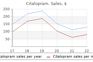
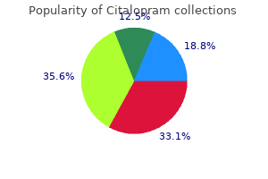
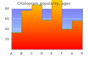
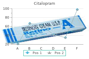
Citalopram
Citalopram dosages: 40 mg, 20 mg
Citalopram packs: 30 pills, 60 pills, 90 pills, 120 pills, 180 pills, 270 pills, 360 pills

Generic citalopram 20 mg visa
Tumor cells could kind sheets symptoms rheumatic fever citalopram 20 mg cheap with amex, brief fascicles medications blood donation 20 mg citalopram discount fast delivery, free storiform arrays, or a variety of different unusual patterns. This morphology might cause confusion with a variety of different myxoid neoplasms, significantly extraskeletal myxoid chondrosarcoma. Chronic inflammatory cells are sometimes identified throughout the stroma, as is hemosiderin. Stromal Sclerosis Hemangioendothelioma-Like Foci (Left) In extreme cases, extensive stromal sclerosis could lead to diagnostic difficulties. In specific, tissue separation artifact may lead to an appearance suggestive of a vascular neoplasm or maybe a nonneoplastic sclerosing process. Erythrocytes may be seen within these clefts, furthering mimicking a vascular lesion. Most of the constituent inflammatory cells are lymphocytes and plasma cells, however eosinophils may be current. In some reported cases, this discovering has been related to focal nuclear palisading, further mimicking schwannoma. A background of stromal sclerosis or fibromyxoid change can also complicate the problem. Given the standard age of the affected person, care should be taken to keep away from a misdiagnosis of rhabdomyosarcoma. Close inspection of the fibrous rind and inflammatory infiltrate typically reveals small nodules of diagnostic tumor cells. Small Tumor Nodules Desmin Expression (Left) Small nodules or nests of tumor cells could appear isolated in zones of sclerosis or throughout the fibrous pseudocapsule. The stroma shows variable composition ranging from myxoid to fibromyxoid to hyalinized. This diploma of bone formation is usually 1st noticeable during gross examination and sectioning. There are also several smaller extracapsular satellite tv for pc nodules in this picture extending into subcutaneous fats. When a myxoid morphology is prominent and diffuse, other tumors, similar to extraskeletal myxoid chondrosarcoma and myoepithelioma, must be excluded. Some longstanding tumors might show intensive hyalinization and overall paucicellularity. Unlike S100 protein, desmin expression is often restricted to focal or patchy positivity, as depicted. Myoepithelioma of Soft Tissue Myxoid Matrix (Left) A myxoid matrix is very common in myoepithelioma and is often quite outstanding. The lesional cells have eosinophilic cytoplasm, range from epithelioid to spindled in shape, and will form a wide selection of architectural patterns. Nuclei are relatively uniform and vesicular and may or could not present small nucleoli. Myoepithelioma of Soft Tissue Tumors of Uncertain Differentiation Corded Growth Reticular Pattern (Left) Cords or thin trabeculae of cells is a typical pattern in myoepithelioma. Micronodular Pattern Variable Cellularity (Left) the lesional cells may type small nests or aggregates and impart an general micronodular look. Spindled Morphology Nodular or Nested Growth (Left) Spindled myoepithelial cells might kind unfastened or tight fascicles and may raise the risk of a clean muscle, neural, or myofibroblastic neoplasm. Clear Cell Change Clear Cell Change (Left) this picture of myoepithelioma reveals sheets of epithelioid cells, a lot of which present cytoplasmic clearing. Clear cell change in a soft tissue neoplasm should all the time elevate the potential of a myoepithelial neoplasm. Lesional myoepithelial cells are arranged in a selection of patterns, including cords or trabeculae, much like predominantly myxoid examples. Hyalinized Stroma Pseudovascular Spaces (Left) In some hyalinized myoepitheliomas, irregular nests and cords of cells present central cellular loss or dyscohesion resembling vascular spaces. Aggregates of Clear Cells 654 Myoepithelioma of Soft Tissue Tumors of Uncertain Differentiation Storiform Architecture Hypocellularity (Left) Spindled myoepithelial cells in a collagenous stroma might often adopt a loose storiform architecture. Scattered cords and small aggregates of neoplastic myoepithelial cells are evident in scattered patches. Osteoid-Like Hyalinization Sclerotic Matrix (Left) this delicate tissue myoepithelioma reveals loose aggregates of plasmacytoid myoepithelial cells inside a dense osteoid-like sclerotic matrix paying homage to osteosarcoma. Myoepithelial Immunophenotype Rare Osteoclast-Like Giant Cells (Left) Osteoclast-like large cells are a really uncommon finding in delicate tissue myoepithelioma. A metastatic carcinoma from a visceral website ought to be thoroughly excluded on this scenario. These tumors are generally referred to as "parachordoma," however are currently thought-about a morphologic variant of myoepithelioma. In distinction, diffuse nuclear atypia, notably with distinguished nucleoli, correlates properly. Malignant Myoepithelioma Malignant Myoepithelioma (Left) this malignant myoepithelioma with a predominant plasmacytoid morphology exhibits distinguished nucleoli imparting a rhabdoid look. Malignant Myoepithelioma Malignant Myoepithelioma (Left) Malignant forms of myoepithelioma are sometimes extra cellular than benign varieties, and the mitotic rate is typically elevated. A attribute discovering is variable-sized deposits of amorphous, frivolously basophilic calcification. The reduce surface varies from tan to white or pink and will show focal hemorrhage or cystic change. Sheet-like or fascicular progress is typical of this form, and the cells are at all times cytologically uniform. In some circumstances, fascicles are well-formed and fairly distinguished and can even present a focal "herringbone" sample of development, as demonstrated in this image. Some areas are hypocellular secondary to stromal edema, myxoid change, or fibrosis. Variably conspicuous irregular, thin collagen fibers are often described as "wiry" but can appear as thicker bundles. However, this morphology is rather more frequent following radiation remedy or chemotherapy. The cellularity of this variant is often less than ordinary, and may therefore could additionally be potentially misdiagnosed as a benign neoplasm. Identification of areas of more typical morphology or utilization of ancillary research can be very helpful. On biopsy, this look can lead to confusion with lowgrade fibromyxoid sarcoma or myxofibrosarcoma. Notably, in poorly differentiated forms with a spherical cell morphology, membranous expression of this antigen can lead to consideration of Ewing sarcoma. Given the extra function of elongated and wavy nuclei, as seen on this image, a tumor of neural origin may be thought-about. In occasional tumors, the presence of extravasated pink blood cells among uniform spindled cells can impart an appearance somewhat harking again to Kaposi sarcoma. The typical spindled element is basically always current however could additionally be extremely focal.
Buy cheap citalopram 20 mg on-line
Third clamp consists of round ligament medications management generic 40 mg citalopram overnight delivery, fallopian tube symptoms low potassium 20 mg citalopram discount visa, mesosalpinx and the ligament of ovary. The sutures of the pedicle containing the uterosacral, Mackenrodt ligaments are passed by way of the vaginal vault cross-wise and are held temporarily. Redundant vaginal flaps are excised and the margins are approximated by interrupted sutures. The cross-wise handed sutures of the lowermost pedicles are now tied, thus fixing the ligaments with the vaginal vault. Preliminaries Preliminaries are the same as in vaginal hysterectomy Important steps To proceed the steps as that of vaginal hysterectomy, upto pushing up the bladder and to see the peritoneum of the uterovesical (U-V) pouch (see p 441). Preliminaries Preliminaries are the same as in vaginal hysterectomy (see p 441) Principal steps A pair of Allis tissue forceps are positioned one on both sides at the decrease end of the labium minus and a third of Allis forceps is placed on the posterior vaginal wall within the midline properly above the rectocele bulge. A horizontal incision is made on the mucocutaneous junction joining the 2 Allis tissue forceps beneath. Through the midpoint of this incision, another vertical incision is made upto the third Allis tissue forceps of the apex. Two triangular vaginal flaps are dissected off laterally from the perineal physique and the rectum. The reduce margins of the posterior vaginal walls are approximated ranging from the apex. When the sutures attain the perineal physique, the knots for the sutures of the levator ani muscle tissue are positioned. The rest of posterior vaginal wall and the skin margins are apposed using interrupted sutures. Large Loop Excision of transformation zone is a straightforward process with minimal problems. Tissue excised, is to be sent for histology examination Operative Gynecology 449 18. Complications: (a) As observed following belly hysterectomy (b) Ureteric fistula (c) Vesico-vaginal fistula (d) Urinary tract an infection (e) Bladder dysfunction (f) Lymphocyst formation Q. Patients of cancer cervix die of problems when left untreated a) Uremia due to urteric obstruction b) Sepsis c) Cachexia d) Metastasis (lung and lymph nodes) Q. It is feasible to visualize the abdominal cavity with laparoscopy and the uterine cavity by doing hysteroscopy. Laparoscopy and hysteroscopy can be accomplished both for the aim of diagnosis in addition to for surgical procedures. Discuss briefly the pneumoperitoneum: Ans: Pneumoperitoneum is created with a specially designed needle known as Veress needle. Endoscopic visualization of the cervical canal in addition to the uterine cavity, is named hysteroscopy. Imaging system includes a telescope, gentle supply, fiberoptic twine, camera units and monitors. Laparoscopic surgical procedures need fundamental instruments (graspers, scissors, aspirator, irrigator, morcellator, uterine manipulator and electrosurgical energy sources) and the imaging system. Advantages of Laparoscopy Over Laparotomy No giant belly incisions Less blood loss Rapid postoperative restoration Less postoperative ache and decreased need of postoperative analgesia Shorter hospital keep and lowered concomitant price Quicker resumption of day-to-day exercise Less adhesion formation Minimal stomach scars (cosmetic value) Reduced risk of incisional hernia Increased patient satisfaction. From anterior to posterior, buildings are: (a) Round ligament (arrow in determine 15. Benefits of laparoscopy are: (a) Confirmation of analysis, (b) staging the disease, (c) biopsy if wanted and (d) remedy on the same time. How laparoscopy could probably be helpful in the prognosis and management of unruptured tubal ectopic pregnancy Advantages are: (a) confirmation of analysis and (b) treatment might be done at the same time. Methylene blue dye is seen to come out of the stomach ostium of the proper fallopian tube. What are some great benefits of laparoscopic chromopertubation over hysterosalpingography Mullerian Anomalies Subseptate, septate, bicornuate uterus or uterus didelphys may be identified. Tubal sterilization (essure) Complications of Hysteroscopy Complications could also be due to any of the following components: A. Distension Media Fluid overload (due to absorption) Pulmonary edema and cerebral edema Electrolyte imbalance Embolism. Operative Procedures Uterine perforation Hemorrhage - throughout or after the operation Injury to intra-abdominal organs. Menstrual abnormality in the form of hypomenorrhea, oligomenorrhea or amenorrhea relying upon the extent of adhesions (partial or total). Left-sided uterine ostium is seen at a depth Common issues associated with a septate uterus are: A. Thorough investigation with laparoscopy and hysteroscopy revealed a complete uterine septum. Presently she is beneath supervision for her sixth pregnancy (following septum resection) 464 Bedside Clinics and Viva-Voce in Obstetrics and Gynecology Q. Open Surgical (i) Minilaparotomy (commonly done) or (ii) Laparotomy process (Ligation of the tubes using sutures see p 427) B. Laparoscopic Methods for tubal occlusion using rings or clips (i) Fallope ring method (commonly utilized in India), (ii) Filshie clip method (Titanium lined with Silicon rubber), (iii) Hulka Clemens clip (Spring loaded) C. Laparoscopic strategies utilizing electrodiathermy (less commonly done) (i) Bipolar electrocautery, (ii) Monopolar electrocautery D. Hysteroscopic method of tubal block (i) Essure (microcoil) inserted inside the tubes, (ii) Insertion of quinacrine pellets, (iii) Adiana- procedure of blocking the tube using radiofrequency power and also with silicon palette Q. It is best carried out within the interval interval or 6 weeks following delivery or termination of being pregnant. What are the opposite laparoscopic procedures that could be carried out for tubal sterilization What methodology of is superior by way of tubal reversal (recanalisation) with greater success rate It depends upon the extent of tubal harm and the fibrosis throughout the tube after the procedure. It occludes a segment of the fallopian tube together with the mesosalpingel blood vessels. The middle and the index fingers go throughout the lower two rings and the thumb via the higher ring. This end maneuvers the instrument to grasp the tube and to release the fallope rings Operative Gynecology 467. Similar to laparoscopy most gynecological surgical procedures may be carried out with this robotic approach. From technological viewpoint, robotic surgical procedure in superior to laparoscopy specifically when it comes to accuracy, dexterity, time (suturing), and general performance. Robotic surgery is found to have less number of surgical complications in comparison with laparoscopy. The devices in robotic surgery permits complex maneuvers inside a small space with seven degrees of freedom in comparison with four degrees in laparoscopy. This is specially important in delicate areas (major vessels, ureter) to dissect tissues. For endocervical cells-the cytobrush goes throughout the cervical canal and is rotated to acquire cells. Methods of slide preparation and fixation: the supplies collected is instantly unfold over a slide.
Generic 20 mg citalopram with mastercard
Discussion No preoperative mechanical bowel preparation (orthograde lavage or enema) is carried out as a outcome of the current incomplete obstruction increases the risk of finishing the obstruction and inducing belly pain symptoms 5-6 weeks pregnant 40 mg citalopram cheap free shipping. Furthermore treatment urinary incontinence 20 mg citalopram discount, current information from the literature question the worth of a preoperative bowel preparation. For carcinomas of the upper third of the rectum, a partial mesorectal excision (5 cm of mesorectum under the primary) is adequate from the oncological viewpoint, as a end result of distal lymphatic unfold occurs not often and by no means past four cm distally to the first. Cytotoxic washout of the rectal stump cleans the remaining rectum of tumor cells and tumor items mobilized during the surgical manipulation, and thus could prevent anastomotic recurrence. Because the anastomosis has good blood provide with no rigidity or air leakage and is positioned at about eight cm from the anal verge, no protective ileostomy is created. The pelvic omentoplasty may cut back the prevalence of clinically evident anastomotic leaks, and therefore, the necessary re-exploration. Finally, you will need to protect the autonomic hypogastric nerves and the superior hypogastric plexus, especially on this younger affected person, as a outcome of injury might lead to retrograde ejaculation. Surgical Approach With the affected person in a supine position, the abdomen is explored by a median laparotomy after a normal combination of an antibiotic single shot has been given. The major tumor is cellular, and at the peritoneal reflection, a cautious palpation of the colon reveals no synchronous second colon neoplasm and no proof of peritoneal carcinomatosis. No lymph node involvement is grossly suspected, particularly not at the root of the inferior mesenteric artery or alongside the aorta. In the United States, the standard administration involves adjuvant 5-fluorouracil-based chemoradiation. In addition to the lymph node involvement, the poor differentiation of the tumor and the microscopic invasion of veins are threat elements for tumor recurrence. In Europe, adjuvant radiotherapy is in all probability not offered if a complete mesorectal excision and an R0-resection have been achieved in rectal cancer of the higher third of the rectum. Complete colonoscopy should be done 3 months after the preliminary surgery to exclude additional neoplastic lesions. Tumor (Tu) has infiltrated by way of all layers of the colonic wall and into the perirectal fat tissue. Left hemicolon with a quantity of small tubulovillous adenomas (low grade) and minor diverticular disease are noted. Total mesorectal excision preserves male genital function compared with conventional rectal cancer surgery. Nelson H, Petrelli N, Carlin A, et al, for the National Cancer Institute Expert Panel. Digital rectal examination is painful, and permits detection of a strong and stuck rectal mass 4 cm above the anal verge. Anal sphincter pressures at relaxation and underneath squeezing are clinically inside the regular range. Any suspicion ought to be verified by genetic testing of the affected person and, if genetic testing results are optimistic, all first-degree family members. The consequence of a positive genetic test in family members is colonoscopy beginning on the age of 25 and repeated within brief intervals (1 to 2 years). In distal rectal carcinoma, modifications in stool type, anorectal tenesmus, or painful defecation are typical. Other symptoms such as pelvic or back pain, malaise, or complete mechanical obstruction are much less frequent and infrequently point out superior illness. Digital rectal examination is the initial diagnostic tool and is essential to estimate tumor size, location, and relation to the encircling constructions such as the sphincter muscle. Rigid or flexible proctosigmoidoscopy is used to visualize the tumor, to take biopsies for histologic confirmation of the suspected analysis, and to give an correct measurement of the gap of the tumor from the anal verge. However, the liver can additionally be evaluated effectively with intraoperative ultrasound. In many patients, hemorrhoidal disease may be discovered to be the supply of bleeding, with constipation having developed secondary to painful defecation. Recommendation Biopsies of the primary tumor ought to be despatched for histologic evaluation. Complete flexible colonoscopy ought to be performed to exclude a synchronous secondary colon most cancers. Case Continued Histology of the biopsies of the rectal primary reveals reasonably differentiated carcinoma of the rectum. Colonoscopy reveals a polypoid, partially ulcerated tumor that includes one third of the rectal circumference and is positioned on the left aspect. No evidence of tumor spread into the prostate, vesicles, pelvic flooring, or anal sphincter. To offer a sphincter-sparing method to the rectal most cancers, the patient is introduced with the choice of neoadjuvant chemoradiation remedy over 6 weeks (50. The lately revealed randomized German rectal cancer trial evaluating the standard U. Although the hypofractionated 1-week schedule of irradiation (5 5 Gy over 1 week) with instant surgical procedure is the favorite preoperative therapy in Europe, on this case we determined to choose a standard long irradiation protocol (50. At the extent of the vesicles (V), distal rectal tumor (Tu) with tumor infiltration into the mesorectum, on the left-hand facet near the mesorectal fascia (visceral fascia of the pelvis). Surgical Approach Following a mechanical bowel preparation the day before surgical procedure, the stomach is explored by a median laparotomy with the affected person in a supine place with straddled legs. As expected, the primary tumor is below the peritoneal reflection and therefore not detectable yet. Lymph node involvement is grossly suspected alongside the inferior mesenteric artery, however not alongside the aorta. Approach After complete evaluation, the patient is discovered to have a distal rectal adenocarcinoma 4 cm from the anal verge (cT3 cN2 cM0 G2) with out infiltration Case 34 143 utterly mobilized, detaching the omentum from the transverse colon, and dissected on the left colonic flexure. The corresponding left mesocolon, together with the inferior mesenteric artery, is dissected about 1. Further sharp dissection of the rectosigmoid follows the mesorectal fascia (visceral pelvic fascia) precisely right down to the pelvic ground and the anorectal junction. The autonomous superior hypogastric nerve plexus, the hypogastric nerves, and the inferior hypogastric nerve plexus on both sides are visualized and punctiliously preserved. Then, the rectum is closed distally to the tumor via an oblong clamp, the anal canal is rinsed with a cytotoxic transanal answer. A transverse coloplasty is constructed on the proximal colonic stump by an 8-cmlong antimesenteric colostomy and transverse closure by a two-layer working suture. For the transanal anastomosis, 12 sutures are put as preparation for the coloanal hand-sewn suture, grasping the inside sphincter muscle and the anoderm. Drains are put in the pelvic cavity, a protecting Brook ileostomy is created, and the abdomen is closed. Photographs are taken as high quality management for the surgeon regarding completeness of mesorectal excision. Minimal distances from the tumor to the circumferential resection margin and the distal resection margin are assessed histologically. Discussion Some of the small print of the surgical approach are mentioned in the chapter on proximal rectal most cancers (Case 33). Rectal tumors of the distal third of the rectum are dissected radically with a minimal bowel resection margin, so lengthy as a complete mesorectal excision has been carried out and the frozen part of the distal margin is negative for tumor unfold.
Order citalopram 40 mg on line
To carry out minor operations like punch biopsy symptoms esophageal cancer 20 mg citalopram purchase with mastercard, floor cauterization or snipping a small polyp symptoms nasal polyps citalopram 20 mg buy discount line. It is the alternative of squamous epithelium of the ectocervix by columnar epithelium of endocervix by the method of metaplasia. It ought to be used solely when the operation is completed underneath common or regional anesthesia as the instrument is heavy. A second layer of (musculofascial) suture with the identical materials is used to reinforce the first layer. It was initially a delicate rubber catheter with a balloon sleeve of rubber across the shaft beneath the eye of the catheter. Currently, the catheter is made from latex which is much less irritating to urethral mucosa. To assess the patency of the fallopian tube during laparotomy, catheter is introduced within the uterocervical canal (vaginally) and the balloon is inflated then dye is pushed. To dilate the cervix to facilitate drainage of intrauterine collection - pyometra, hematometra or lochiometra. Ans: (a) Senile endometritis (b) Endocervical carcinoma (c) Tubercular endometritis (d) Infected lochiometra (obstetrical). To maintain the fundus of the uterus and to give traction whereas the clamps are positioned during complete abdominal hysterectomy for benign lesion. Usually the anterior lip is held in conditions like D and C, D and E, but in some situations, the posterior lip is to be held. Such situations are: (a) During amputation of cervix or vaginal hysterectomy when the posterior cervico-vaginal mucous membrane is incised. Ans: Subtotal hysterectomy is completed in certain conditions the place complete hysterectomy Self-assessment 484 Bedside Clinics and Viva-Voce in Obstetrics and Gynecology is found to be troublesome or time-consuming. Difficult tubo-ovarian mass with obliteration of the anterior and posterior pouches. In non descent vaginal hysterectomy as an alternative alternative to laparoscopic hysterectomy. To detect proof of ovulation-by seeing the secretory adjustments within the endometrium. Cervical mucus examine: Disappearance of fern sample beyond twenty second of the cycle suggests ovulation. Vaginal cytology: Maturation index shifts to the left because of the impact of progesterone. Serum Progesterone: A rise in serum levels of progesterone within the secretory phase of the cycle (D-21) when compared to D-8 of the cycle suggests ovulation 5. To plug the uterine cavity with gauze twigs in continued bleeding after removal of polyp. Uses To remove the merchandise of conception in D and E after its separation partially or utterly. The merchandise are caught and then with twisting movements and simultaneous traction, the products are removed. Dangers: It could produce harm to the uterine wall to the extent of even perforation. Not infrequently, a section of intestine or omentum could even be pulled out via the rent. This forceps is used to hold delicate tissues for a very long time with minimal tissue damage. Uses To maintain the margins of the vaginal flaps in colporrhaphy operation To hold the peritoneum or rectus sheath during repair of the stomach wall To hold the margins of the vagina in stomach hysterectomy To hold the anterior lip of the cervix in D and C operation To catch the torn ends of the sphincter ani externus in full perineal tear repair To remove a small polyp To take out the tissue in wedge biopsy. The frequent symptoms are genital organs protruding out of the vaginal opening, problem in walking, sitting, urination or defecation. Prolapse could interfere with sexual intercourse or could cause vaginal bleeding due to ulceration of mucosa. Uses To fix and steady the uterus when conservative surgical procedure is completed on the adnexae (tuboplasty operation). Cervix is occluded with the instrument and methylene blue dye is injected into the uterine cavity through the fundus utilizing a syringe and a needle. Important issues earlier than myomectomy (prerequisites of myomectomy) Submucosal polyp, submucous fibroid, any tubal block or endometrial carcinoma should be excluded before performing myomectomy. History: Bonney, William Francis Victor (1872-1953): Gynecologist on the Middlesex and Chelsea Hospitals, London. Simultaneous, bilateral clamping of the infundibulopelvic ligaments by rubberguarded sponge holding forceps could additionally be employed. The clamp is eliminated after suturing the myoma mattress however earlier than closing the peritoneal layers. The medication instilled are-dexamethasone four mg with gentamicin eighty mg in 10 ml normal saline. Ans: It is a radiographic study to assess the interior anatomy of the uterus, cervical canal and the tube. It entails instillation of saline within the uterine cavity to study the uterine cavity and the tubes with trans vaginal sonography. Mention the completely different sites the place the clamps are placed in total abdominal hysterectomy (see p 432). To introduce it by way of the opening of the uterovesical pouch and to retract the bladder while the clamps are placed. Conservative treatments include: (i) to keep away from aggravating factors (obesity, chronic cough, constipation) (ii) pelvic floor train, (iii) Estrogen replacement remedy, (iv) pessary in some instances 498 Bedside Clinics and Viva-Voce in Obstetrics and Gynecology Q. Mention the completely different websites the place the clamps positioned during vaginal hysterectomy Ans. Use: To know the patency of the tubes in infertility investigation or following tuboplasty. Self-assessment Ideal time of operation It is completed in the early proliferative section, usually 2 days after the menstrual bleeding stops. Retractors are held in place both by an assistant (manual retractor) or by counter pressure with some gadget (self-retaining retractor). Manual retractor can be used alone or in combination with a self-retaining retractor. A pack may be positioned between the viscera and the retractor to keep away from direct trauma to the viscera. Uses It is utilized in abdominal operation to retract the viscera as and when required so as to facilitate the operative procedures like stomach hysterectomy. Uses It is used as a clamp in hysterectomy, salpingectomy or salpingo-oophorectomy operation. Paired clamps are placed on the infundibulopelvic ligament on the lateral facet and one other paired clamps are placed medially over the medial end of the fallopian tube together with the ovarian ligament and the mesosalpinx.
40 mg citalopram proven
Previous uneventful first and second stage could turn out to be irregular within the third stage and will lead to medicine grand rounds order citalopram 40 mg amex maternal death medicine 93832 buy citalopram 20 mg with amex. To stop such complications active management of third stage of labor is helpful. The important benefits of energetic administration of third stage of labor are: (i) Third stage blood loss is lowered approximately to one-fifth (ii) Duration of third stage is reduced to its half. Active management therefore wants extra skilled nursing personnel in the labor ward to give the injection in time. These circumstances are girls with coronary heart illness, extreme pre-eclampsia and in instances with twins until the 2nd child is born. Considering all the advantages, active management must be done in nearly all instances in the third stage of labor. In India life time risk of dying for a lady during being pregnant is 1 in 70 compared to one in forty eight,000 in developed nations. The necessary social components related are illiteracy, ignorance, unregulated fertility, poor socioeconomic situation, beneath utilization of existing health care companies and lack of communication and referral services. The necessary steps to cut back maternal mortality are: Utilization of primary antenatal, intranatal and postnatal care. Availability of emergency obstetric care, protected abortion companies and household planning companies. Improvement of legislative and policy action to take away social inequalities ongrounds of gender. Infection (labor and puerperium): Clean supply practices, expert start attendant, use of antibiotics-when infection is clear. Medical disorders in pregnancy (diabetes, chickenpox)-appropriate intervention or referral for optimum care. Combining all the above factors (health, social and coverage actions) and by proper implementation of interventions against the major causes, maternal mortality can be prevented significantly in India. Prenatal counseling means evaluation after which counseling a girl about being pregnant, its course and the doubtless consequence properly earlier than the time of precise conception. The goal of prenatal counseling is that girl should enter the being pregnant in an optimal state of well being which might be protected both to herself and the fetus. Otherwise many antagonistic factors begin to exert their effects by the point woman is seen in the antenatal clinic. Generally girls are first seen in the antenatal clinic at round 14 weeks of gestation. At the same it helps to manage care to cut back or to get rid of risk factor so that being pregnant end result is improved. Folic acid supplementation (4 mg a day) starting 4 weeks before conception and continued upto 12 weeks of pregnancy. Women with medical problems (hypertension and diabetes) in being pregnant, need schooling and remedy earlier than conception. Many medicine used through the nonpregnant state must be avoided throughout pregnancy because of fetal hazards. Warfarin, oral antidiabetic medicine are replaced with other drugs like heparin and insulin respectively for the protection of the fetus. This can only be accomplished as quickly as the woman is seen and endorsed before being pregnant (prenatal counseling). External cephalic model is a maneuver carried out externally to change the fetal presentation and to deliver the fetal head to the lower pole of uterus. These are fetal misery, placental abruption, premature rupture of membranes, and so forth. Considering all the advantages and the risks, it seems that each case must be selected fastidiously excluding the contraindications. Cardiotocography must be carried out before and after the process to assess fetal well-being. Facilities for cesarean delivery have to be there, should any issues develop during process. Therefore it seems on crucial analysis that external cephalic version has received a spot within the management of breech presentation in a well-selected case. High-risk pregnancy is outlined as one which is difficult with issue (s) that adversely impacts the pregnancy outcome-maternal or perinatal or each. The fetal hazards are miscarriage, vanishing twin, fetus papyraceus, preterm start, fetal anomalies, discordant progress, intrauterine dying of 1 fetus, twin transfusion syndrome, wire prolapse, locked twins and elevated perinatal mortality. Considering all these issues affecting the mother, fetus and the neonate, twin being pregnant is taken into account as a "high-risk pregnancy". The significance of defining the high-risk scenario is to anticipate the problems. For example antenatal supplementation of increased amount of iron and folic acid can meet up the increased demand and thereby can prevent issues because of anemia. So twin being pregnant needs careful antenatal care and intrapartum care to stop all these issues. All these are depicted in a single sheet of paper, against the length of labor in hours. The components of a partograph are designed to assess the progress of labor and the well-being of the mom and the fetus. Labor is said to be abnormal when cervicograph crosses the alert line and falls on zone 2. The major advantage is that it could detect deviation from normal course of labor early. Secondary arrest could be diagnosed when the energetic part of labor commences normally however stops or slows down significantly for two hours or extra prior to full dilatation of the cervix. Obstructed labor may be diagnosed when the progressive descent of the presenting part is arrested inspite of fine uterine contraction. This is commonly related to maternal features of dehydration, exhaustion and sepsis. Partograph can detect each the extended labor or obstructed labor early earlier than any opposed effect on the mom or the fetus units in. Partograph abnormality suggests either early referral to an outfitted middle or an early intervention. When intervention is done timely, majority of cases result in successful vaginal delivery. So introduction of partograph has lowered the incidence of extended labor and cesarean supply. Breastfeeding has received a number of benefits whereas synthetic feeding has received several disadvantages.
Tu Sizi (Dodder). Citalopram.
- What is Dodder?
- Are there safety concerns?
- Dosing considerations for Dodder.
- Bladder, liver, and spleen problems.
- How does Dodder work?
Source: http://www.rxlist.com/script/main/art.asp?articlekey=96067
40 mg citalopram with mastercard
This histologic picture could be seen in all kinds of other spindle cell sarcomas medicine used to treat bv discount citalopram 20 mg without a prescription, including fibrosarcoma and malignant peripheral nerve sheath tumor symptoms jaw pain and headache discount citalopram 20 mg otc. Herringbone Pattern: High Power Monophasic Synovial Sarcoma (Left) Monophasic synovial sarcoma of the mediastinum with a prominent herringbone pattern of development exhibits quick fascicles of monotonous atypical spindle cells intersecting one another. This image is nearly indistinguishable from that of a malignant peripheral nerve sheath tumor. Bcl-2 Immunostaining 778 Synovial Sarcoma Mediastinum: Neoplasms, Malignant, Primary Hemangiopericytic Vascular Pattern Branching Vessels (Left) Scanning magnification of monophasic synovial sarcoma of the mediastinum with a outstanding hemangiopericytic development pattern reveals a dense spindle cell inhabitants flanking small, dilated vascular spaces with open lumina. Hemangiopericytic Vascular Pattern Branching Vessels (Left) Monophasic synovial sarcoma of the mediastinum with a prominent hemangiopericytomatous progress pattern exhibits vascular areas that appear to department at proper angles. Hemangiopericytic Vascular Pattern Vimentin Immunostaining (Left) Monophasic synovial sarcoma of the mediastinum with a distinguished hemangiopericytic growth pattern exhibits multiple small vessels with open lumina and a fancy branching luminal sample. Tumors with these features are simply mistaken for malignant fibrous histiocytoma. Higher Magnification Storiform Pattern (Left) Scanning magnification of monophasic synovial sarcoma of the mediastinum with distinguished storiform pattern shows atypical spindle cells that seem to be radiating from a central spoke in a pinwheel trend reminiscent of fibrohistiocytic tumors. The uniformity of the tumor cells, absence of pleomorphism, and important mitotic exercise bear a superficial resemblance to dermatofibrosarcoma protuberans on this tumor. Storiform Pattern 780 Synovial Sarcoma Mediastinum: Neoplasms, Malignant, Primary Palisading of Nuclei Palisading of Nuclei (Left) Scanning magnification shows monophasic, spindle cell synovial sarcoma of the mediastinum characterized by hanging nuclear palisading of the cells. Tumors with these options can closely resemble malignant peripheral nerve sheath tumors. The tumor cells are quite monotonous and merge imperceptibly with extra conventional fascicular areas in the neighborhood. Neural-Like Appearance Neural-Like Wavy Nuclei (Left) Scanning magnification of monophasic synovial sarcoma of the mediastinum with hanging palisading of tumor cells reveals structures which might be reminiscent of Verocay our bodies. Tumors with these features could be simply mistaken for malignant peripheral nerve sheath tumors. Nesting Pattern Nesting Pattern (Left) Monophasic synovial sarcoma of the mediastinum with distinguished nesting sample shows discrete nests of tumor cells separated by thin fibrovascular septa. Tumors with these options can carefully resemble metastatic malignant melanoma to the mediastinum. Round Cells Lobular Configuration (Left) Scanning magnification of poorly differentiated synovial sarcoma of the mediastinum shows ill-defined lobules composed of spherical, epithelioid tumor cells admixed with a few spindle cells within the background. The tumor bears a superficial resemblance to epithelioid malignant peripheral nerve sheath tumors. Poorly Differentiated Synovial Sarcoma Rhabdoid Cells (Left) Poorly differentiated synovial sarcoma of the mediastinum, rhabdoid cell variant, reveals sheets of small spherical blue cells displaying prominent eosinophilic cytoplasmic inclusions indistinguishable from these seen in rhabdoid tumors. Rhabdoid Cells: High Power 782 Synovial Sarcoma Mediastinum: Neoplasms, Malignant, Primary Cystic Changes Areas of Necrosis (Left) Monophasic synovial sarcoma of the mediastinum with secondary cystic modifications shows a big cystic space flanked by a dense spindle cell population. The tumors can present huge, gross cystic modifications and be mistaken clinically for cystic teratomas. Myxoid Stromal Changes Myxoid Changes: High Power (Left) Prominent myxoid stromal modifications are seen in this monophasic synovial sarcoma of the mediastinum. Tumors with these options could also be confused with fibromyxoid sarcomas and myxoid malignant fibrous histiocytoma. Artifactual Clefting Stromal Collagenization (Left) Monophasic synovial sarcoma of the mediastinum exhibits artifactual clefting and separation of the stroma resulting in a pseudovascular look which might be mistaken for a malignant vascular neoplasm. Tumors with these features could additionally be mistaken for fibrosarcoma or solitary fibrous tumors. The glandular cells in biphasic synovial sarcoma stain very strongly for cytokeratin, in distinction to the spindle cells that generally display only focal positivity. Glands: High Power Biphasic Synovial Sarcoma (Left) Biphasic synovial sarcoma of the mediastinum exhibits massive glandular constructions with collapsed lumina lined by tall columnar cells. Notice the residual small lumen filled with proteinaceous materials in one of the glands. Cases like this may be simply mistaken for metastases from adenocarcinomas of enteric origin, except for the fact that the glands are surrounded by a skinny layer of atypical spindle cells. Biphasic Synovial Sarcoma Papillary Structures (Left) Scanning magnification of biphasic synovial sarcoma of the mediastinum with intestinal-type glands shows focal papillary formations protruding into the lumen of the dilated glands. Notice the spindle cell stroma in the cores of the papillae and the mucinous content material in the lumen. Biphasic Synovial Sarcoma Epithelioid Elements: High Power (Left) Scanning magnification of surprising variant of biphasic synovial sarcoma of the mediastinum shows areas characterised by clear cells surrounded by a dense, monotonous, atypical spindle cell proliferation. Fibrocollagenous Component Neurotized-Like Areas (Left) Neurofibroma shows a "globular" architecture composed of in depth areas of what appear to be WagnerMeissner bodies. Spindle Cell Proliferation Lobular Pattern (Left) In some circumstances, neurofibromas might undertake a quite lobular progress sample during which lobules are separated by fibrocollagenous tissue. Degenerative Changes Crush Artifact and Spindle Cells (Left) Mediastinal neurofibroma shows in depth dense fibrocollagen, and the neurofibroma consists of a quite strong spindle cellular proliferation. Vascular Proliferation 788 Neurofibroma Mediastinum: Neoplasms, Malignant, Primary Vascular Hyalinization Absence of Mitotic Activity (Left) Neurofibroma reveals compressed blood vessels. Note the perivascular collagenization of the vessels as well as the spindle cellular proliferation admixed with some inflammatory cells. Solid Spindle Cell Component Wagner-Meissner Bodies (Left) Mediastinal neurofibroma reveals a spindle cellular proliferation in a more solid growth sample. Focal Pigmented Areas Melanin Pigment (Left) Mediastinal neurofibroma is proven with focal areas of melanin pigment. The hypocellular areas present dense fibrocollagenous tissue, while the hypercellular areas present a extra epithelioid cellular proliferation. Note the concentric association of tumor cells around the blood vessels and the adjacent tumor necrosis. The neoplastic cellular proliferation around the glandular structure shows myxoid adjustments, in addition to nuclear atypia and mitotic figures. Bland Spindle Cells Rope-Like Collagenous Pattern (Left) Characteristic histologic appearance of solitary fibrous tumor shows parallel linear arrays of rope-like collagen strands flanked by small, bland-appearing spindle cells. The morphologic appearance of this tumor is harking again to a fibrohistiocytic neoplasm. Rope-Like Strands of Collagen Neural-Like Wavy Nuclei (Left) High magnification of solitary fibrous tumor of the mediastinum shows the attribute histologic look composed of bland spindle cells flanked by thin, rope-like strands of collagen disposed in parallel linear arrays. Notice the outstanding wavy appearance of the spindle cells, reminiscent of that which is often noticed in schwannian tumors. Rope-Like Pattern of Collagenization Parallel Rows of Collagenized Stroma (Left) Characteristic lowpower appearance of a solitary fibrous tumor exhibits parallel arrays of skinny, ropelike strands of keloidal collagen flanked by small spindle cells. This has often been referred to because the patternless sample of development in these tumors.
Syndromes
- Has a decreased appetite
- Use of medications that suppress the central nervous system (such as barbiturates or benzodiazepine tranquilizers)
- Poor feeding or irritability in children
- Upper endoscopy
- Sweating
- Low blood pressure
20 mg citalopram order fast delivery
The lymphoma is composed of predominantly small lymphocytes that have ample cytoplasm symptoms 9 days after embryo transfer cheap 40 mg citalopram mastercard. The gastritis accommodates a superficial plasma cell infiltrate in the pit compartment and nodular lymphoid aggregates within the deep mucosa medicine urology quality citalopram 20 mg. Unlike the basal aggregates of simple gastritis, the lymphoma within the deeper mucosa is diffuse and encroaches on epithelial constructions. There is disruption of the fibers of the muscularis mucosae and lymphocytes infiltrating gastric glands. The germinal heart is a part of the underlying gastritis, however the expanded marginal zone is part of the lymphoma. In addition to being confused with easy gastritis, this appearance may be mistaken for different malignancies similar to plasma cell myeloma. Much of the lymphoma has plasmacytic differentiation, but a few monocytoid lymphocytes may be seen across the edge of a residual germinal middle. Note the ulcer exudate, disruption of regular architecture with lymphoma cells infiltrating between epithelial buildings, and areas of energetic irritation within pits and glands. Some cells have 1 outstanding nucleolus (immunoblast-like), and others have multiple inconspicuous nucleoli (centroblast-like). Takahashi H et al: Prognostic impression of extranodal involvement in diffuse large B-cell lymphoma in the rituximab era. Keratin negativity excludes the possibility of a poorly differentiated epithelial neoplasm, similar to carcinoma. Bcl-6 Immunohistochemistry Bcl-2 Immunohistochemistry (Left) Positive Bcl-6 stain highlights the lymphoma cells with nuclear positivity on this double-hit lymphoma, proof of a germinal heart phenotype. Lymphomatous Polyposis Mantle Cell Lymphoma (Left) At high magnification, mantle cell lymphoma consists of a monotonous population of cells with irregular, angulated nuclei. Salar A et al: Gastrointestinal involvement in mantle cell lymphoma: a potential clinic, endoscopic, and pathologic study. Kodama T et al: Lymphomatous polyposis of the gastrointestinal tract, together with mantle cell lymphoma, follicular lymphoma and mucosa-associated lymphoid tissue lymphoma. Okazaki K: Multiple lymphomatous polyposis kind is common but not particular for mantle cell lymphoma within the gastrointestinal tract. This occurs rarely in mantle cell lymphoma and may potentially trigger diagnostic confusion. Duplication Cyst Cross Section of Duplication Cyst (Left) Light microscopy exhibits a low-power view of an intestinal duplication cyst. This finding helps distinguish a duplication cyst from an enterogenous cyst (which has disorganized smooth muscle and lacks ganglion cells). Gastric-Type Mucosa Within Meckel Diverticulum Gastric and Pancreatic Epithelia (Left) High-power view shows gastric mucosa on the top and pancreatic acinar tissue on the bottom. In some circumstances, the gastric epithelium inside the diverticulum may be colonized by Helicobacter pylori. Berber U et al: Peptic ulcer and intestinal metaplasia associated with Helicobacter pylori colonization in gastric heterotopia of the tongue. Ectopic Pancreas With Islets Acinar Cells Within Muscularis Propria (Left) High-power view exhibits pancreatic acinar cells inside deep muscularis propria. These vacuoles correspond to the microvillous inclusions seen on electron microscopy. Ischemic Damage Reactive Epithelium (Left) this high-power view shows regenerating epithelium overlying an ulcer bed. Primary Lymphangiectasia Dilated Lacteals (Left) this high-power view highlights the dilated lacteals. Fully Developed Sprue-Like Changes Surface Damage With Lymphocytosis (Left) High-power view exhibits damaged floor epithelium with quite a few intraepithelial lymphocytes and increased lamina propria plasma cells. Mansfield-Smith S et al: Including duodenal bulb histology should be standard of care when evaluating Celiac Disease in kids. Note that whereas the villi are absent, the overall thickness of the mucosa stays the same. Surface Damage With Lipid Hang-Up Marsh 3B Lesion (Left) this fully flat biopsy has broken surface epithelium with increased intraepithelial lymphocytes and vacuolated floor cells, indicative of lipid hang-up. Partially Developed Sprue-Like Changes Partially Developed Sprue-Like Changes (Left) this small bowel biopsy shows reasonable villous blunting (Marsh 3B). Marsh 1 Lesion Normal Architecture With Increased Intraepithelial Lymphocytes (Left) High-power view of this villus shows an increased variety of intraepithelial lymphocytes. This patient was so sick that she was positioned on complete parenteral vitamin, hence the shortage of lymphocytes in the floor epithelium. Collagenous Sprue 224 Celiac Disease Small Intestine: Nonneoplastic Normal Architecture With Increased Intraepithelial Lymphocytes Common Variable Immunodeficiency (Left) High-power view of the tip of a villus shows increased intraepithelial lymphocytes. Note the presence of plasma cells in the lamina propria (compared to the adjacent image). Autoimmune Enteropathy Autoimmune Enteropathy (Left) this part of small bowel reveals marked villous atrophy with a densely mobile lamina propria. Peptic Duodenitis Peptic Duodenitis (Left) Low-power view of this small bowel biopsy exhibits a whole lack of villi with elevated lamina propria inflammation, mimicking fully developed celiac disease. The floor epithelium has neutrophils rather than lymphocytes, indicative of peptic damage. Increased Intraepithelial Lymphocytes Increased Intraepithelial Lymphocytes (Left) High-power view of the tip of a villus highlights the big numbers of intraepithelial lymphocytes, just like what one might see in celiac disease. Pretreatment Biopsy of Tropical Sprue Post-Treatment Biopsy of Tropical Sprue (Left) this picture reveals a pretreatment biopsy of tropical sprue with mild villous blunting and increased intraepithelial lymphocytes. Pretreatment Biopsy of Tropical Sprue Post-Treatment Biopsy of Tropical Sprue (Left) Higher power view of a pretreatment biopsy highlights the rise in intraepithelial lymphocytes. Prokinetic agents for motility issues Mild Nonspecific Inflammatory Changes (Left) Low-power view of small intestinal mucosa exhibits delicate nonspecific villous blunting and thickening. Increased Intraepithelial Lymphocytes Fibrosis of Muscularis Propria (Left) Full thickness part of the small bowel shows fibrosis replacing the sleek muscle of the muscularis propria. Collagenous Sprue-Like Changes Subepithelial Collagen Deposition (Left) this stomach biopsy shows subepithelial collagen deposition equivalent to collagenous gastritis. Crypt Destruction Crypt Destruction (Left) the bottom of the mucosa usually bears the brunt of the injury in autoimmune enteropathy. Gambineri E et al: Clinical and molecular profile of a model new sequence of sufferers with immune dysregulation, polyendocrinopathy, enteropathy, X-linked syndrome: inconsistent correlation between forkhead box protein 3 expression and disease severity. Al Khalidi H et al: Enteropathy with loss of enteroendocrine and paneth cells in a patient with immune dysregulation: a case of adult autoimmune enteropathy. Crypt Abscess Crypt Distortion (Left) Some cases of autoimmune enteropathy have crypt abscesses which are reminiscent of lively ulcerative colitis. Celiac Mimic Lymphocytic Colitis Pattern (Left) There is marked villous blunting with a complete lack of goblet cells. Although the villous morphology and intraepithelial lymphocytosis might mimic celiac illness, the complete lack of goblet cells should help establish the proper prognosis. There is a superficial plasmacytosis with floor epithelial injury and elevated intraepithelial lymphocytes.
Citalopram 40 mg cheap with amex
Smooth Muscle Bundles Smooth Muscle Cytology (Left) Deep leiomyoma exhibits traditional cytologic features of easy muscle differentiation symptoms 5-6 weeks pregnant 40 mg citalopram cheap free shipping, including plentiful eosinophilic cytoplasm and uniform nuclei with blunt ends ("cigar shaped") treatment urinary incontinence 20 mg citalopram discount. Miettinen M: Smooth muscle tumors of soft tissue and non-uterine viscera: biology and prognosis. Calcification Stromal Edema (Left) Scattered, separated, eosinophilic easy muscle cells are evident at decrease magnification in this picture from a deep leiomyoma with areas of distinguished stromal edema. Myxoid Stromal Change Stromal Hyalinization (Left) Stromal hyalinization and hypocellularity is another widespread finding in leiomyoma and may be focal or diffuse. This finding is typical of longstanding tumors and may be associated with stromal calcification. It is commonly a focal finding when present, however rare instances have been described with diffuse palisading. Nuclear Palisading 326 Deep Leiomyoma Smooth Muscle Tumors Macrotrabecular Architecture Microtrabecular Architecture (Left) Macrotrabecular progress in deep leiomyoma is characterised by thick, elongated bundles of clean muscle cells inside a unfastened myxoid or edematous stroma. Microtrabecular Architecture Epithelioid Morphology (Left) Deep leiomyoma with a microtrabecular growth sample can also present areas with a variably complicated reticular look. Clear cell change may be seen but is likewise usually focal and, in some circumstances, related to calcification. Epithelioid Morphology Mature Adipose Tissue (Left) this picture of a retroperitoneal deep leiomyoma shows a mix of conventional spindled smooth muscle cells and epithelioid easy muscle cells with extra prominent eosinophilic cytoplasm. Hussein K et al: Clinico-pathological traits of various varieties of immunodeficiency-associated clean muscle tumours. Not occasionally, fascicles appear to be organized perpendicularly to one another, both longways and en face, as depicted. In truth, some observers have instructed diagnosing such superficial lesions as "atypical intradermal easy muscle neoplasm" on condition that they might recur but basically by no means metastasize. At times, the collagen could resemble osteoid deposits and lift issues of extraskeletal osteosarcoma. Tumor cells on this variant show ample eosinophilic cytoplasm and cytologically malignant nuclei. It is histologically characterised by sheets and nests of uniform, round to epithelioid cells arranged around a conspicuous vasculature. Mravic M et al: Clinical and Histopathological Diagnosis of Glomus Tumor: An Institutional Experience of 138 Cases. Some zones of outstanding hyalinization or sclerosis can feature tumor cells organized singly or in small mixture or cords (right). Sheet-Like Growth Vasculature (Left) Glomus tumor may characteristic a strikingly strong growth pattern with a less prominent vasculature, as depicted. This morphology can lead to consideration of a cellular solitary fibrous tumor (previously termed hemangiopericytoma). Prominent Dilated Vessels Cavernous Vascular Pattern (Left) As proven in this image, vascular areas in glomangioma could additionally be markedly expanded and carefully resemble these of cavernous hemangioma. In the latter state of affairs, immunohistochemistry is commonly wanted to assist the prognosis. Well Circumscribed Concentric Perivascular Growth (Left) A characteristic feature of myopericytoma is multilayered, concentric perivascular growth by the lesional cells, as depicted. The prominence of this discovering varies from case to case, however is commonly easy to spot. Fisher C: Unusual myoid, perivascular, and postradiation lesions, with emphasis on atypical vascular lesion, postradiation cutaneous angiosarcoma, myoepithelial tumors, myopericytoma, and perivascular epithelioid cell tumor. Solid Growth Ectatic Vascular Channels (Left) Ectatic, "staghorn" vascular channels are a standard discovering in myopericytoma and may be prominent. Hemangiopericytoma-Like Growth Angioleiomyoma-Like Areas (Left) Some cases of myopericytoma contain areas which are cytomorphologically much like the graceful muscle cells of angioleiomyoma, which is taken into account to be one other perivascular neoplasm related to myopericytoma. Intravascular Growth Peritumoral Vessels (Left) Intravascular and intramural growth is an occasional discovering in myopericytoma. Malignant Myopericytoma Malignant Myopericytoma (Left) Malignant forms of myopericytoma are extremely uncommon and are normally characterized by significant nuclear atypia, pleomorphism, and mitotic activity. Of notice, a perivascular association of the tumor cells is normally maintained, at least focally. Note that the myoid nodules might project or extend into thin-walled vascular channels. Myoid Nodules Cytologic Features (Left) Cytologically, the cells of myofibroma are bland and comparatively uniform. Linos K et al: Myofibromas With Atypical Features: Expanding the Morphologic Spectrum of a Benign Entity. Myoid Morphology Myoid Zones (Left) In some cases, the myoid zones are paucicellular and include a continual inflammatory infiltrate, resembling a reactive myofibroblastic process. Identification of a low-power nodular progress sample or presence of a pericytomatous vascular part is helpful. Stromal Hyalinization Stromal Hyalinization (Left) Myoid zones may show intensive stromal hyalinization. When these areas are bigger, it might be tough to acknowledge the lesion as a myofibroma, particularly if a more mobile pericytomatous component is inconspicuous or absent. Smooth Muscle Actin 350 Myofibroma and Myofibromatosis Pericytic (Perivascular) Tumors Multinucleated Giant Cells Necrosis (Left) Multinucleated large cells are not often seen in myofibroma, but are extra doubtless to be seen in intraosseous cases. When current in soft tissue lesions, a analysis of nodular fasciitis may be thought-about. Prominent Vascularity Vascular Intimal Involvement (Left) Myofibroma is presently thought to be related to different pericytic tumors, such as myopericytoma, glomus tumor, and angioleiomyoma, and should present overlapping features with 1 or more of these entities. Tumors with a conspicuous myoid component are in all probability finest categorised as myofibroma. Increased Cellularity Hypercellularity (Left) Rare circumstances of myofibroma present a relatively vital increase in cellularity and should mimic a sarcoma. In these cases, quite lots of spindle cell sarcomas, similar to childish fibrosarcoma and spindle cell rhabdomyosarcoma, must be diligently excluded first. Perivascular Growth Mature Smooth Muscle Cells (Left) the well-differentiated clean muscle cells of angioleiomyoma present traditional cytologic options including prominent eosinophilic cytoplasm and elongated, blunt cigar-shaped nuclei. They are additionally usually arranged in bundles and fascicles, as seen in different clean muscle neoplasms. Liu Y et al: Angioleiomyomas within the head and neck: A retrospective medical and immunohistochemical evaluation. The cytoplasm is eosinophilic and finely vacuolated with vague cell borders imparting a syncytial appearance. Lasota J et al: Nuclear expression and gain-of-function -catenin mutation in glomangiopericytoma (sinonasal-type hemangiopericytoma): insight into pathogenesis and a diagnostic marker. A reassessment with electron microscopy, immunohistochemistry, and long-term follow-up.
Order 20 mg citalopram
Afterwards medicine zithromax buy generic citalopram 40 mg, if blood glucose continues to lower symptoms 20 weeks pregnant citalopram 40 mg cheap with mastercard, neuropsychiatric manifestations occur. Glucagon and adrenaline (epinephrine) are secreted instantly and act rapidly, whereas the actions of cortisol and development hormone are sluggish Hypoglycaemia 73 and turn into evident several hours after onset of hypoglycaemia. Stimulation of a-adrenergic receptors suppresses insulin secretion, and stimulation of b-receptors stimulates glucagon secretion. Glucagon then promotes glycogenolysis and gluconeogenesis within the liver and inhibits glucose disposal from the plasma. In contrast, epinephrine secretion is preserved and is the main compensatory hormone within the case of hypoglycaemia. This course of known as gluconeogenesis and uses major substrate amino-acids, derived from proteolysis in the muscular tissues. Decreased provide of glucose to the brain causes neuroglycopenic symptoms that start as the lack to focus and sluggishness of reflexes, and find yourself in tonic-clonic convulsions and coma. In this explicit case might be because of a combination of things: the delay or omission of a meal, along with alcohol consumption, most likely led to the hypoglycaemic coma. This primarily happens in individuals with very good glycaemic management and in these with long-standing disease (hypoglycaemia unawareness). The same can even occur when the affected person has had multiple hypoglycaemic episodes (usually throughout the last few hours). Management of hypoglycaemia unawareness is tough: often loosening of strict glycaemic control for a quick time can restore awareness of hypoglycaemias. Central nervous system symptoms are potentially dangerous, particularly when the affected person performs delicate and responsible acts, drives a automobile, and so forth. If this episode is considered unintended, no adjustments in his insulin routine are essential. If, nevertheless, it has occurred before, alterations in his routine may be considered. His mother reports that he at all times consumes goodies and sweets after such an episode, which later results in extremely high blood sugar levels. The finest therapy regimen for hypoglycaemias is prevention and training of the patient. These foods comprise quite a lot of fat, which supplies further needless calories, and on the same time slows absorption of contained carbohydrates. The risk of intentional hyperinsulinism so as to justify additional sweets should be thought-about in young youngsters. Glucagon injection should and must be administered by a relative or pal of the diabetic patient, as long as the mandatory education and training has been supplied. If the patient is in a semi-comatose situation, marmalade, honey or glucose gel can additionally be given within the mouth adopted by massage of the cheeks, which helps with absorption from the buccal mucosa. She additionally suffers from hypertension, handled with slow-release nifedipine tablets, 30 mg twice a day. She was instantly given an intravenous infusion of dextrose in water 10 percent. Around an hour later the patient was in full restoration, with none neurologic sequelae. A image of stroke is probably considered one of the possible clinical manifestations of severe neuroglycopenia that accompanies hypoglycaemia in these folks. If, regardless of restoration of blood glucose levels, the neurologic abnormality persists, a computed tomography of the mind and further examinations for the presence of a stroke or other neurologic abnormality is warranted. A seek for a potential suppressive drug effect or alcohol impact should also be initiated. Hypoglycaemia due to antidiabetic tablets usually persists for a few days in these folks and thus shut monitoring with a steady glucose supply is required. In these individuals with renal failure, each the delayed motion of dispensed antidiabetic tablets and the reduced gluconeogenesis (around 30 p.c of gluconeogenesis is derived from the kidneys) contribute to hypoglycaemia occurrence. Glitazones are additionally not contraindicated in cases of renal failure and could be thought-about as an alternative (either in monotherapy or higher together with a small dose of an insulin secretagogue). He makes use of a rapid-acting insulin injection before meals thrice a day and one injection of isophane insulin at bedtime each night time. He has seen that whenever he will increase his evening insulin dose, things worsen. Isophane insulins, used before bedtime, exert their peak action early within the night. These two components (peak insulin activity and elevated sensitivity) create a high risk for hypoglycaemia at these hours. This night-time hypoglycaemia is often adopted by a phenomenon of compensatory hyperglycaemia (rebound phenomenon), contributing on this method to the early morning high glucose ranges (Somogyi phenomenon). At the identical time, the nighttime insulin dose must be transferred as late as potential (10 to 11 p. A good different is to substitute isophane insulin with a long-lasting insulin analogue (insulin glargine), in this means decreasing significantly the danger of night-time hypoglycaemias. Night-time hypoglycaemias frequently go unnoticed and are to be suspected by the morning fatigue and nightmares that they trigger. High morning blood glucose ranges are sometimes because of such episodes of night-time hypoglycaemia. One ought to consider different options, although, such as inadequate dose of insulin, large meal the earlier evening, very high blood sugar degree the previous evening and poor insulin injection approach. Her insulin regimen includes pre-prandial administration of insulin lispro and once a day administration of insulin glargine. When requested by her physician, she stories that her stomach feels full even many hours after a meal. The important delay of gastric motility results in the delayed switch of food to the gut and consequently delayed absorption of eighty Diabetes in Clinical Practice carbohydrates within the blood. Delayed and unpredictable propulsion of meals in the intestine renders the synchronization of injected insulin with food absorption difficult. A gastric prokinetic agent, like metoclopramide (at a dose of 10 mg before meals), might assist with food propulsion. At the identical time, changing the sort of insulin from a fast analogue to regular insulin can be effective in ameliorating the problem (regular insulin acts a bit later than the insulin analogue). Another alternative could presumably be the administration of the insulin analogue after the meal and even fragmentation of its dose, with administration of some items in small intervals after the meal, and monitoring of blood glucose ranges. Administration of insulin by way of a steady insulin pump is also indicated in people with this downside. This hypoglycaemia is as a end result of of hypersecretion of insulin and probably denotes these individuals are potential diabetics (decreased initial first section of insulin secretion, together with delayed hypersecretion at a second phase, comprise a premature detectable abnormality of carbohydrate metabolism in these people). Further reading Diabetes Control and Complications Trial Research Group (1993) the effect of intensive therapy of diabetes on the development and progression of longterm problems in insulin-dependent diabetes mellitus. During the earlier couple of weeks she had skilled polydipsia, polyphagia and polyuria, with important weight reduction (12 kg). She reported pain in her left flank space, with dysuria, for the earlier forty eight hours.