Coreg
Coreg dosages: 25 mg, 12.5 mg, 6.25 mg
Coreg packs: 10 pills, 20 pills, 30 pills, 60 pills, 90 pills, 120 pills, 180 pills, 270 pills, 360 pills
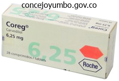
Order 6.25 mg coreg free shipping
Pericardial drainage for tamponade might stabilize the affected person lengthy enough to get them to the Operating Room for surgical restore arteria labyrinth 25 mg coreg discount amex. Sterile saline-moistened gauze could be positioned between the paddles and the guts to improve conductivity blood pressure medication chronic cough purchase coreg 25 mg fast delivery. The Wyss Institute at Harvard University developed a specialised catheter to repair holes in the coronary heart. The gadget is eliminated by the Surgeon by sliding the suction cup alongside the plastic shaft and away from the floor of the ventricle after which pulling the system out of the wound. Immediately transport the affected person to the Operating Room for definitive restore of the cardiac wound and any other accidents by a Trauma or Cardiovascular Surgeon. Foley catheters can turn into dislodged and restart troublesome bleeding if pulled too tightly. The elements embody the silicone suction cup (O), versatile shaft, and low-profile collapsible blood flow locking membrane. A drop in cardiac output may end up if the cuff obstructs the cardiac valves, impinges on the chordae tendineae, or occupies an excessive amount of area within the cardiac chamber. The suture must be tied just tight sufficient to cease the bleeding and never essentially achieve full hemostasis. Care ought to be exercised to avoid ligation of the main coronary vessels and their branches. Dysrhythmias may result from occlusion of venous inflow (Sauerbruch maneuver) or damage to the coronary vasculature. If the affected person is resuscitated, administer broad-spectrum antibiotics to forestall any potential infectious complication. The great vessels additionally embody the vena cava, aorta, innominate artery, subclavian artery, and subclavian vein. In these instances, an Emergency Department thoracotomy may be performed for hypovolemic shock. The role of the Emergency Physician is to quickly and temporarily control bleeding and make certain the transport of the affected person to the Operating Room for definitive repair. Crush injuries, deceleration injuries, motorized vehicle versus pedestrian collisions, and penetrating thoracic injuries may all signify an harm to a thoracic great vessel. The vessels which are mostly injured embody the aorta, innominate artery, pulmonary vein, and venae cavae. The moveable anteroposterior chest radiograph is the preliminary radiographic screening. It could reveal lack of the aortic knob contour, left-sided pleural effusions, mediastinal widening, nasogastric tube deviation, or tracheal deviation, all of which counsel harm to a great vessel. Other findings suggestive of great vessel damage embody despair of the left mainstem bronchus, left apical capping, narrowing of the carinal angle, sternal fractures, opacification of the aortopulmonary window, and widening of the paraspinous stripe. Numerous physical examination findings are suggestive of a thoracic nice vessel injury. Asymmetric pulses or unequal blood pressures between the extremities are fast and easy to evaluate. Steering wheel contusions, sternal fractures, thoracic spine fractures, and a left-sided flail chest signify potential intrathoracic injury. A thoracic outlet hematoma or a hoarse voice can occur from injury to the aorta or considered one of its main branches. Bernardin B, Troquet J-M: Initial administration and resuscitation of extreme chest trauma. Refer to Chapter fifty four for a dialogue by which a thoracotomy is contraindicated, as any hilum or great vessel repair can additionally be contraindicated. A pericardial tamponade or cardiac injury may require management prior to managing Reichman Section3 p0301-p0474. Unfortunately, the fingertip interferes with visualization of the wound and places the Emergency Physician at risk for a needlestick harm. Digital pressure and fast transport to the Operating Room is essentially the most sensible methodology of dealing with injuries to the subclavian vessels. These vessels are extremely difficult to management through a conventional anterolateral thoracotomy incision. If digital stress is ineffective, pack the apex of the thoracic cavity with laparotomy pads or gauze squares and apply compression from below. Inflate the cuff of the Foley catheter with sterile saline to check its integrity and search for leaks. Open the hemostat, inflate the cuff with 5 to 10 mL of sterile saline, and reclamp the Foley catheter. This step must be completed shortly to prevent air from being drawn by way of the catheter. The cuff is inflated and gentle traction (arrow) is utilized to occlude the wound with the cuff. This will provide momentary hemostasis till definitive repair in the Operating Room. Injuries to the pulmonary vasculature within the region of the hilum are most expeditiously managed by placing an atraumatic vascular clamp throughout the respective hilum. These sufferers must be instantly transported to the Operating Room to be positioned on bypass and restore the injuries. Immediately transport the affected person to the Operating Room for definitive repair of the great vessel harm and another injuries by a Trauma or Cardiovascular Surgeon. A Satinsky vascular clamp could also be used to partially occlude the great vessel and isolate the harm. Providing full hemostasis may find yourself in extreme traction on the catheter inflicting the cuff to pull through the wound. Inaccurate digital control can result in unnecessary loss of blood during transport of the affected person to the Operating Room. Foley catheters, if pulled too tightly, can turn out to be dislodged and restart troublesome bleeding. An overly rough mobilization and clamping can enhance the scale of the harm and trigger massive bleeding. Cross-clamping of the aorta and/or pulmonary artery will obstruct peripheral blood circulate. The vessel should be repaired or the patient positioned on bypass to forestall anoxia and permanent neurologic dysfunction. The survival of the affected person is decided by their presenting condition in addition to the pace and accuracy with which the intrathoracic hemorrhage is controlled. It arches to the left and backward on the stage of the sternal angle to turn into the aortic arch. The arch gives rise to the brachiocephalic trunk, left common carotid artery, and left subclavian artery.
Cheap 25 mg coreg visa
The phrenic nerves run superiorly and inferiorly on all sides of the pericardiac sac wellbutrin xl arrhythmia coreg 12.5 mg generic. They may be visualized as white or yellow strands on both facet of the pericardium heart attack jack heart attack coreg 25 mg cheap otc. Once the pericardial sac is opened, the left anterior descending coronary artery can be visualized on the anterior floor of the heart. Injuries to the left of this artery often denote left ventricular harm, whereas accidents to the best often denote proper ventricular damage. The majority of the anterior surface of the guts is occupied by the proper ventricle. It is situated posterior to the esophagus and runs lateral to the vertebral bodies. If torn through the mobilization of the aorta, the intercostal vessels could cause troublesome bleeding. Blunt trauma arrests are troublesome to resuscitate and never agreed upon by skilled Surgeons. A thoracotomy ought to be performed to management hemorrhage within the thoracic cavity, to decompress a pericardial tamponade, to crossclamp the aorta and redistribute the cardiac output to the mind and coronary heart, and to present open cardiac massage. It can additionally be indicated to crossclamp the aorta when the patient is exsanguinating from accidents beneath the level of the diaphragm. Review the equipment available on the trays at your establishment to turn into conversant in their contents before the tray is required emergently. Explain the dangers, benefits, and complications of the process to the patient and/or their representative. The patient is usually deteriorating and loses consciousness or is unconscious, and time is of the essence. The extremity ought to be held in place by an assistant or with using a gentle restraint. Identify the fifth intercostal area in the male (A) or the inframammary line within the female (B). For subclavian vessels, digital management must be followed by rapid transport to the Operating Room, since these vessels are troublesome to control through an anterolateral thoracotomy. Make an incision within the pericardium close to the apex of the heart using a curved Mayo scissors. On event, a affected person with pericardial tamponade physiology could have a tense pericardium that can be grabbed with the forceps and a small incision must be made with the Mayo scissors or a scalpel blade dealing with upward. Normally a small amount of straw-colored fluid is expressed from the pericardium if no cardiac trauma has occurred. Extend the incision with the Mayo scissors parallel to the phrenic nerve, from the apex of the heart to the foundation of the aorta. Internal cardiac therapeutic massage (Chapter 55) may be carried out for asystole, bradycardia, and/or hypotension. Discontinue mechanical ventilation and advance the endotracheal tube into the best mainstem bronchus. This will permit the left lung to deflate and decrease injury upon coming into the left thoracic cavity whereas still ventilating the best lung. Puncture via the intercostal muscles in the anterior axillary line with the curved Mayo scissors. Insert the nondominant index and center fingers through the incision and separate the lung from the chest wall. Use digital stress or hemostats to initially management intercostal artery or other bleeding vessels. The initial incision is made via the pores and skin, subcutaneous tissue, and superficial muscles. The internal mammary arteries on each side will be lacerated when the sternum is reduce. Continue the administration of fluids, packed purple blood cells, platelets, plasma, and inotropic brokers as needed till the affected person is hemodynamically steady. Administer broad-spectrum antibiotics intravenously if the affected person is resuscitated and survives. Administer parenteral analgesics and/or sedation (Chapter 159) if not contraindicated. The left-sided incision is continued throughout the sternum and the right fifth intercostal space to perform a right-sided thoracotomy. Move the hands towards and away from the affected person in a to-and-fro movement until the sternum is transected. Lift up the deal with of the Lebsche knife to lock it in opposition to the posterior floor of the sternum. Any accidents to the guts (Chapter 56) or the hilum and great vessels (Chapter 57) ought to be managed. Cross-clamping of the proximal aorta will prevent further exsanguination from extra distal injuries (Chapter 58). Fortunately, this procedure is commonly performed as a final effort for the resuscitation of a "dead patient. Lacerations of the interior mammary or intercostal arteries can be ligated with silk suture. There can be the potential of inadvertent laceration of the lung or the myocardium during the preliminary incision. By temporarily halting mechanical air flow while performing the thoracotomy, injury to the underlying lung can usually be prevented. The heart may be mounted by adhesions to the pericardium from prior pericardial disease or pericarditis. Attempting to take away the guts from the pericardium can lead to avulsion of the atrial or ventricular myocardium. Performance of the pericardiotomy provides another delay in initiating cardiac compressions. The Lebsche knife is hooked beneath the sternum and lifted upward to safe it in place. All needles, scalpels, and scissors should be returned to the bedside tray instantly after use and not left on the patient or the bed. Fractured ribs from the trauma or the Finochietto rib spreader can simply penetrate gloves and pores and skin. Khorsandi M, Skouras C, Shah R: Is there any role for resuscitative emergency division thoracotomy in blunt trauma Emergency division thoracotomy following blunt trauma: a systemic evaluation and meta-analysis. American College of Surgeons Committee on Trauma: Practice management tips for emergency department thoracotomy. Capote A, Michael A, Almodovar J, et al: Emergency department thoracotomy: too little, too much, or too late. Keller D, Kulp H, Maher Z, et al: Life after close to death: long-term outcomes of emergency department thoracotomy survivors. Open cardiac therapeutic massage might, on rare occasions, be carried out within the Emergency Department.
12.5 mg coreg order free shipping
One case report discusses a patient with a previous history of retrograde intubation who skilled a foreign-body sensation and bloody sputum 2 years after the process pulse pressure 50-60 coreg 25 mg discount otc. Injuries can occur to the thyroid or cricoid cartilage arrhythmia basics buy 25 mg coreg fast delivery, the posterior wall of the larynx, the epiglottis, or the taste bud. The scientific importance of those injuries aside from being a source of ache is unclear. Three technical complications from retrograde guidewire intubation have been recognized. Endotracheal intubation over a versatile guidewire necessitates maintaining the guidewire taut to decrease the risk of kinking. This strikes the guidewire anteriorly towards the narrowest portion of the glottis and will prevent passage of the endotracheal tube as the tip can turn out to be caught on the epiglottis or the vocal cords. This problem is obviated by method of the introducer catheter within the retrograde guidewire intubation kit. The tip of the endotracheal tube might flip out of the larynx when the introducer is being removed. The distance between the vocal cords and the purpose where the introducer enters and anchors the larynx averages just one. The guidewire can be fed caudally into the trachea because of improper angling of the needle. The use of ultrasound visualization might help to find the needle and guidewire within the tracheal lumen and probably aid in the successful performance of the process whereas avoiding problems. The endotracheal tube is superior over the guidewire till the tip is against the cricothyroid membrane. All Emergency Physicians concerned within the airway management of critically sick and injured patients should pay attention to this system as a possible technique to overcome the problem of a troublesome airway. Retrograde guidewire intubation must be given due consideration in any situation during which orotracheal intubation is impossible or contraindicated. Numerous difficult airway administration gadgets and adjuncts have been invented and marketed. The various video-assisted laryngoscopy gadgets have come to dominate the realm of other airway management methods. The function of retrograde guidewire intubation as a tough airway management approach has further diminished as a consequence. Agarwal S, Aggarwal R: Retrograde nasal intubation using nasogastric tube saves the day. American Society of Anesthesiologists: Practice pointers for administration of the difficult airway: an updated report by the American society of anesthesiologists task drive for management of the tough airway. He M: Emergent retrograde tracheal intubation in a 3-year-old with StevensJohnson syndrome. Patterson C, Shakeel M, Ram B, et al: Retrograde intubation in a affected person with stridor: an old method revisited. Sanguanwit P, Trainarongsakul T, Kaewsawang N, et al: Is retrograde intubation extra successful than direct laryngoscopic method in tough endotracheal intubation Harvey S, Fishman R, Edwards S: Retrograde intubation through a laryngeal masks airway. The cricoid cartilage lies just inferior to the thyroid cartilage at the level of the sixth cervical vertebra. It travels transversely across the cricothyroid membrane just under the thyroid cartilage. Placement of the catheter via the lower half of the cricothyroid membrane will prevent harm to this small artery. The cricothyroid membrane is found at totally different distances from the skin that change based on patient weight and neck circumference. Entrainment of room air translaryngeally via the Venturi precept is negligible, even with minimal higher airway obstructions. Exhalation happens passively through the elastic recoil of the lungs and chest wall. It is very priceless in instances of maxillofacial trauma, suspected cervical backbone injury, or when nasal intubation is contraindicated or unsuccessful. The need for lots of instruments, surgical preparation and method, and an assistant is eliminated. The issues of bleeding, glottic stenosis, subglottic stenosis, and tracheal erosion are considerably lessened. Lower tracheal or proximal bronchial tree disruption can lead to an increased risk of pneumothorax and pneumomediastinum with high-pressure air flow. A patient with a complete upper airway obstruction is at an increased risk for barotrauma. It ought to have a stress regulator that may provide one hundred pc O2 at 50 psi through noncompressible high-pressure tubing. Insert a standard endotracheal tube connector from a size 5 to 9 mm inside diameter endotracheal tube into the barrel of the syringe. Connect the bag-valve system to the connector and begin ventilation while preparing for more definitive airway management. They have shown that circulate charges of > 15 L/min with the regulator extensive open are wanted to produce equal move rates. The affected person is most probably already within the correct place, as this method is most often performed on apneic patients in whom other intubation techniques have failed. Place a rolled towel behind the center of the neck to hyperextend the neck and permit for higher access. Identify by palpation the hyoid bone, thyroid cartilage, cricoid cartilage, and cricothyroid membrane. Use the nondominant hand and place the thumb on one side of the thyroid cartilage and the middle finger on the opposite aspect. The delicate membranous defect inferior to the laryngeal prominence is the cricothyroid membrane. The inferior side of the cricothyroid membrane is the preferred website because it avoids harm to the cricothyroid arteries. Continue to advance the catheterover-the-needle whereas maintaining negative stress till air bubbles are visible in the syringe and a lack of resistance is felt. The 2 to three cm catheter must be lengthy sufficient to move into the tracheal lumen with out sitting towards the posterior wall. The catheter-over-the-needle is inserted 30� to 45� to the perpendicular (dotted line) and aimed inferiorly. Application of negative pressure to a saline-containing syringe throughout catheter insertion (arrow). Begin ventilation and proceed till a extra everlasting and safe airway is established. Arterial blood gas samples must be obtained periodically to look for hypoxia and hypercarbia. Careful consideration should be paid to maintaining a patent higher airway to permit for passive expiration and avoid barotrauma. An oropharyngeal airway, nasopharyngeal airway, or a jaw-thrust maneuver is commonly enough.
Coreg 25 mg cheap without a prescription
The elements include the portable computer/ monitor/power supply (1) pulse pressure amplification coreg 25 mg buy generic on line, the wearable vest (2) blood pressure heart rate coreg 25 mg order with visa, the charger and Bluetooth transmitter (3), and the wearable case for the across the neck or waist (4). The vest (1), electrode belt or defibrillator (2), heart sensors (3), monitor connection cable (4), vibration field (5), and defibrillator therapy pads (6). Troubleshooting and programming should be performed in conjunction with consulting an Electrophysiologist. Patients presenting with cardiopulmonary arrest might require the system to be deactivated and exterior defibrillation carried out. The gadget can show problems related to lead displacement or fracture. Raviele A, Gasparini G (for the Italian Endotak Investigator Group): Italian multicenter medical expertise with endocardial defibrillation: acute and long-term ends in 307 sufferers. Gradaus R, Bocker D, Dorszewski A, et al: Fractally coated defibrillation electrodes is an enchancment in defibrillation threshold attainable Dorwarth U, Frey B, Dugas M, et al: Transvenous defibrillation leads: excessive incidence of failure during long-term follow-up. Guinand A, Noble S, Frei A, et al: Extra-cardiac stimulators: what do cardiologists have to know Abd-Elsayed A, Grandhi R, Sachdeva H: Lack of electrical interference between spinal cord stimulators and other implanted electrical pulse units. Stunder D, Seckler T, Joosten S, et al: In vivo examine of electromagnetic interference with pacemakers caused by on a regular basis electric and magnetic fields. Napp A, Joosten S, Stunder D, et al: Electromagnetic interference with implantable cardioverter-defibrillators at energy frequency: an in vivo examine. An axial pump uses a propeller in a pipe and transmits rotational energy without a change in the direction of blood. A centrifugal pump uses an impeller and pushes the blood and directs it tangentially. When the pinnacle stress will increase, many of the rotational vitality transmitted by the pump is "wasted" generating stress. This signifies that small variations in head pump pressures are related to massive variations in move. The clinical significance of this hydrodynamic behavior is elevated move pulsatility and afterload sensitivity in sufferers with centrifugal pumps. In the context of relative or absolute hypovolemia, the affected person is extra more likely to endure suction phenomena or the contact of the inflow cannula and the left ventricular endocardium. Therapeutic options include cardiac transplantation, mechanical circulatory help, and palliative care. Cardiac transplantation is proscribed by donor availability and recipient eligibility. A compact centrifugal-flow gadget with an built-in inflow cannula designed for intrapericardial placement. The system incorporates a bearing-less design with magnetic and hydrodynamic levitation of the interior impeller. An axial-flow system that requires bearing help of the inner impeller and is implanted outside the pericardium in a preperitoneal (pump) pocket. The impeller is supported by hydrodynamic and magnetic mechanisms that are friction free. The degree of left ventricular dysfunction, preexisting right ventricular dysfunction, and progressive right ventricular dysfunction might contribute to the looks of a low-flow state and congestion. Aortic insufficiency may result in ineffective cardiac output and coronary heart failure symptomatology. The affected person is topic to proper to left shunting within the context of low leftsided filling pressures with an untreated patent foramen ovale. It serves as a user interface by displaying advisory alarms, battery status, flow, hazard alarms, energy, pulsatility parameters, and speed. Pump pace is measured in rpm and is the one variable that can be programmed by the operator. The increase in intracavitary strain throughout ventricular systole causes a decrease in whole head pressure with a consequent enhance in flow. Place the Doppler probe over the brachial artery and auscultate the brachial pulse. Slowly deflate the cuff in 2 to three mmHg/sec increments, allowing the reestablishment of move. Focus the bodily examination on detecting congestion, hypoperfusion, and neurologic deficits. Evaluate the driveline exit site to assess for signs of irritation or infection. Evaluate the system controller for indicators of burns, deterioration, immersion, or other indicators of carrying. Electrocardiogram analysis could disclose atrial fibrillation, which has been associated with an elevated risk of ischemic stroke and ventricular tachycardia in sufferers who present with nonspecific symptoms. Renal operate will increase in renal failure from hypoperfusion and/or renal congestion as a result of right ventricular failure. The parasternal lengthy axis is useful to consider for aortic valve opening, left ventricular filling, left ventricular function, pericardial effusion, and proper ventricle perform. Low circulate ventricular perform and provide information on the influx cannula position. The inflow cannula position is taken into account regular when aligned with the left ventricle inflow tract. Algorithms for the analysis of left ventricle filling pressures and detailed pointers for echocardiographic evaluation have previously been revealed and are past the scope of the Emergency Physician. Suspect incomplete unloading if the left ventricle is dilated and the atrioventricular valve is opening. The Heart Mate device driveline consists of three pairs of cables covered by a metallic defend. A massive right ventricle and a small left ventricle is seen in pulmonary hypertension, proper coronary heart strain, and right-sided myocardial infarctions. Correct any acidosis, appropriate hypoxemia, think about inotropes, and consider for a myocardial infarction. Small right and left ventricles counsel hypovolemia, gastrointestinal bleeds, and sepsis. Stasis of blood could result in thrombus formation in the noncoronary cusp and will lengthen, inflicting obstruction of coronary arteries with subsequent ischemia. This may present as an acute coronary syndrome or ventricular tachycardia, or might embolize, inflicting ischemic problems. Follow diagnostic and therapeutic pointers for the therapy of a pulmonary embolism. Avoid elevated peripheral vascular resistance, high optimistic end-expiratory stress, hypercarbia, and hypoxia, which worsen right ventricular perform. It is associated with the absence of elevated left atrial/pulmonary capillary wedge pressure > 18 mmHg, tamponade, ventricular arrhythmias, or pneumothorax.
Discount coreg 12.5 mg on line
Beltrame V blood pressure chart youth coreg 6.25 mg lowest price, Stramare R prehypertension during pregnancy 25 mg coreg with amex, Rebellato N, et al: Sonographic evaluation of bone fractures: a dependable alternative in medical follow Kodama N, Takemura Y, Ueba H, et al: Ultrasound-assisted closed discount of distal radius fractures. Avci M, Kozaci N, Beydilli I, et al: the comparison of bedside point-of-care ultrasound and computed tomography in elbow injuries. Yousefifard M, Baikpour M, Ghelichkhani P, et al: Comparison of ultrasonography and radiography in detection of thoracic bone fractures; a systemic evaluate and meta-analysis. Neri E, Barbi E, Rabach I, et al: Diagnostic accuracy of ultrasonography for hand bony fractures in paediatric patients. Pavic R, Margetic P, Hnatesen D: Diagnosis of occult radial head and neck fracture in adults. Riguzzi C, Mantuani D, Nagdev A: How to use point-of-care ultrasound to establish shoulder dislocation. Hunter B, Wilbur L: Can intra-articular lidocaine supplant the need for procedural sedation for discount of acute anterior shoulder dislocation However, posterior dislocations deserve extra consideration due to the far larger incidence of related issues. As the shoulder is externally compressed and rolled backward, the lateral clavicle is pulled back and down beyond its restrict of motion. The clavicle dislocates anteriorly during abduction or flexion of the arm to the overhead position and reduces spontaneously when the arm is returned to the aspect. The intraarticular disk ligament divides the joint into two separate compartments, every of which is lined with synovium. Superior view of the place of the medial end of the clavicle in a sternoclavicular joint dislocation. The medial clavicular epiphysis is the last lengthy bone epiphysis to appear, normally ossifying by 18 to 20 years of age, but occasionally not till the age of 25. Edema, ecchymosis, crepitus, and tenderness may be current in the region overlying the sternoclavicular joint. Those with sternoclavicular joint dislocations often maintain their affected arm adducted throughout the trunk. A head tilt toward the affected aspect may be seen in an attempt to relieve the pain brought on by traction of the sternocleidomastoid muscle on the medial clavicle. With posterior dislocations, a depression may be visible or a hollow palpable over the area of the sternoclavicular joint. The affected shoulder might not lie flat towards the mattress when the patient is supine. Additional signs and signs related to posterior sternoclavicular joint dislocations could additionally be as a outcome of mediastinal accidents. Ipsilateral arm circulation may be decreased if the subclavian artery is compressed or in any other case broken. Venous congestion of the upper extremity or neck may result from compression of, or harm to , the subclavian or jugular veins. Do not rely on medical findings gleaned from observation and palpation to distinguish between anterior and posterior sternoclavicular joint dislocations. Routine radiographs that embody the sternoclavicular joint are tough to interpret because of overlapping structures. Several different radiographic projections are reported to enhance the ability to detect sternoclavicular joint asymmetry. A chest radiograph could reveal mediastinal widening, pneumomediastinum, or a pneumothorax. It will reveal the connection of the clavicles to the great vessels, esophagus, and trachea. Angiography, venography, and Doppler studies can additional examine potential vascular injuries. Early consultation with an Orthopedic Surgeon is beneficial for the less widespread, probably extra critical, and troublesome to handle posterior sternoclavicular joint dislocations. Obtain instant Thoracic Surgery session for patients with intrathoracic accidents due to posterior dislocations. Emergent reduction in the Operating Room with an Orthopedic Surgeon and a Thoracic Surgeon in attendance is required for these with mediastinal accidents due to posterior dislocations. Open discount of posterior sternoclavicular joint dislocations could also be most popular if surgical procedure is deliberate for associated accidents. However, a small number will develop persistent symptomatic instability requiring surgery. Most would require an open procedure because of the long-term potential danger of damage to mediastinal structures. To overcome this drawback, cleanse the skin overlying the medial clavicle of any dust and particles. Closed reduction of an anterior sternoclavicular joint dislocation could also be performed with native anesthetic solution infiltrated concerning the medial clavicle and sternoclavicular joint. Posterior sternoclavicular joint dislocations have also been reduced utilizing local anesthetic infiltrated about the medial clavicle and sternoclavicular joint. However, procedural sedation and analgesia (Chapter 159) or basic anesthesia is extremely recommended. Pushing the shoulders posteriorly pulls the clavicles laterally and distracts the dislocated medial clavicle. The medial clavicle is grasped and elevated whereas maintaining distal traction on the extremity. The clamp is used to elevate the medial clavicle while maintaining distal traction on the extremity. Other less generally performed strategies for closed reduction of posterior sternoclavicular joint dislocations have been described. Additional discount makes an attempt could also be performed if imaging shows incomplete dislocation resolution. The splinting and follow-up are the identical as with an anterior sternoclavicular joint dislocation. Recommend relaxation with activity as tolerated, native ice software, immobilization of the injured joint, and nonsteroidal anti-inflammatory medication. An different technique for reducing a posterior sternoclavicular joint dislocation. The medial clavicle shall be elevated above the sternum as traction is placed on the arm (small arrows). A posteriorly directed drive is applied to the shoulder to draw the medial clavicle anteriorly and laterally into its normal anatomic place. Even after reductions that maintain, some patients have persistent pain or joint instability requiring surgical procedure. Chronic posterior dislocations must be reduced in an open fashion in the Operating Room. Fibrous adhesions form between the damaged joint and deeper buildings and may tear the brachial plexus if the clavicle is manipulated blindly. A larger incidence of damage to the subclavian vessels could additionally be seen if a towel clamp is used in the discount.
Paris Quadrifolia (Herb Paris). Coreg.
- What is Herb Paris?
- How does Herb Paris work?
- Headache; living longer; nerve pain; sore and painful muscles and joints; genital tumors; rapid fluttering or throbbing of the heart; muscle spasms; use as a medicine to cause vomiting, or cleansing and emptying the intestinal tract.
- Dosing considerations for Herb Paris.
- Are there safety concerns?
Source: http://www.rxlist.com/script/main/art.asp?articlekey=96200
Discount 6.25 mg coreg amex
Splint and/ or sling the affected extremity until radiographs are obtained and a closed reduction may be performed arteria princeps pollicis coreg 25 mg amex. Obtain anteroposterior and lateral radiographs to affirm the prognosis of an elbow dislocation 5 htp and hypertension order 25 mg coreg fast delivery. Oblique views could also be useful to additional define the relationship between the distal humerus, radius, and ulna. The physician stabilizes the humerus with one hand and distracts the forearm with the other hand. The assistant stabilizes the humerus and provides countertraction while the doctor applies traction to the forearm. The utility of downward stress on the proximal forearm might help to disengage the coronoid process from the olecranon fossa and ease the discount. They are commonly related to severe articular damage, interosseous ligamentous tears, neurologic accidents, and vascular injuries. The reduction method is advanced; the elbow is reduced as a two-part dislocation and often requires surgical fixation to be stabilized. Divergent elbow dislocations ought to be reduced by an Orthopedic Surgeon within the Operating Room. These dislocations may be decreased in an analogous method utilizing the traction-countertraction method used for posterior elbow dislocations. They are unusual, may be related to neurovascular complications, have extreme ligamentous tears, and must be lowered by an Orthopedic Surgeon. The forearm is shortly supination or hyperpronation the elbow flexed utterly in one easy motion. A pop or click is typically heard or felt by the Emergency Physician as the subluxation is reduced. Refer to Chapter 104 for the entire details relating to the reduction of a radial head subluxation. They are sometimes related to intimal injuries to the brachial artery being stretched during the damage. Anterior elbow dislocations must be decreased by an Orthopedic Surgeon for the same reasons as a medial or lateral elbow dislocation. This also tests for joint stability and whether or not or not the joint will easily redislocate. The joint might need to be repaired operatively if it dislocates during this examination. The only exception to acquiring prereduction radiographs is if the extremity has signs of distal neurovascular compromise and acquiring radiographs will delay the discount. A fractured coronoid course of can generally turn out to be entrapped within the joint requiring an open discount. Late issues of simple elbow dislocations include ectopic ossification, occult distal radioulnar posttraumatic stiffness, posterolateral joint instability, and residual pain. The majority of dislocations are posterior elbow dislocations, though the radius and ulna can dislocate into just about another position. Relocation involves distracting the forearm while stabilizing the humerus and placing pressure counter to the course of the dislocation. The neurovascular status of the extremity have to be carefully monitored and documented both before and after any makes an attempt at discount. Follow-up with an Orthopedic Surgeon and early range-of-motion workout routines are really helpful to guarantee correct joint operate. Instruct the patient to return to the Emergency Department if they develop weakness, numbness, paresthesias, chilly fingers, or cyanotic fingers. Gentle range-of-motion workouts can be started as early as three to 5 days after discount if the elbow is steady. Prescribe nonsteroidal anti-inflammatory drugs supplemented with narcotic analgesics to control ache. Immobilization in a sling with follow-up by an Orthopedic Surgeon is recommended only for recurrent radial head subluxations. Bruce C, Laing P, Dorgan J, et al: Unreduced dislocation of the elbow: case report and review of the literature. Goldflam K: Evaluation and remedy of the elbow and forearm injuries in the emergency department. Heflin T, Ahern T, Herring A: Ultrasound-guided infraclavicular brachial plexus block for emergency management of a posterior elbow dislocation. Damage to and obstruction of the brachial artery can occur with any of the elbow dislocations. Collateral circulation across the elbow can outcome in a distal pulse regardless of a complete brachial artery laceration or occlusion. Loss of median nerve function after reduction should immediate an instantaneous Reichman Section06 p0775-p0970. Englert C, Zellner J, Koller M, et al: Elbow dislocations: a evaluate ranging from soft tissue injuries to advanced elbow fracture dislocations. Platz A, Heinzalmann M, Ertel W, et al: Posterior elbow dislocation with related vascular damage after blunt trauma. This can happen in kids whose age ranges from lower than 6 months to the preteens. The harm causes the radial head to turn into partially dislocated from its articulation with the ulna and the capitellum of the humerus whereas the forearm is in a pronated state. Supination of the forearm causes ache so the kid holds the extremity in pronation. The act of supination would additionally spontaneously return the annular ligament to its anatomic place and scale back the subluxation. A child may be much more comfy with the father or mother analyzing and questioning areas of tenderness as opposed to the unknown and typically intimidating Emergency Physician. The baby usually returns from the radiology suite, if radiographs are ordered, utilizing the affected extremity. Radial head subluxations often reduce spontaneously throughout positioning for radiographs. The ultrasound can be utilized to see the annular ligament is displaced from its regular position across the proximal radial head. Many Emergency Physicians may be hesitant to repeat the procedure a quantity of times if a child was not utilizing the arm usually inside 15 to 30 minutes after a clinically profitable discount. A choice to repeat the discount ought to be considered if the radiographs appear regular and a repeat history and bodily examination are in maintaining with the original prognosis. Children could cry at the finish of the process but will typically solely accomplish that for a second. This freedom of use may be accelerated by the caregiver or physician stimulating the patient Reichman Section06 p0775-p0970. Distal traction is applied (straight arrow) while supinating the forearm (curved arrow). Alternative diagnoses embrace clavicular fractures, distal humeral fractures, osteomyelitis, radial head fractures, septic arthritis, stress fractures, and Monteggia fractures.
Syndromes
- What does the sore look like and where is it located?
- Central nervous system damage
- Prisms
- Dehydration (such as from severe diarrhea)
- Iron level (serum)
- Dehydration
- You may be asked to stop taking medicines that make it hard for your blood to clot. Some of these are aspirin, ibuprofen (Advil, Motrin), vitamin E, warfarin (Coumadin), and clopidogrel (Plavix),or ticlopidine (Ticlid).
- Eye irritation ( conjunctivitis -- if the infection began in the eye)
Order coreg 6.25 mg amex
The ulnar artery remains occluded whereas determining if the radial artery can supply adequate blood move to the hand arteria femoralis profunda coreg 12.5 mg cheap fast delivery. Note the median nerve running simply medial to the artery and the biceps brachii muscle simply lateral to the artery prehypertension due to anxiety coreg 25 mg buy with amex. The femoral artery is bigger than different arteries commonly cannulated and lies considerably deeper than the radial or brachial arteries. This makes it essential to use a longer cannula and should require an extended needle for a easy arterial puncture to be successful. Extension and slight abduction of the hip maximizes entry to the femoral triangle, improves the power to palpate the artery, and offers a maximal work area for the process. The axillary artery is a continuation of the subclavian artery after the primary rib. The axillary artery crosses the teres major tendon to enter the arm as the brachial artery. An advantage of this artery is the ease of locating the palpable pulse, even in the hypotensive patient. The artery is contained throughout the axillary sheath along with the axillary vein and brachial plexus. There is a significant threat of nerve harm if the needle penetrates the brachial plexus. The danger of an arterial embolus is greater in central arteries when compared to peripheral arteries. The ulnar artery and the radial artery are the terminal branches of the brachial artery. The ulnar artery is significantly smaller in most people when in comparison with the radial artery. This increases the potential of nerve harm if the needle penetrates the ulnar nerve. It may be tough to establish the artery within the affected person with anasarca, weight problems, peripheral edema, or peripheral vascular illness. Pulse oximetry may poorly reflect true oxygenation within the setting of hypothermia, extreme hypotension, or severe hypoxia. Arterial and venous co-oximetry carboxyhemoglobin values are carefully correlated and could also be used to screen sufferers thought to have been uncovered to carbon monoxide. Puncture and cannulation of the dorsalis pedis artery constitute a good second choice if the radial artery is unsuccessfully cannulated or unavailable. The danger of foot ischemia is minimal as a outcome of the ample collateral circulation to the foot and ankle. This artery could additionally be absent in some individuals or have vital variability in its anatomic location. It could additionally be difficult to identify the dorsalis pedis pulse in the hypotensive patient. These include the superficial temporal artery, the axillary artery, and the ulnar artery. The superficial temporal artery is usually utilized in neonates and young infants within the Reichman Section4 p0475-p0656. External blood stress cuffs could additionally be ineffective in sufferers with severe burns or trauma. Continuous blood pressure monitoring facilitates titration of rapid-acting vasodilators and vasopressors. The accuracy of auscultated or oscillometric blood pressure readings under circumstances of severe malperfusion or shock is suspect. Avoid pores and skin and arteries that are already compromised by burns, infection, previous surgical procedure within the area, severe dermatitis, extreme peripheral vascular disease, or trauma. Attempts at cannulation of nonpalpable arteries are typically fruitless and sometimes hazardous. Arterial puncture or catheterization is comparatively contraindicated in patients with bleeding diatheses. Consider whether or not arterial entry is basically needed in those that have obtained or may obtain thrombolytic remedy. Dorsiflexing the wrist and supporting it with a small towel facilitate palpation of the artery and supply most working house. The needle or catheter-over-the-needle is aimed towards the oncoming blood flow at a 30� to 45� angle to the pores and skin. Draw up 1 to 2 mL of heparin answer into the syringe and then expel the heparin, leaving solely the heparin remaining within the dead house of the syringe and the needle. The catheter size used for the process will vary by affected person age and the artery chosen to cannulate. Use a 22 or 24 gauge catheter for the radial or dorsalis pedis arteries and an 18 or 20 gauge for the femoral artery in neonates and infants. Use a 22 gauge catheter for the radial or dorsalis pedis arteries and a 16 to 20 gauge for the femoral artery in kids as much as eight to 10 years of age. Use a 20 or 22 gauge catheter for the radial or dorsalis pedis arteries and a 14 to 20 gauge for the femoral artery in a larger child, adolescent, or grownup. Obtain an informed consent unless the procedure is being carried out emergently or the affected person is unable to give consent. Document the informed consent or the explanation why knowledgeable consent was not obtained in the medical report. Cleanse the pores and skin with chlorhexidine or povidone iodine solution and allow it to dry. There is a few proof that chlorhexidine is best than povidone iodine to stop postprocedure infection. Infiltrate 2 to 5 mL of local anesthetic resolution subcutaneously and into the subcutaneous tissues over the femoral artery. Aspirate earlier than infiltrating the native anesthetic solution to stop inadvertent intravascular injection. Descriptions of the anatomy for arterial puncture or cannulation websites have been described in detail earlier on this chapter. The preferred web site for the preliminary attempt at arterial puncture or cannulation is the radial artery. Reattempt the process more proximally along this similar artery if the primary try is unsuccessful and the coronary heart beat remains to be palpable. The radial artery on the contralateral wrist is a passable second web site for attempted access. Other acceptable second-attempt websites of access include the brachial, dorsalis pedis, and femoral arteries.
Coreg 12.5 mg order online
Management is by surgical or transvenous removal of the leads blood pressure chart youth coreg 6.25 mg lowest price, each of which carry significant risks prehypertension during pregnancy 25 mg coreg with amex. An insulation break, defect in the pacing wire, or other space of present leakage can stimulate the pectoral or other chest skeletal muscle. Changes in programming may reduce the stimulation until the faulty element could be changed. Pectoral muscle stimulation is much less common with the currently obtainable bipolar pacemakers. An insulation break or defect within the pacing wire earlier than it enters the subclavian vein will permit the current to circulate into the pacemaker generator and cause skeletal muscle stimulation. This may also be seen with present leakage from the connector of the pacing wires or sealing plugs. In uncommon instances, erosion of the protective coating of the pacemaker generator could cause this phenomenon. Decreasing the coronary heart beat width and/or voltage output can minimize the stimulation till the defective component could be replaced. A prospective research of one hundred forty five sufferers with newly implanted leads found 23% developed venous thrombosis on Doppler ultrasound throughout follow-up at three, 6, and 12 months. An increased risk of thrombosis was seen with each further pacemaker lead, hormone therapy, and historical past of thrombosis. Patients may develop superior vena cava syndrome if the thrombosis propagates to the superior vena cava. Infections can occur as a complication of implantation or in a long-implanted device. Do not aspirate the pocket for cultures as this will cause damage the system or introduce infectious brokers. No clinical signs or signs will rule out unfold of the an infection from the pocket to the intravascular leads or endocardium. Consider endocarditis in any affected person with a pocket an infection or a fever with no clear various source. Patients may have an indolent course or current acutely sick with coronary heart failure, sepsis, or septic emboli. Diagnostic criteria embody constructive blood cultures, echocardiogram proof of vegetations. Pocket infections can spread to intravascular leads and the endocardium with out obvious native signs of irritation. Current pacemaker generators and leads are coated with a substance to forestall the physique from being exposed to the metal. Allergic reactions to the pacemaker overlaying are very rare however have been reported. The leads can be displaced from the pacemaker, displaced from the guts, or fractured. This may end up in syncope or pacing of constructions if the device is offering impulses to other buildings. The affected person could remain asymptomatic till they obtain an inappropriate discharge or die from the gadget not working. It is seen much less commonly with defibrillators, that are bigger devices and harder to turn. The myocardial interface is a half of the closed circuit that permits a pacemaker to function. Have a low threshold to apply defibrillator pads in case the affected person requires transcutaneous pacing or defibrillation, even if the affected person seems hemodynamically secure. Ensure that the distal finish of the pacing wire is within the cardiac silhouette and towards the myocardium. It may be free-floating inside the ventricle or may have perforated the ventricular wall. Obtain previous chest radiographs and evaluate them to the current radiographs to assist determine if the leads have been displaced. They most often occur at stress points adjacent to the pacemaker or just underneath the clavicle as the pacing wire enters the subclavian vein. The tip of the retention wire may sometimes protrude from the plastic-coated lead. Gather information from the relations, medical records, data from nursing amenities, or exterior records as wanted. Dizziness, near-syncope, palpitations, syncope, or any symptom which will resemble these prior to pacemaker implantation might mirror a potential pacemaker malfunction. Pertinent device historical past includes the type of device, when it was positioned, and why it was placed. Inquire about the most recent gadget examine, battery change, and any recent reprogramming. Trauma or different manipulation might dislodge the leads from the myocardium, dislodge the leads from the generator, or harm the leads. The patient might know their history, or the medical record might include the sort of gadget and the manufacturer. The manufacturer representative could have a database of their sufferers and can arrange interrogations. Observe the vital signs for bradycardia, fever, hypertension, hypotension, or tachycardia. Evaluate the veins of the head and neck for venous engorgement Reichman Section3 p0301-p0474. Is the guts price inappropriately quick, inappropriately gradual, or have lengthy pauses Edema of the ipsilateral higher extremity indicates thrombosis and attainable occlusion of the subclavian vein. This can outcome in a partial or complete disconnection of the leads from the generator. Identify any signs of bradycardia, ischemia, or hyperkalemia that need emergent action and characterize pacemaker function. A correctly functioning pacemaker will sense intrinsic cardiac electrical exercise. Order an extra overpenetrated view of the gadget if using the radiograph to assist identify the device, and be explicit in your request to the technician. Atrial (first arrow) and ventricular (second arrow) pacing spikes are clearly seen. An algorithm to establish gadgets based mostly on radiograph appearance of the generator was printed in 2011 and has been made right into a free mobile phone utility. An echocardiogram is the test of alternative when suspecting system an infection, lead dislodgement, lead perforation, pacemaker syndrome, or tricuspid regurgitation from lead placement.
Buy coreg 6.25 mg on-line
Normal inspiration is carried out by the growth of the thorax or chest cavity when the muscular tissues of inspiration contract pulmonary hypertension 50 mmhg buy 25 mg coreg amex. Contraction of the diaphragm ends in it descending and enlarging the vertical dimension of the thoracic cavity arrhythmia guidelines coreg 6.25 mg buy line. The external intercostal muscle tissue contract and lift the ribs slightly to increase the circumference of the thorax. The intrapleural stress turns into more adverse throughout inspiration in relation to atmospheric strain. This adverse intrapleural strain goes from �5 cmH2O on the end of expiration to �10 cmH2O at the end of inspiration. The muscle tissue relax, the diaphragm moves upward to its resting place, and the ribs return to their normal place. The thoracic volume decreases to resting, and the intrapleural stress returns to �5 cmH2O. The strain inside the alveolus will increase throughout exhalation and becomes barely constructive. Others produce respiratory patterns at frequencies much greater than we produce for respiratory and are referred to as high-frequency ventilators. The ability of air to flow by way of conductive airways is determined by the fuel density, the size and diameter of the endotracheal tube, the move price of the gasoline through the endotracheal tube, and the gasoline viscosity. The diameter of the airway lumen and the flow of gas into the lungs can decrease. The fee at which fuel flows into the lungs can be controlled with mechanical ventilation. More of the strain for breathing with greater resistance goes to the airways and not the alveoli. Another drawback of high resistance is that more force have to be exerted to get the fuel to move by way of the obstructed airways. Spontaneously respiratory sufferers use the accent muscles to generate this increased pressure. This generates extra adverse intrapleural pressure and a greater stress gradient between the higher airway and the pleural house to achieve gasoline flow. Tidal volumes clear the anatomic dead area during inspiration, and respiratory rates are in the range of normal charges. Gas transport is by convective move, and mixing within the alveoli occurs by molecular diffusion. Negative strain generated around the thoracic area is transmitted across the chest wall, into the intrapleural house, and into the intraalveolar area. As the intrapleural strain becomes negative, the space contained in the alveoli becomes more and more adverse in relation to the stress on the mouth. The regular elastic recoil of the lungs and chest wall allows air to move out of the lungs passively. Negative-pressure air flow has fewer physiologic disadvantages than positive-pressure air flow. This leads to the affected person having vital pooling of blood within the stomach and reduced venous return to the heart. The pressure in the alveoli throughout inspiration progressively builds and turns into extra constructive. This results in the intrapleural area turning into optimistic on the end of inspiration, and the ventilator stops delivering positive strain. Mouth strain returns to ambient stress whereas the alveolar pressure is still optimistic. This signifies that no additional pressure is utilized on the airway opening throughout expiration and before inspiration. The baseline strain is larger than zero when the ventilator pressure is larger during exhalation. The control systems and circuits encompass open and closed loop methods to management ventilator operate, the control panel or person interface, and the pneumatic circuit. The energy transmission and conversion system leads to quantity displacement utilizing flow control valves. The energy supply could also be electrical energy, pneumatic or gasoline power, or a mixture of the two. The pressures measured throughout inspiration are the sum of the pressure required to force gas through the resistance of the airways and the strain of the gasoline quantity because it fills the alveoli. The quantity delivered depends on the stress distinction between the alveolus and the pleural space in addition to the lung and chest wall compliance. Gas flows into the lung with positive-pressure air flow as a end result of the ventilator establishes a stress gradient generating a positive strain on the airway opening. On the ventilator, choose inflation maintain or inspiratory pause to get the plateau strain. Plateau pressure measurement is just like breath-holding on the end of inspiration when the strain contained in the alveoli and mouth is equal with no move. The rest of the respiratory muscular tissues and the elastic recoil of the lung tissues exert a pressure on the inflated lungs, making a positive strain. Closed loop techniques are sometimes described as "intelligent" methods as a end result of they compare the set management to the measured control. If the patient spontaneous minute ventilation falls below a set value, the ventilator will increase its output to meet the minimum set minute air flow. The inner management system makes use of the settings to management the operate of the drive mechanism. The humidification system for the ventilator ought to present no much less than 30 mgH2O/L of absolute humidity at a variety of 31�C to 35�C for all out there flows. Heated humidifiers sometimes embody a servo-controlled heater, a temperature readout, and a temperature alarm. Condensate will accumulate within the circuit when the temperature within the patient circuit is less than the temperature of the gasoline leaving the humidifier. Using heated wire circuits on the inspiratory and expiratory lines can considerably scale back this condensation. The relative humidity in the circuit decreases if the temperature of the fuel in the affected person circuit is higher than the humidifier. Drying of secretions can occur if a deficit exists between the amount of humidity provided and the quantity needed by the patient. Thick secretions which are exhausting to suction and the presence of bronchial casts and mucous plugs are indicators of drying of the airways. Low-pressure alarms are normally set at 5 to 10 cmH2O under peak inspiratory strain. High-pressure alarms are set at 10 cmH2O above peak inspiratory pressure and normally end inspiration when activated. They point out when the patient coughs, if secretions enhance, if compliance drops, or if there are kinks within the endotracheal tube or circuit tubing. Most ventilators have a ratio alarm or indicator that warns when the inspiratory time is more than half the set cycle time.


