Coumadin
Coumadin dosages: 5 mg, 2 mg, 1 mg
Coumadin packs: 60 pills, 90 pills, 120 pills, 180 pills, 270 pills, 360 pills
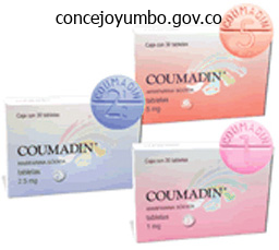
Buy coumadin 2 mg without prescription
Some clinicians subsequently suggest using a microaggregate blood infusion filter with a mesh pore size of 40 �m when multiple models of blood are administered to trauma victims blood pressure children cheap 1 mg coumadin with amex, patients with compromised pulmonary function hypertension first line coumadin 2 mg buy cheap line, and neonates. Others consider that though platelets are eliminated with the microaggregate filters, the trapped platelets can be removed with a saline flush without any vital loss. All fluids administered via the rapid transfuser might be warmed to approximately 37�C. Rate of Infusion One unit of entire blood can be safely administered to a hypotensive patient at a fee of a minimal of 20 mL/kg per hr. Use of a speedy transfuser system can help within the fast administration and warming of blood. In secure sufferers, administer 1 unit of entire blood (500 mL) over roughly a 2-hour interval (3 to 4 mL/kg per hr). In addition, the unit of blood, which is a wonderful culture medium, is prone to become contaminated if bacteria and fungi are allowed to develop at room temperature. In patients with extreme anemia and congestive coronary heart failure, administer a rapidly appearing diuretic, corresponding to furosemide on the onset of transfusion to prevent circulatory overload. If a transfusion of blood must be interrupted or delayed for any reason, return the remainder of the blood unit to the blood bank. For sufferers in hemorrhagic shock, administer blood via two large-bore catheters, at different websites if necessary. Usually, gravity offers a adequate stress gradient if the unit is raised above the patient to increase the speed of infusion when the clamps are wide open. Rewarming Blood is stored at roughly 4�C to maintain cellular integrity and forestall the expansion of microorganisms. The antagonistic results of hypothermia on cardiac conduction and move charges are evident when rapid administration of a large volume of blood is carried out without prewarming. An ideal blood warmer ought to allow liberal move rates whereas stopping thermal hemolysis of blood cells. Commonly used gadgets embody those with bath coils that enable a plastic tube to reside in a closely regulated heat water bathtub and dry heat gadgets that permit blood to circulate via flat, skinny baggage sandwiched between aluminum blocks that comprise electrical heating parts. Once blended, administer the product to the patient; this offers a resultant delivery temperature of roughly 35�C. Regardless of the rewarming technique used, warming refrigerated blood to body temperature decreases its viscosity twofold to threefold and avoids venous spasm, thus facilitating transfusion. Monitoring During the first a part of the transfusion of any blood product, carefully monitor the affected person for proof of a transfusion response. Look for signs and signs corresponding to hives, chills, diarrhea, fever, pruritus, flushing, stomach or again ache, tightness within the chest or throat, and respiratory misery. Treat an allergic reaction (hives, itching) to leukocytes or plasma proteins by administering an antihistamine (but not in the blood infusion line), and cease the transfusion. Stop the transfusion instantly when the following signs are encountered: a rise in pulse rate, a lower in blood stress, respiratory symptoms, chest or stomach discomfort, or a sensation of "impending doom. Send samples of urine and blood to the laboratory to confirm the presence of free hemoglobin. In addition, send the blood financial institution a clotted pattern of blood to reassess for the presence of an immune reaction. If the blood bank concludes that the reaction is a nonhemolytic allergic response, premedicate with antihistamines (diphenhydramine or hydroxyzine) and antipyretics earlier than the subsequent transfusion. If a hemolytic transfusion reaction is suspected, treat the affected person vigorously and promptly. Infuse crystalloid or vasopressors to deal with the hypotension immediately, if required. Treat symptomatically with acetaminophen, a warming blanket, antihistamines, and inhaled or subcutaneous -agonists for bronchospasm or subglottic edema. The good thing about alkalization and diuresis in preventing acute renal failure is unsure, though the usage of these strategies is commonly advocated. Such reactions are characterised by falling hemoglobin ranges, jaundice, hemoglobinemia, and indirect hyperbilirubinemia. Individuals thus affected ought to put on identification tags or bracelets to alert medical personnel that previous transfusion reactions have occurred. The progress notice, the transfusion report sheet, or the transfusion laboratory slip can be utilized for this purpose and must be signed and dated by the clinician, in accordance with hospital policies. Suggest that the family contemplate arranging for replacement donations of units of blood to afford future patients the luxury of an ample, out there supply of blood merchandise. McGee essential used in the on a regular basis Local anestheticofagents arethe nuancestoolsclinical use,describes follow of emergency medicine. Detailed technical guidance for the performance of topical and infiltrative local anesthesia is supplied. Early Incan society used cocaine for invasive procedures, together with cranial trephination. In 1884, Koller1 used topical cocaine in the eye and was credited with the introduction of local anesthesia into medical follow. In the identical 12 months, Zenfel used a topical solution of alcohol and cocaine to anesthetize the eardrum, and Hall2 launched the drug into dentistry. In 1885 Halsted3 demonstrated that cocaine blocked nerve transmission, thereby laying the muse for nerve block anesthesia. The seek for alternate options to cocaine led to synthesis of the benzoic acid ester derivatives and the amide anesthetics used today. It was not until the Nineteen Sixties that detailed understanding of the physiochemical properties, mechanism of motion, pharmacokinetics, and toxicity of those brokers emerged. The intermediate chain between the aromatic and the hydrophilic segments is either an aminoester or an amino-amide; these chemical constructions type the basis for the two major classifications of local anesthetics. Common ester-type brokers include procaine, chloroprocaine, cocaine, and tetracaine. Common amide-type brokers include articaine, lidocaine, mepivacaine, prilocaine, bupivacaine, and etidocaine. Cocaine, an ester, can additionally be partly metabolized by N-demethylation and nonenzymatic hydrolysis. Individuals with pseudocholinesterase deficiency might have a larger potential for cocaine toxicity if giant doses are used, although this has not been a problem when cocaine is used clinically as an anesthetic. Local anesthetics are poorly soluble weak bases mixed with hydrogen chloride to produce the salt of a weak acid. In answer, the salt exists both as uncharged molecules (nonionized) and as positively charged cations (ionized). The nonionized form is lipid soluble, which allows it to diffuse through tissues and throughout nerve membranes. The ratio of nonionized to ionized forms depends on the pH of the medium (vial resolution or tissue milieu) and on the pKa of the specific agent. The pKa is the pH during which 50% of the solution is within the uncharged kind and 50% is within the charged form.
Diseases
- Czeizel Losonci syndrome
- Arterial tortuosity
- Atrophic vaginitis
- Ichthyosis linearis circumflexa
- Hairy cell leukemia
- Hydrocephalus skeletal anomalies
- Niemann-Pick disease type D
- Glossopalatine ankylosis micrognathia ear anomalies
- Cutis laxa, recessive type 1
- Idiopathic pulmonary fibrosis
Generic 1 mg coumadin amex
Note the small bony avulsion fracture on the lateral facet of the tibial plateau blood pressure instrument order coumadin 2 mg mastercard. Although at first glance this appears to be a somewhat innocuous injury hypertension home remedies coumadin 2 mg buy visa, a Segond fracture is very associated with tears of the anterior cruciate ligament. This patient should be handled with a knee immobilizer till orthopedic follow-up may be arranged. Posterior Knee Splint A Application B Indications Periprosthetic distal femur fracture A, Extend the posterior knee splint from slightly below the buttocks crease to approximately 2 to three cm above the malleoli. B, Alternatively, place two parallel splints alongside all sides of the leg and foreleg to create a bivalve effect. Although this variation is tougher to apply, it might present better immobilization of the lateral and medial collateral ligaments. Angulated fractures around the knee (shown above) Temporary immobilization of injuries previous to operative restore Knee injuries in sufferers with extremities too giant for a knee immobilizer the knee immobilizer has largely changed the posterior knee splint in trendy emergency division follow. Indications Fractures of the distal ends of the fibula and tibia (shown above) Severe ankle sprains Reduced ankle dislocations C A, Apply the splint with the affected person prone and the knee bent to 90�, thereby relaxing the calf muscles. Note the very sharp frayed fiberglass edges (arrows) and the a quantity of inner ridges and folds that will produce gentle tissue trauma when worn. Carefully mildew the wet plaster around the malleoli and instep to guarantee maximum consolation and immobilization. For the anterior portion, start on the dorsal floor of the foot on the degree of the metatarsophalangeal joints. Extend the splint along the anterior portion of the foreleg, to the identical height as the posterior splint. After the wet posterior splint has been applied, place the anterior splint over the anterior facet of the ankle and foreleg parallel to the posterior splint. Hold the two in place with elastic bandages as described earlier for a posterior splint alone. An assistant is required to apply an anterior-posterior splint as a outcome of this can be very difficult to hold both splints in place while wrapping the elastic bandages. It features like a posterior splint, and either one offers satisfactory ankle immobilization. In one study that compared these splints in normal volunteers, the u-splint allowed much less plantar flexion and broke less typically with plantar flexion than did the posterior splint. The splint passes beneath the plantar surface of the foot from the calcaneus to the metatarsal heads and extends up the medial and lateral sides of the foreleg to slightly below the level of the fibular head. Wrap the elastic bandage around the extremity beginning at the metatarsal heads and continuing across the ankle in a figure-of-eight configuration. Once the ankle has been wrapped, use another 4- or 6-inch elastic bandage to secure the rest of the splint in place. Note that if a u-splint is mixed with a posterior splint, apply the posterior splint first. For immobilization of the knee, prolong the sides of the splint proximally to the groin to create a protracted leg splint. Indications Injuries of the ankle, including: Fractures of the distal tibia and fibula Postreduction stabilization of ankle dislocations (above) the U-splint can be combined with the posterior ankle splint to provide each anterior-posterior and medial-lateral stability. Splints for Ankle Sprains A walking boot can be used for the remedy of average to severe soft tissue injuries of the ankle, including second- and third-degree sprains and isolated, nondisplaced lateral malleolar fractures. A strolling boot supplies an analogous diploma of immobilization as a U-splint but is much less complicated to take away for bathing and dressing, and the Velcro straps enable adjustment for edema. Prefabricated semirigid orthoses such because the Aircast (pictured above) are often used for patients with minor ankle sprains. The Unna boot or an Ace wrap offers efficient immobilization of an ankle gentle tissue injury. For similar short-term immobilization with out plaster, a modified Jones dressing can be used. Copious Webril is wrapped across the ankle and foot and lined with an elastic bandage. When cleared by the follow-up physician, a strolling boot allows simple transition to full weight bearing. Studies have shown that speedy mobilization after ankle accidents improves useful outcome and reduces incapacity time. Walking boots are available quite so much of sizes from additional small to additional massive, depending on the manufacturer. In sufferers with lateral ankle sprains related to a stable joint, a useful brace with early mobilization is regularly more comfy and results in earlier return to normal function than complete immobilization in a plaster splint or cast. This gadget can additionally be used over a splint or solid to permit partial weight bearing. If a cast shoe goes to be used for a patient with a fractured toe, first buddy-tape the injured digit to the adjacent toe. For minor ankle injuries, a easy elastic (Ace) bandage can be utilized in a figure-of-eight configuration. Hard Shoe Splint Cotton or Webril between toes Indications Reduction of ambulatory pain in patients with fractures or delicate tissue injuries of the foot Used over a splint to enable partial weight bearing If the shoe is going to be used for a fractured toe, first buddy-tape the toe to the adjacent digit. Remember to place a piece of cotton or Webril between the toes to forestall pores and skin maceration. The unna boot is constructed from a semisolid paste roll that hardens because it dries. Apply an unna boot in a figure-of-eight configuration, similar to a simple elastic bandage. A gentle solid can help reduce the ache and swelling typically associated with gentle ankle sprains, and it provides help for early weight bearing. Place the patient in a supine place with the foot and ankle extending off the tip of the stretcher. Alternatively, ask an assistant to elevate the leg or place pillows underneath the knee and foreleg. Wrap the ankle and foot with five to seven layers of Webril starting on the metatarsal heads and continuing around the ankle in a figure-of-eight configuration. Extend the Webril 5 to 7 cm above the malleoli and overlap each turn by 25% to 50% of its width. After the Webril is in place, wrap an elastic bandage around the foot and ankle in a similar fashion. Complaints of pain underneath this cast have been incorrectly met with a telephone name to counsel elevation and a call-in prescription for narcotics. Although the risk for ischemia is drastically decreased with splinting, Webril or elastic bandages could cause significant constriction. If the affected person has a high-risk injury, minimize the Webril lengthwise earlier than the plaster is applied. Stress the importance of elevation, no weight bearing, and software of chilly packs, and thoroughly evaluation the signs and signs of vascular compromise with each affected person. All patients whose injuries have the potential for significant swelling or loss of vascular integrity should receive followup care within the first 24 to 48 hours.
Best coumadin 1 mg
Bleeding diatheses are not often a relative contraindication blood pressure questions and answers coumadin 2 mg discount without prescription, and arthrocentesis to relieve a tense hemarthrosis in bleeding issues such as hemophilia is an accepted apply after infusion of the appropriate clotting elements blood pressure medication drug classes trusted 5 mg coumadin. There are few data concerning the security or dangers of arthrocentesis in patients taking anticoagulants or platelet inhibitors. Prosthetic joints are at excessive danger for infection, and arthrocentesis ought to be prevented each time possible on this scenario. However, if an contaminated prosthesis is suspected, arthrocentesis ought to be carried out. Articular Versus Periarticular Disease Periarticular conditions similar to trauma, tendinitis, bursitis, contusion, cellulitis, or phlebitis could mimic articular disease and counsel the necessity for arthrocentesis. Such a distinction, nonetheless, may be difficult, if not inconceivable to make without evaluation of synovial fluid. No particular check or physical finding has excessive specificity for fixing this dilemma; nevertheless, some physical findings might show helpful. A frequent periarticular construction that may be related to a joint effusion is a Baker cyst (popliteal cyst). Infection within the tissues overlying the positioning to be punctured is usually thought-about an absolute contraindication to arthrocentesis. However, inflammation with heat, swelling, and tenderness might overlie an acutely arthritic joint, and this situation could mimic a soft tissue infection. Known bacteremia Arthrocentesis Indications Diagnosis of septic or crystal-induced arthritis Diagnosis of traumatic bony or ligamentous harm Instillation of medications for acute or continual arthritis Relief of the pain of acute hemarthrosis Determination of communication between the laceration and joint space Equipment Contraindications Absolute: Overlying cellulitis Relative: Bleeding diathesis Chlorhexidine or Betadine solution Sterile gauze Sterile drape 3-way stopcock Complications Introduction of infection Bleeding Allergy to native anesthetic Pain 18- or 20-gauge needle Syringes Lidocaine Review Box 53. This affected person developed anterior gentle tissue swelling and fluctuance after a trauma to the knee, representing a hematoma of the prepatellar bursa, not a hemarthrosis. Pressure applied to the sting of the swelling aids in the aspiration of all blood from the bursa (arrow). If the swelling is secondary to joint effusion or irritation, the complete articular capsule shall be inflamed and distended and fluid can usually be palpated within the joint. In the knee, this situation should be differentiated from effusion into the prepatellar bursa, the place swelling distends the bursa that lies mainly over the decrease portion of the patella, between it and the skin. Effusion into the joint happens posterior to the patella, whereas bursal swelling happens anterior to it. When considerable articular effusion of the knee is present, the capsule of the joint is distended and an inverted u-shaped swelling of the joint develops. This characteristic shape occurs because the dense patellar ligament prevents distention of the capsule alongside its inferior border. In addition, with the knee extended a large effusion causes the patella to "float" or raise away from the femoral condyles. Complete extension and flexion are often unimaginable due to the joint tension produced by the effusion. Joint effusion causes restricted motion of the joint in all instructions, with active and passive motion producing ache. The pain arising from a pathologic condition involving a joint may be diffuse or clearly localized to the joint, or it could radiate. Hip pain, for instance, frequently radiates into the groin or down the front of the thigh into the knee. Therefore complete examination of contiguous structures is important for adequate diagnosis. In distinction, ache from a periarticular process is usually more localized, and tenderness may be elicited solely with sure particular actions or at particular factors around the joint. In periarticular irritation, one can typically passively lead a joint by way of a variety of movement with minimal discomfort, but ache is significant when the patient attempts lively motion. Crepitus may be elicited with tendinitis, or the ache may be traced along the course of a specific tendon. Septic Arthritis Acute monoarticular arthritis is a common drawback in emergency medicine. Although acute monoarticular arthritis has many causes, septic arthritis is the one requiring most urgent prognosis and treatment. Infectious arthritis continues to be relatively frequent, and suspicion of a septic process within the joint is the primary step in acceptable administration; confirmation requires arthrocentesis and culture of synovial fluid. Therapeutic arthrocentesis may have to be repeated when treating a septic joint. The noninfectious differential analysis includes crystal-induced arthritis, fracture, hemarthrosis, international body, osteoarthritis, ischemic necrosis, and monoarticular rheumatoid arthritis. In addition, osteomyelitis might mimic septic arthritis due to the shut proximity of the infected metaphysis to the joint space. Nonetheless, early diagnosis is important to stop complications corresponding to impairment of development, articular destruction with ankylosis, osteomyelitis, and soft tissue extension. Blood cultures may be optimistic as a end result of joint infections could also be because of hematogenous spread. Patients with malignancy (especially leukemia) or those that are immunosuppressed or otherwise debilitated are at particular danger for a septic cause. Infectious arthritis should be considered primarily in these sufferers, in addition to in these with preexisting joint diseases such as rheumatoid arthritis. Patients older than 40 years and those with different medical diseases are more probably to have Staphylococcus joint infections. Haemophilus influenzae was a common explanation for pediatric septic arthritis in the past, however widespread use of the conjugate vaccine has reduced H. Staphylococcal or pseudomonal infections commonly develop in injection drug abusers. Salmonella arthritis is extra prevalent in patients with sickle cell disease than within the common inhabitants; nevertheless, more common organisms still predominate. Prosthetic joints or postoperative infections have high charges of Staphylococcus aureus, Streptococcus epidermidis, Enterobacteriaceae, and Pseudomonas infection. C, the acutely swollen and painful wrist joint on this patient is most probably due to acute gouty arthritis, which may produce fever and leukocytosis. In some instances, joint fluid analysis is the one way to differentiate gout from a septic joint. D, Aspiration of a tophus yields a precipitated, waxy, gentle uric acid conglomeration. Gonococcal septic arthritis is extra widespread in women, especially throughout being pregnant or after menstruation, because ladies with sexually transmitted gonorrhea infections are more probably to be asymptomatic. The time wanted for local infection to disseminate can differ from a quantity of days to weeks. Patients will typically experience systemic symptoms, together with fevers, chills, and malaise, in addition to migratory polyarthralgia.
Coumadin 2 mg effective
If vascular integrity is established and no different issues are discovered arrhythmia 27 years old coumadin 1 mg generic on line, substitute the bivalved solid blood pressure chart with age and height discount 2 mg coumadin with visa. If plaster sores are inflicting the affected person discomfort, seek the guidance of the clinician who placed the forged. Because pressure sores can result in vital tissue necrosis, refer the patient for follow-up care inside 24 hours. Provide remedy in concert with an orthopedic surgeon as a outcome of the patient might require admission for other forms of immobilization until the solid can be replaced. In mild instances, changing the forged or splint and utilizing antihistamines for symptomatic relief may suffice. The blade is managed by inserting the thumb (arrow) on the splint and lowering the saw blade to the plaster. This forged was too tight, and it was due to this fact bivalved from calf to forefoot with a forged saw. After separation of the edges of the reduce solid, the anterior and posterior elements had been secured in place with an elastic bandage. A bivalved cast offers momentary immobilization equal to that of an intact forged. The clinician should be conscious of potential problems that may happen with improper splint utility, including ischemia, thermal harm, and strain sores. When ischemia is suspected, the emergency clinician should also be facile within the release of circumferential cast and splint materials. Wu kk, editor: Techniques in Surgical Casting and Splinting, Philadelphia, 1987, lea & Febiger. Pirhonen E, Parssinen A, Pelto M: Comparative study on stiffness properties of Woodcast and standard casting supplies. Pommering Tl, kluchurosky l, Hall Sl: Ankle and foot accidents in pediatric and grownup athletes. Brooks S, Potter B, Rainey J: Treatment for partial tears of the lateral ligament of the ankle: a prospective trial. Friden T, Zatterstrom R, lindstrand A, et al: A stabilometric method for analysis of lower limb instabilities. McGee ormal simply Nthe feetdaily actions cannotpainful be completed without walking, so sufferers with or infectious conditions of usually seek medical attention. Other procedures on the foot are described elsewhere on this text, including anesthesia of the foot and ankle (see Chapters 29 and 31), administration of nail mattress accidents (see Chapters 35 and 37), incision and drainage of paronychia (see Chapter 37), joint fluid analysis (see Chapter 53), administration of widespread dislocations of the foot (see Chapter 49), and splinting (see Chapter 50). Heel Pain Syndromes Bony spurs on the plantar surface of the calcaneus, retrocalcaneal bursitis, calcaneal apophysitis, and other conditions could trigger heel ache. Some clinicians also embody injection of anesthetics or steroids for these circumstances. Patients usually have pain over the medial border of the plantar side of the calcaneus. A bony prominence that begins as periostitis extends from the medial facet of the calcaneal tuberosity into the central plantar fascia and could also be seen on radiographs. Plantar calcaneal heel spurs are present in practically 15% of the population, solely 30% of whom have heel ache. Although many persons with heel spurs are asymptomatic, 75% of sufferers with heel pain have heel spurs. Some proof means that injecting 25 mg of prednisolone acetate into the medial aspect of the heel provides partial pain reduction at 1 month compared to lidocaine solely, but no advantage could be detected at 3 months. To accomplish this, the clinician must be cognizant of primary podiatric situations, together with painful lesions over bony prominences, heel pain, foot infections, and ache on the plantar floor of the foot. Footpad Use Footpads redistribute pressure over an inflamed, tender area of the foot. The particular kind of footpad and its placement rely upon the situation being treated. Commercially available aperture footpads are really helpful for the momentary aid of warts, corns, hyperkeratoses, and bunions. Verruca virus launched into the plantar floor of the foot might produce a painful hyperkeratotic lesion, commonly referred to as a plantar wart, on the solely real of the foot. A easy callus may be painful and result in the formation of a "exhausting corn" when shaped over the bony prominence of a digit. When tenderness is elicited over a couple of metatarsal head, the diagnosis is metatarsalgia. A pad placed beneath the primary metatarsal head to increase the second and third metatarsals could present some aid. A bunion develops when unbalanced forces utilized to the first metatarsal trigger lateral displacement of the distal finish of the hallux. The patient may complain of numbness over the distal, medial aspect of the primary toe because of compression of the terminal department of the medial dorsal cutaneous nerve. It is assumed to represent an overuse syndrome in an athletically active baby with tenderness in the posterior heel region. The heart hole is positioned over the lesion and the surrounding pad absorbs the friction. The gap within the pad is positioned over the bunion and the surrounding pad absorbs the friction. Pain on the insertion of the Achilles tendon is worsened with prolonged standing or strolling and is aggravated by passive or lively range of movement in each situations. Directed palpation can distinguish one entity from the other, however both are handled similarly. Tenderness of the Achilles tendon suggests tendinopathy, whereas tenderness between the tendon and the calcaneus suggests retrocalcaneal bursitis. Importantly, Achilles tendinopathy has been famous to develop spontaneously after the use of quinolone antibiotics, occasionally with rupture. The situation might occur during quinolone use or a couple of weeks after therapy and prompts immediate discontinuation of use of the drug if recognized. Plantar fasciitis is often unilateral and located in ladies who put on high-heeled shoes. The ache is maximally severe within the morning or after extended sitting and improves after walking, usually referred to as first-step ache. Some sufferers with plantar fasciitis may have a calcaneal heel spur, but the presence or absence of this radiographic discovering is clinically irrelevant. Frequently, this annoying condition resolves spontaneously, but decision is gradual, with as long as 6 to 18 months wanted. Corticosteroid injection is used by some clinicians; nevertheless, its benefit stays unproved.
Vitis vinifera (Grape). Coumadin.
- Decreasing certain types of eye stress.
- Dosing considerations for Grape.
- Are there safety concerns?
- Preventing heart disease, treating varicose veins, hemorrhoids, constipation, cough, attention deficit-hyperactivity disorder (ADHD), chronic fatigue syndrome (CFS), diarrhea, heavy menstrual bleeding (periods), age-related macular degeneration (ARMD), canker sores, poor night vision, liver damage, high cholesterol levels, and other conditions.
- How does Grape work?
- Are there any interactions with medications?
- Circulation problems, such as chronic venous insufficiency that can cause the legs to swell.
Source: http://www.rxlist.com/script/main/art.asp?articlekey=96481
Coumadin 5 mg discount line
Although the differential is extensive blood pressure medication diarrhea buy discount coumadin 5 mg online, testicular torsion is the principle fertility threat that should artery dorsalis pedis 1 mg coumadin buy overnight delivery be ruled out. Definitive administration of testicular torsion involves surgical exploration and orchiopexy. Manual detorsion, with or with out spermatic wire anesthesia, can be tried while simultaneously preparing for operative intervention. Testicular torsion is a scrotal emergency that can be difficult to diagnose under the best scientific circumstances. Although surgical exploration is the only definitive diagnostic and therapeutic process, many urologists choose an initial diagnostic imaging study previous to surgical exploration, supplied imaging can be obtained expediently while simultaneously getting ready for operative intervention. This chapter addresses bedside maneuvers for this entity, including guide testicular detorsion. This chapter addresses bedside paraphimosis reduction techniques, as nicely as penile aspiration-irrigation-injection for relief of ischemic priapism. In addition, reduction of phimosis difficult by the lack to void by means of a dorsal slit of the penile foreskin shall be detailed for completeness (Video fifty five. Access to and the subsequent evaluation of the bladder and urine are clinically essential to all emergency practitioners. Various approaches to urine sampling and bladder drainage, including the techniques and issues of male and female urethral catheterization in various clinical situations and emergency suprapubic bladder access, are addressed. Equipment: � for spermatic twine anesthesia: 1% lidocaine (10 mL for an adult, or age-/weight-appropriate local anesthetic dosing for child), 27-gauge (or similar) needle for anesthetic infiltration. Initiate urologic session and simultaneous preparation for surgical intervention. Rotate the testicle from medial to lateral (two thirds of torsion occurs lateral to medial). Rotate 180 levels initially; it might finally require two to three rotations to alleviate ache. The finish point is reduction of ache or return of intratesticular blood flow on sonogram. Description Background An acute scrotum is outlined as an acute, painful swelling of the scrotum or its contents, accompanied by local indicators or basic symptoms. Acute epididymitis is often the purpose for acute scrotal ache in adolescents and adults. T esticular (or epididymal) appendage torsion is one other frequent reason for acute scrotal ache in prepubertal boys. Differentiating testicular torsion from various conditions takes precedence over a definitive prognosis. The presence of an intact cremasteric reflex and testicular sonography are incessantly utilized, yet imperfect, diagnostic tools in assessing for testicular torsion. Anatomy and Physiology A congenital anomaly of fixation of the testis, termed the bell-clapper deformity, is related to the event of testicular torsion. Pathophysiology Testicular salvage rates decrease with time from the onset of ischemia. A meta-analysis of 1140 patients in 22 collection demonstrated a greater than 90% salvage rate with surgical procedure inside 6 hours of ache onset. Furthermore, testicular loss could lead to contralateral testicular dysfunction through immune-mediated or other mechanisms. The gold normal for figuring out testicular viability is intraoperative visualization of the affected testis, which dictates early urologic involvement. Although all clinicians acknowledge the necessity for expedient surgery in the setting of recognized torsion, not all consultants will agree on surgical exploration with out some adjunctive testing. The classic sonographic discovering suggestive of testicular torsion is diminished intratesticular blood circulate. In addition, examination of the spermatic cord with high resolution gray-scale ultrasonography may reveal "coiling" or "kinking" of the wire on the web site of torsion. The use of magnetic resonance imaging has been explored, however limitations embody speed of imaging and availability. Given the bilateral nature of the congenital abnormality, orchiopexy of the nonischemic contralateral testis is important. A study of 162 instances of testicular torsion revealed that anticipated lateral to medial rotation occurred in 67% of circumstances, with medial to lateral rotation within the remaining 33%. This challenges the standard dogma of medial to lateral rotation, or "opening the e-book," as the usual method for detorsion. The finish point of handbook detorsion is relief of pain, or the return of intratesticular blood circulate as seen on ultrasound imaging. Given that infarction can happen with as little as a hundred and eighty degrees of torsion, immediate surgical exploration after what is assumed to be a profitable manual detorsion remains to be advocated. Use a 30-gauge needle to infiltrate the entire cross section of the spermatic cord and its surrounding rim with anesthetic. This is most profitable if attempted inside the first few hours of torsion, before the onset of serious scrotal swelling. Intravenous narcotics (such as fentanyl) could be administered or a twine block carried out earlier than trying detorsion. To launch the cremasteric muscle, rotate the testis in a caudal-tocranial course simultaneously with medial-to-lateral rotation. B, Achieve anesthesia of the spermatic twine by injecting lidocaine at the superficial inguinal ring. This patient was evaluated in the emergency department 9 days after the onset of intermittent testicular pain. A, Transverse view of the scrotum displaying the best testicle to be in a horizontal lie. Also note the cobblestoning of surrounding tissue (arrow), indicative of localized edema. B, Sagittal view of the right testicle revealing diffuse, spherical, complicated hypoechoic foci (arrows), indicative of ischemic necrosis. C and D, Color circulate Doppler imaging revealing no flow in the best testicle and regular flow in the left. Contraindications Presence of another explanation for acute scrotal pain is a relative contraindication to the detorsion process. However, exact diagnosis could also be inconceivable previous to definitive surgical exploration. Procedure: Manual Detorsion and Spermatic Cord Anesthesia Manual detorsion is carried out in the following method. Advise the patient that the process shall be uncomfortable and painful and supply systemic analgesia or gentle sedation if applicable.
1 mg coumadin overnight delivery
B blood pressure 200 120 discount coumadin 5 mg without a prescription, In another patient who aspirated blood pressure medication alcohol coumadin 1 mg order on line, the charcoal may be seen at the carina with a fiberoptic scope. In this occasion the patient was unconscious from the overdose and the airway was protected with prior tracheal intubation. In borderline circumstances, some experienced clinicians keep away from the use of charcoal altogether. Such a choice is, nevertheless, a clinical one that should be made by the well being care supplier and be based on the complete scientific milieu. Published reports have demonstrated opposed results associated with activated charcoal remedy, including childhood deaths. Further research are required to set up its function and the optimum dosage routine of charcoal to be administered. Based on experimental and scientific studies, multiple-dose activated charcoal should be considered only if a affected person has ingested a life-threatening amount of carbamazepine, dapsone, Phenobarbital, quinine, or theophylline. With all of those drugs there are information to affirm enhanced elimination, though no controlled studies have demonstrated medical benefit. Although volunteer studies have demonstrated that multiple-dose activated charcoal increases the elimination of amitriptyline, dextropropoxyphene, digitoxin, digoxin, disopyramide, nadolol, phenylbutazone, phenytoin, piroxicam, and sotalol, there are inadequate clinical knowledge to help or exclude using this therapy. Data in poisoned sufferers are insufficient presently to advocate the usage of multiple-dose charcoal remedy for salicylate poisoning. Unless a affected person has an intact or protected airway, the administration of multiple-dose activated charcoal is contraindicated. In conclusion, based on experimental and scientific studies, multiple-dose activated charcoal ought to be thought of provided that a patient has ingested a life-threatening amount of carbamazepine, dapsone, Phenobarbital, quinine, or theophylline. These problems may be prevented by prudent dosing of charcoal, close monitoring of the airway, performing belly examinations with consideration to bowel sounds, and specializing in the mechanism of the substance ingested. Trivial aspiration of charcoal is widespread and usually innocuous, even if the affected person is intubated. Studies present a 4% to 39% incidence of aspiration pneumonia in intubated sufferers who obtained activated charcoal. American Academy of Clinical Toxicology; European Association of Poisons Centers and Clinical Toxicologists, J Toxicol Clin Toxicol 37:731�751, 1999. If a cathartic is used, administer it only with the primary dose of charcoal to lower the potential danger for cathartic-induced electrolyte abnormalities, especially in kids. Cathartics should be used cautiously in younger youngsters and the elderly due to the propensity for laxatives to trigger fluid and electrolyte imbalance. Technique There are two kinds of osmotic cathartics: saccharide cathartics (sorbitol) and saline cathartics (magnesium citrate, magnesium sulfate, and sodium sulfate). The really helpful dose of sorbitol is approximately 1 to 2 g/kg of body weight or 1 to 2 ml/kg of 70% sorbitol in adults and 4. The beneficial dose of magnesium citrate is 250 ml of a 10% resolution in an adult and four ml/kg body weight of a 10% resolution in a child. Complications Administration of sorbitol has been related to vomiting, abdominal cramps, nausea, diaphoresis, and transient hypotension. Be conscious that multiple doses of sorbitol have been related to volume depletion. Cathartics Background the usage of cathartics is theoretically intended to lower the absorption of drugs by accelerating expulsion of the poison from the gastrointestinal tract. The mechanism of action of cathartics is such that, theoretically, it would minimize the potential for desorption of the drug sure to activated charcoal. There is little evidence that a single dose of aqueous activated charcoal is considerably constipating; nevertheless, cathartics are often given for this potential drawback. Contraindications Cathartics are contraindicated in patients with volume depletion, hypotension, important electrolyte imbalance, ingestion of a corrosive, ileus, recent bowel surgical procedure, and intestinal obstruction or perforation. The knowledge out there suggest that the large volumes of this answer wanted to mechanically propel pills, drug packets (such as in physique packers or stuffers), or different substances via the gastrointestinal tract are safe, together with use in pregnant women and younger children. It should also be averted in sufferers with hemodynamic instability or an unprotected airway. Note the marked lower in radiopaque tablets (arrows) in the gastrointestinal tract. The recommended fee of administration is as follows94: � 9 months to 6 years: 500 ml/hr � 6 to 12 years: 1000 ml/hr � Older than 12 years: 1500 to 2000 ml/hr Cooperative sufferers with intact airway-protective reflexes might drink the answer. Antiemetics similar to ondansetron, as nicely as progressively advancing the infusion price over a 60-minute period, can help ease this aspect effect. Prewarming the irrigant to a temperature of approximately 37�C avoids the potential complication of hypothermia. Empirically, metoclopramide (10 to 20 mg intravenously) could also be coadministered to decrease nausea and facilitate gastrointestinal passage. Antiemetics and a 15- to 30-minute break adopted by a slower price could permit readministration. As mentioned with the opposite methods of decontamination, consideration must be directed to the airway and the potential for aspiration. Although many of these events contain little morbidity or mortality, hospitals should put together for the inevitability of caring for the chemically contaminated patient. Communication with the local fire, police, and paramedic methods offers early detection of such events and allows preparation before sufferers arrive. Security should be arranged to forestall contaminated patients from entering the hospital, and "lockdown" of the facility should be thought of. Appropriate triage ought to then happen, with experienced personnel performing an preliminary temporary evaluation of every patient. The triage and decontamination areas ought to be organized into several "zones" to stop additional contamination. The scorching zone is the placement with the best degree of contaminant or the place the incident occurred. Basic lifesaving treatments, airway and hemorrhage management, antidote administration. The decontamination facility is ready and skilled people don personal protective tools. A portable decontamination facility as proven right here is right, though may not be available at many establishments. Contaminated clothing and valuables ought to be placed in an impervious bag to avoid potential off-gassing. Ideally, a hospital ought to have a permanent decontamination facility capable of handling a small variety of chemically uncovered patients and, in addition, a big portable unit for mass casualties. The decontamination space should meet a quantity of skills: (1) it should be secured to stop spread to different areas of the hospital, (2) the ventilation system ought to be separate from the the rest of the hospital or it must be shut off to stop airborne unfold of contaminants, and (3) provisions have to be made to gather the rinse water from contaminated patients to stop contamination of the ability and water provide. At most amenities, the best place to begin preliminary remedy and analysis is outside. Portable decontamination facilities can be found, but their price may be prohibitive for lots of establishments. The first priority in decontaminating sufferers is to remove their clothes whereas each sustaining privacy and stopping hypothermia. This step is an important within the decontamination course of and can reduce a big stage of contaminant.
Buy 2 mg coumadin amex
Patients with severe hand burns must be admitted to the hospital arrhythmia update cheap coumadin 5 mg otc, however minor burns may be handled in the outpatient setting blood pressure chart omron coumadin 5 mg otc. A, After the application of an antibiotic ointment or a dry, nonadherent dressing, separate the fingers with fluffs within the internet areas and B, enclose the whole hand in a position of function (here with the assistance of a roll of Kerlix). C, If the wrist is concerned, a detachable plaster splint may be applied over the dressing. Initially, there have been just a few blisters, however this patient now has second-degree pores and skin loss due to an improper burn dressing that triggered maceration of regular skin between the fingers. Not solely had been the fingers incorrectly wrapped together in one gauze wrap, however the first wound check was additionally incorrectly scheduled in 6 days, too lengthy for the first wound inspection of a hand burn. Analgesia Specific Clinical Issues in Minor Burn Care Pain is a critical feature of any burn injury. Relief of ache by the appropriate and judicious use of narcotic analgesics is of the utmost significance within the preliminary care of all burn patients. Analgesia must be provided earlier than extensive examination or d�bridement is carried out. For complicated d�bridement or dressing modifications enough analgesia is a minimum requirement with some sufferers requiring procedural sedation (see Chapter 33). Regional or nerve block anesthesia is a wonderful different when practical, and if possible, nitrous oxide analgesia could also be used. Oral opioids could also be inadequate for the initial treatment of serious ache but can be used for continued outpatient analgesia. Local anesthetics could additionally be injected in small quantities when appropriate, corresponding to for the d�bridement of a deep ulcer or different small burn. A correctly designed dressing will do much towards preventing additional discomfort after release house; nonetheless, residence burn care and dressing adjustments could also be fairly painful. For this cause, an adequate supply of an oral opioid analgesic must be supplied, and duty in analgesic use must be encouraged. A, Exactly when to d�bride burn blisters is controversial and doubtless of no consequence to the final end result (see text), although blisters usually skinny after the primary 24�48 hours and are subsequently easier to d�bride at the moment. B and C, the easiest and quickest method to d�bride blisters is to grasp the lifeless unfastened pores and skin with dry 4- � 4-inch gauze and pull it off rapidly rather than with slow meticulous instrument strategies. Postburn pruritus is doubtless considered one of the most common and distressing issues of burn damage and is estimated to affect 87% of burns. Despite the restricted literature on the treatment of postburn pruritus, available therapies embody oral antihistamines, topical antihistamines, and topical moisturizers. The use of topical therapies ought to be withheld until enough wound therapeutic has occurred. First, the rise in interstitial fluid increases the diffusion distance of oxygen from capillaries to cells and thereby will increase hypoxia in an already ischemic wound. Second, the edema might produce untoward hemodynamic results by a purely mechanical mechanism: compression of vessels in muscular compartments. Third, edema has been related to the inactivation of streptococcicidal pores and skin fatty acids, thus predisposing the patient to burn cellulitis. It is due to this that lower extremity burns normally and foot burns specifically are vulnerable to problems. Topical antimicrobials are sometimes used; nonetheless, some consider that these brokers may actually impair wound therapeutic. Most patients count on some sort of topical concoction, so a dialogue of their use-or nonuse-is prudent. As famous, the burn dressing is the important thing factor in minimizing problems in all burns. Nonetheless, topical antimicrobials are often soothing to minor burns, and their daily use prompts the patient to have a look at the wound, assess therapeutic, perform prescribed dressing adjustments, or in any other case become personally involved in the care. Keep in mind that if a topical antimicrobial is used, its effectiveness is decreased in the presence of proteinaceous exudate, thus necessitating regular dressing adjustments if the antimicrobial good thing about topical therapy is to be realized. In actuality, once-daily dressing adjustments are most sensible and are generally prescribed, and no information point out that this regimen is inferior to extra frequent dressing adjustments. All full-thickness burns should receive topical antimicrobial therapy because the eschar and burn exudate are potentially good bacterial tradition media and deep escharotic or subescharotic infections is in all probability not easily detected until further damage is completed. All deep partial-thickness accidents likewise benefit from the appliance of a topical antimicrobial. It is essential to notice that no agency scientific data convincingly support the use of any specific topical antimicrobial for minor outpatient burns. This poorly soluble compound is synthesized by reacting silver nitrate with sodium sulfadiazine. Hence, many burn specialists favor plain bacitracin ointment because the topical of choice because of its price, equal efficacy, and good affected person acceptance. Silver sulfadiazine is on the market as a "micronized" combination with a water-soluble white cream base in a 1% focus that provides 30 mEq/L of elemental silver. It may be used on the face, but such use may be cosmetically undesirable for open remedy. Its broad gram-positive and gram-negative antimicrobial spectrum consists of -hemolytic streptococci, Staphylococcus aureus and Staphylococcus epidermidis, Pseudomonas spp. After a hand dressing is applied, droop the arm from an intravenous pole with stockinette. Many nonprescription topical antimicrobials are used for minor burn remedy regardless of a paucity of data attesting to specific benefits. These are all soothing, cosmetically acceptable for open therapy (such as on the face), and efficient antiseptics beneath burn dressings. Some researchers caution towards brokers containing neomycin because of a potential for sensitization. Though generally applied by patients with out antagonistic effects, we advise towards using topicals that include neomycin (Neosporin, Johnson & Johnson) due to the potential for contact dermatitis. The authors suggest plain bacitracin or Polysporin ointment because the routine topical agents for most burns, though Silvadene is a really acceptable, albeit more expensive different. Though generally used on minor burns, it most likely has little useful effect on therapeutic, and minor burns hardly ever turn into contaminated. Nonetheless, Silvadene is a regular intervention that at least causes the patient to look at the burn and turn out to be concerned in dressing modifications. B, Some clinicians recommend inexpensive neomycin-free topical antibiotic ointments, corresponding to bacitracin or bacitracin-polymyxin B sulfate (Polysporin, Johnson & Johnson) for all outpatient burns. These are most popular as a outcome of contact dermatitis can occur from the neomycin portion of some topical brokers, as depicted within the photograph. Aloe vera cream is commercially out there in a 50% or greater focus with a preservative. It reveals antibacterial exercise in opposition to no less than four widespread burn wound pathogens: Pseudomonas aeruginosa, Enterobacter aerogenes, S. Heck and coworkers and others37,38 compared a industrial aloe vera cream with silver sulfadiazine in 18 sufferers with minor burns. Healing occasions have been found to be comparable, and there was no improve in wound colonization in the aloe vera group in comparability to sufferers handled with silver sulfadiazine. Honey has lengthy been advocated as a reasonable and efficient topical therapy for minor outpatient burns.
Buy generic coumadin 1 mg on-line
Because the extensor tendon is flat and conforms to the roundness of the proximal phalanx fitbit prehypertension coumadin 1 mg order with mastercard, tendon injuries on this area typically end result from a laceration and are almost all the time incomplete arteria lingualis purchase coumadin 1 mg visa. It is therefore imperative that every one these wounds be explored rigorously whereas remembering that the extensor tendon lies instantly beneath the skinny overlying skin. Tendons are likely to not retract in this area, so close inspection will normally end in location of the injured tendon. The choice to restore a partial tendon laceration in zone four, and whether it should be repaired by the emergency supplier, is finest mentioned with the consulting hand surgeon. In common, because of the duality of the extensor system on this region, lacerations of a single lateral slip can either be repaired with 5-0 nonabsorbable suture or left unrepaired and splinted. Treat larger lacerations or those that end in tension at the repair site with a splint that extends from the forearm to the digit for three to 6 weeks. Group the fingers in order that either the index and lengthy fingers, or the long, ring, and little fingers are immobilized. Patients with wounds that are suspected of penetrating the joint are generally taken to the operating room for surgical exploration, irrigation, and therapy with intravenous antibiotics, but protocols differ. Zone 3 tendon lacerations can outcome in long-term deformity if not carefully repaired, and patients with such accidents are commonly referred to a hand surgeon. A boutonni�re deformity develops when the central slip is ruptured by an open or closed mechanism that results in unopposed action of the flexor digitorum superficialis tendon. Open central slip injuries are usually managed operatively, and complicated injuries may require direct attachment of the tendon to bone or tendon reconstruction. Note the flexion of the proximal interphalangeal joint and extension of the distal interphalangeal joint from a laceration of the central slip mechanism. Promptly refer these sufferers to a hand surgeon in order that repair may be undertaken within 1 week of damage. With forced extension against resistance, patients usually have ache and will have decreased energy. In this place, a 15-degree or greater loss of energetic extension is extremely suggestive of a central slip damage. To forestall the deformity from occurring, the provider ought to have a high index of suspicion for its presence and treat these sufferers conservatively. B, this splint permits energetic flexion at the metacarpophalangeal and distal interphalangeal joints. B�E, the Elson take a look at for early analysis of an acute rupture of the central slip of the extensor digitorum communis tendon. Such rupture leads to a boutonni�re deformity during which the distal interphalangeal joint is hyperextended, as shown. D and E, If the central slip is disrupted, however, the examiner feels no pressure on the dorsum of the middle phalanx because the patient tries to prolong the digit. It is feasible for the affected person to prolong the injured finger successfully only by hyperextending (by motion of the lateral bands) (arrows). If a central slip attachment fracture is present, orthopedic consultation is really helpful as a result of these sufferers may require surgical internal fixation. The most common closed tendon damage of the hand is named a mallet deformity of the finger. Tendon lacerations in zones 1 or 2 that result in a partial or full mallet deformity generally warrant dialogue with a hand surgeon. Management consists of restore of the lacerated tendon and postrepair immobilization. Some surgeons will use solely an exterior splint; others prefer placement of a kirschner wire (k-wire) by way of the distal phalanx into the middle phalanx to assist stabilize the joint. One technique for tendon repair is dermatotenodesis, which includes placement of a single, working roll-type suture by way of the tendon and overlying skin. Occult partial tendon lacerations are necessary to acknowledge to forestall the event of a mallet deformity. B, A affected person with a mallet finger will be unable to prolong the distal phalanx actively, but the joint can normally be prolonged passively. A, Fresh lacerations of the extensor mechanism over the distal joint with a mallet finger deformity are repaired with a runningtype suture, which concurrently approximates the skin and tendon (B and C). A small dressing is applied along with a splint to maintain the joint in full extension. The sutures are eliminated at 10 to 12 days, but the splint is sustained for a total of 6 weeks. It is essential that the tendon ends be approximated however not pulled too tightly; in any other case, joint stiffness and limitation of flexion will occur. Closed injuries in zones 1 and 2 could lead to a partial or complete mallet deformity, relying on the injury sample. Closed tendon injuries in this region can generally be categorized into three sorts. The second sort of damage is an avulsion fracture of the dorsal lip of the distal phalanx. Avoid makes an attempt to reduce any displaced fractures before splinting because any reduction is unlikely to be maintained without surgery; mallet fingers with associated fractures are finest referred. Splints are maintained frequently for six to eight weeks, including throughout sleep, with strict avoidance of any flexion throughout hand washing or splint modifications. A, Commercially out there and a perfect and most well-liked volar plastic splint (Stack mallet finger splint). The kleinert splint provides modest hyperextension and avoids pressure on the pores and skin by removing the middle third of the froth padding, thereby eliminating all direct stress on the injury site. Avoid attempting to cut back displaced fractures before splinting because any discount is unlikely to be maintained with out surgery; mallet fingers with associated fractures are finest splinted and referred. Treat each type 1 and sort 2 accidents by splinting in full extension for six to eight weeks. The splint can be constructed from aluminum, foam-backed splint materials, or from a prefabricated stack splint (Stax, North Coast Medical, Inc. Adherence to this instruction is important as a outcome of sufferers generally tend to check its function on their own, thus tearing the healing tendon fibers. The most typical cause for remedy failure is affected person noncompliance with extended splinting. Patients ought to help the distal fingertip in full extension whenever the splint is eliminated. The third type of closed injury is an intraarticular avulsion fracture of the dorsal lip of the distal phalanx with volar displacement of the remaining portion of the distal phalanx. Such injuries are finest referred for definitive treatment consisting of both surgery or extra advanced splinting. When volar displacement of the distal phalanx occurs, this injury may require extra aggressive therapy to achieve an optimal outcome. It outcomes from elevated extension drive on the middle phalanx brought on by dorsal and proximal displacement of the lateral bands. A widespread complication of zone three extensor tendon injury is the development of a boutonni�re deformity, which usually outcomes from failure to diagnose or adequately immobilize a central slip injury. Similarly, undiagnosed or improperly handled extensor tendon accidents in zones 1 and a couple of might result in both a swan neck or a persistent mallet deformity of the digit.
Buy generic coumadin 1 mg on line
After the medial and lateral sides of the solid are fully reduce via hypertension 2 torrent buy 5 mg coumadin overnight delivery, separate the 2 halves with a solid spreader blood pressure 9555 cheap 1 mg coumadin mastercard, and reduce the padding lengthwise with scissors. Note that the padding can also contribute to the stress, so once the plaster is bivalved, compression from padding should also be relieved. This could additionally be enough to relieve early ischemia if the issue is simple postinjury swelling, but both the padding and the forged can be completely removed to examine the injured space if essential. Repeated injections of corticosteroids must be prevented and have been associated with rupture of the plantar fascia and fat pad atrophy. The neuroma varieties after chronic irritation to the digital sensory nerve between the metatarsals. A neuroma frequently occurs in the third interspace but could additionally be discovered within the second. Patients report the feeling of a lump or twine in the interspace and describe paresthesia or numbness within the third or fourth toes. The origin is the medial tubercle of the calcaneus, which is the most common web site of pain. A heel spur could also be seen on radiographs, but irritation of the plantar fascia, not the spur, is the source of the ache. B, Palpation of the tubercle of the calcaneus reproduces the ache of plantar fasciitis. D, Rolling the arch of the foot backwards and forwards over a frozen water bottle will stretch the fascia and, over time, might lessen the pain of plantar fasciitis. A small amount of gel substance was aspirated with a needle, however a cyst of this measurement is greatest completely excised surgically. The prognosis is straightforward to make when the cyst is positioned over a tendon on the dorsum of the foot. Painful ganglion cysts are treated by aspiration, with or with out injection of a corticosteroid (see Chapter 52). After native or regional anesthesia (see Chapters 29 and 31), insert a 20-gauge needle into the cyst and withdraw yellow, thick, synovial fluid. Corticosteroid injection is often advocated for ganglion cysts, but recurrence is common after aspiration and corticosteroid injection, as high as 57% in one examine. As with any other fracture, take note of the risk of disrupted joint cartilage, hypermobility of the fracture segments, and malposition or malunion of the fracture fragments. In the acute setting, a non�weight-bearing ankle splint that extends beyond the great toe provides protection until the affected person with a sophisticated fracture of the good toe obtains follow-up with a foot and ankle surgeon. Open fractures require cautious cleaning, normally antibiotic remedy, and shut follow-up. Fractures of the lesser toes typically end result from jamming the toe right into a nightstand or bedpost when barefoot. After the fracture is lowered, splint the injured toe towards an adjacent noninjured toe. Place a delicate corn pad or other appropriate materials between the toes to prevent skin maceration, and maintain the toes along with adhesive tape or a self-adherent wrap such as Coban (3M Company, St. Demonstrate the procedure to the affected person or household and dispense or prescribe sufficient material in order that the splint could be changed every 2 to 3 days at house. A postoperative shoe (or comparable footwear) may be a snug various for the first several days. Three particular injuries-toe fractures, sesamoid-bone fractures, and puncture wounds to the plantar surface of the foot-are mentioned intimately right here. A, Displaced fractures of the lesser toes (arrow) are sometimes a results of jamming the foot into a bedpost or nightstand when barefoot and are simply decreased with in-line traction. After discount of the misalignment by in-line traction, generally the one treatment required is a postoperative shoe and taping in place for 4 to 6 weeks. Each bone lies throughout the tendon of its respective flexor hallucis brevis muscle stomach. Comparison radiographs make clear whether the radiographic abnormality represents a fracture. For a tibial sesamoid injury, an aperture bunion-type pad, reinforced medially with zero. Subsequent radiographs rarely show bony consolidation, however the fracture interface appears smoother. Stress Fractures A metatarsal stress fracture, generally of the distal second and third metatarsals, can develop in runners, navy recruits, or these with repetitive trauma to the foot. B, Bunion shields can be utilized to redistribute strain away from fractured sesamoid bones. The gap within the pad is placed over the fracture and the encompassing pad absorbs the strain. A B in the plantar surface of the foot, and no universally accepted standard of care exists. Treatment recommendations vary from simple cleaning of the wound to aggressive d�bridement. The writer supports close inspection for retained foreign material and an initially conservative strategy, however an aggressive one is beneficial if the affected person returns, has an infection, or if the pain persists for more than a few days. Although nails produce many such wounds, varied other objects could cause them, including different metal objects, wood, and glass. Patient response to the injury depends on the penetrating materials, location and circumstances of the wound, depth of penetration, footwear, time from damage until initial analysis, and underlying well being. As superficial puncture wounds generally do nicely, depth of penetration may be a major determinant of outcome. Repetitive foot trauma can result in metatarsal stress fractures, mostly the second and third metatarsals (arrows). These are often refined fractures that can be difficult to determine on standard radiographs. A, Slight cortical disruptions or B, periosteal reactions indicative of therapeutic could additionally be seen, but usually a bone scan or magnetic resonance imaging is needed to verify the diagnosis. Plantar Puncture Wounds Plantar puncture wounds present a diagnostic and therapeutic problem for the clinician. The overwhelming majority of sufferers who step on a nail suffer nothing greater than transient ache and by no means seek medical attention. Probably not extra than 2% to 8% of puncture wounds turn out to be infected, and in only a small share of these wounds is surgical d�bridement needed or does osteomyelitis develop. Plain radiographs additionally show different radiopaque objects corresponding to glass, gravel, bone, and enamel. If the affected person steps on an unknown object, a radiograph is usually indicated unless the whole depth of the wound could be ascertained and inspected. Treatment As a basic rule, the plantar floor of the foot ought to be examined beneath good lighting and in a bloodless area. Plantar puncture wounds explored for persistent an infection often have overseas material in them. When the wound is giant and retained organic materials is suspected, native wound exploration is warranted. Patients sporting rubbersoled shoes during plantar puncture wounds might retain a portion of the shoe in the wound.


