
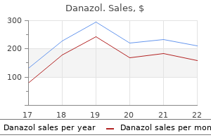
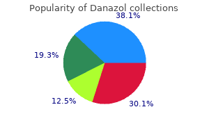
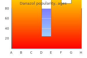
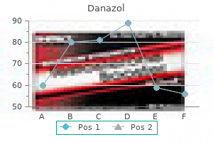
Danazol
Danazol dosages: 200 mg, 100 mg, 50 mg
Danazol packs: 30 pills, 60 pills, 90 pills, 120 pills, 180 pills

Buy 100 mg danazol fast delivery
The clinical evaluation revealed severe trismus breast cancer zumba pants discount 100 mg danazol with visa, which made it very tough to get a spoon in her mouth menstrual spotting causes danazol 200 mg buy discount on-line. Tongue Musculature the examiner asks the affected person to protrude the tongue and move it laterally. Rapid tongue actions could additionally be assessed by asking the affected person to repeat tongue-tip sounds such as "ta" rapidly. Ask the affected person to move the tongue tip to the roof of the mouth, an activity essential throughout bolus transfer. After reviewing the scientific examination of 3919 patients in danger for dysphagia, Leder et al. Protruding the tongue against a tongue blade gives the examiner a gross estimate of tongue strength. Objective measures of tongue energy may be achieved with a cooperative patient as he or she pressures towards a stress transducer. If the patient has had tongue resection due to most cancers, notice how much has been spared. Knowing probably the most delicate area could also be important in food placement throughout treatment. These pathologic reflexes are seen most commonly in patients with bilateral hemispheric or frontal lobe damage. The suck reflex may be elicited both by tapping the upper lip with a reflex hammer or by stroking the lips quickly with a tongue blade. The bite reflex is often elicited in patients with extreme neurologic lesions by touching the lips, teeth, or gums with a tongue blade and observing a strong closure of the jaw. This reflex could be particularly troublesome for the examiner as a end result of it could forestall a good oral examination. The examiner should keep away from robust resistance that might result in fracture or dislocation of the mandible. In some patients, spontaneous mouth opening will occur as a stimulus object, corresponding to a spoon or meals, is seen approaching the mouth. The dentist referred her to the speech pathologist for an analysis of her swallowing. The physical evaluation was regular except for some atrophy on the left lateral border of her tongue. This usually stems from the reality that an lively gag might cause momentary affected person discomfort and in some sufferers really stimulates emesis. The examination is achieved finest if it is done casually as part of the routine oral cavity inspection with a tongue blade. Quickly depress all sides of the tongue dorsum under the level of the palatal curtain. This should take not than 2 seconds for the check and the judgment of the velar response. Patients with xerostomia usually have little moisture throughout the oral cavity and report poor taste. Oropharynx Observations of the velum at relaxation and through duties of phonation ought to be made. The posterior dorsum of the tongue is stimulated on either side with a tongue depressor to assess the gag reflex. If the affected person has a gag response, it is important to notice if the velum is elevated symmetrically and if the patient coughed. The absence of a gag reflex as an isolated abnormal discovering in the examination of the cranial nerves for swallowing may not be essential (see Practice Note 7-3). If left untreated, thrush may trigger odynophagia, which is incessantly seen in these whose immune system has been decompensated by acute or continual illness. In some patients, the activity of the superior pharyngeal constrictor muscle may be noticed after an lively gag reflex as the posterior pharyngeal wall contracts or during the production of a falsetto voice. The exercise of the pharyngeal constrictor muscular tissues is best visualized by endoscopy throughout duties such as producing a falsetto voice. Speech is an especially complicated, overlearned conduct, and as such serves as a barometer from which the examiner can assess the standing of the neuromuscular system that additionally serves swallowing. Patients should be requested to sustain a vowel, with the examiner noting period, quality (hoarseness, breathiness, and harshness), pitch, and depth. The use of oral diadochokinetic duties (forced rapid alternating movements) utilizing consonant-vowel combos is really helpful. The remaining physical examination ought to verify the integrity of the peripheral sensory-motor swallowing mechanism. Test Swallows In a cooperative, alert patient, who as much as this point within the examination has not demonstrated significant neurologic impairment and has been in a position to swallow secretions without vital airway compromise, the examiner could need to grossly assess the swallow response with real meals gadgets. This a part of the examination is beneficial as a end result of it supplies the examiner details about swallowing dynamics. Before this portion of the examination, each cranial nerve should be evaluated in isolation. Test trials present the opportunity to see the coordinated integration of all of the swallowing muscles. Most examiners use an array of things ranging from skinny to thickened liquids, to pudding and softer objects, to items that require mastication. Volumes usually vary from 5 to 10 mL, beginning first with a smaller bolus and, if profitable, transferring towards larger boluses. If successful with 10-mL boluses, the examiner may wish to take a look at the swallow with a 20-mL bolus. Methods of supply, such as cup versus straw, may yield important variations in performance for the rationale that latter requires longer and more coordinated airway closure mechanics. One scientific method of making a judgment of whether or not the swallow response is delayed is the use of cervical auscultation. Preswallow sounds could be heard before the swallow because the bolus size increases,fifty three in all probability as a outcome of the tongue attempting to include a larger bolus. The first two sounds are low-frequency power, whereas the final sound (exhalatory burst) accommodates high-frequency power. Microphones and accelerometers are capable of detecting the total frequency spectrum of those swallowing sounds; however, not all stethoscopes have this functionality. There are two sides to a stethoscope-the flat, or diaphragm, aspect and the concave, or bell, side. Hamlet and colleagues found that the bell surface was best in detecting the sounds related to swallowing. Comparisons ought to be made between the predeglutitory and postdeglutitory patterns. Marked change within the respiratory rate or an increase in respiratory congestion could also be an indication of airway compromise. Within the brief apneic interval, two bursts of sound are markers of the presence of a swallow; these can be heard by cervical auscultation (see Practice Note 7-4).
Discount 100 mg danazol overnight delivery
Involvement of extranodal sites happens quite frequently and is seen in roughly 25% to 40% of sufferers pregnancy portraits buy cheap danazol 50 mg on line. The most frequent extranodal websites concerned are various structures of the head and neck women's health birth control pill generic 200 mg danazol amex, the higher respiratory tract airway, and skin. Skeletal involvement is comparatively unusual and has been documented in solely 25 of 423 instances reported to the Sinus Histiocytosis with Massive Lymphadenopathy registry. The majority of sufferers with skeletal illness additionally have other extranodal involvement, similar to involvement within the upper respiratory or gastrointestinal tract. The imply age of onset for sufferers with skeletal lesions is approximately 23 years. Fever and massive cervical lymphadenopathy are essentially the most frequent symptoms at presentation. Bone involvement within the absence of lymphadenopathy is uncommon, reported in only 2% of instances. Quite typically the disease totally manifests after a short interval of a nonspecific fever and pharyngitis. To date, no proof of a viral or different infectious etiology has been demonstrated. Rosai-Dorfman disease is taken into account a histologically benign, proliferative, histiocytic dysfunction with a variable, however occasionally fatal, outcome. The majority of patients have indolent regressive or clinically secure disease after several years of follow-up. In the Sinus Histiocytosis with Massive Lymphadenopathy registry data, 14 of 423 patients died of or with clinically lively disease. Among the extranodal websites, involvement of the kidneys, liver, and lungs appears to be notably ominous as a result of approximately 30% of sufferers with illness at these websites die of or with the illness. The deadly outcome in these instances is said to the frequent involvement of other extraskeletal sites such as the lungs and kidneys. Radiographic manifestations and clinical symptoms recommend an inflammatory disorder, corresponding to osteomyelitis. Microscopic Findings In typical circumstances, the sinuses of lymph nodes are filled with histiocytes. A, Lateral radiograph of elbow of teenage boy exhibits well-circumscribed, lytic, 2-cm intramedullary focus in lower humeral shaft. B and C, Mixed inflammatory cell infiltrates with distinguished elements of multinucleated histiocytes in addition to lymphocytes and plasma cells. Inset and D, Higher energy magnification exhibits a mixture of lymphocytes and large histiocytes with foamy cytoplasm. Aside from the distinct look of the histiocytic cells, the most striking and diagnostically necessary characteristic of these cells is outstanding emperipolesis or lymphophagocytosis. In addition to lymphocytes, a smaller number of phagocytized plasma cells, neutrophils, and red blood cells are also present. Prominent neutrophilic infiltrates with the formation of microabscesses may be found in some circumstances. The lymph node capsule is usually fibrotic and thickened, and the lymph node exhibits reactive hyperplasia of follicular centers. Extranodal illness has all these options besides that histiocytic cells, instead of rising in sinuses, kind irregular geographic areas separated by other inflammatory cells. The analysis of Rosai-Dorfman disease could be harder in extranodal websites, together with the skeleton. In basic, extranodal illness reveals less prominent emperipolesis, is accompanied by intensive fibrosis, and has fewer histiocytic cells. Therefore the likelihood that it could be misdiagnosed is way higher in extranodal sites than in nodal sites, especially in cervical disease. Some cases follow upper respiratory an infection, but no infectious etiology has been confirmed. No genetic abnormalities have been demonstrated in sporadic Rosai-Dorfman disease. The most common function of H syndrome is pores and skin hyperpigmentation with hypertrichosis, adopted by flexion contractures of the fingers and toes. One study of comparative genomic hybridization in sufferers with concurrent sinus histiocytosis with massive lymphadenopathy and Langerhans cell histiocytosis detected genomic positive aspects and losses in the Langerhans cells but detected no abnormalties within the histiocytes in areas with options of sinus histiocytosis with large lymphadenopathy. The first description of this lesion was in 1853 by King, who used the term chloroma to describe the green shade of the gross mass because of manufacturing of myeloperoxidase. Not all sufferers present with leukemic involvement of the blood and bone marrow; however, the majority go on to develop frank acute myeloid leukemia with a lag time ranging from 1 month to 4 years. In the absence of a historical past of a myeloid neoplasm, the radiographic findings might overlap with quite so much of reactive and neoplastic processes. A, Lateral radiograph of distal femur showing a harmful lytic lesion with moth-eaten sample. B, Sagittal magnetic resonance picture of the same case as proven in A with harmful lesion of the distal femur of intermediate sign depth. C, Anteroposterior radiograph of the proximal femur showing a harmful lytic lesion involving head, neck, and intertrochanteric region. D, Radioisotopic scan displaying diffuse involvement of the axial and proximal appendicular skeleton with a high signal intensity similar to the damaging lesion of the left proximal femur. The mobile composition ranges from a homogeneous inhabitants of blasts to a mixed population of blasts and more mature myeloid precursors, ranging from promyelocytes to neutrophils. Occasional instances could show a couple of line of myeloid differentiation, erythroid differentiation, or megakaryoblastic options. Immunohistochemical Stains and Differential Diagnosis If the analysis is suspected, recent tissue could also be submitted for flow cytometric immunophenotyping, cytogenetic studies, and molecular research. The diagnosis is aided by history in circumstances of known previous acute myeloid leukemia. The analysis of primary myeloid sarcoma, however, may be fairly tough, with misdiagnosis rates starting from 25% to one hundred pc. Flow cytometric immunophenotyping, including lymphoid, myeloid, and blast markers, is carried out in suspected instances. A generous panel of immunohistochemical stains is required to confirm the analysis and rule out different diagnostic prospects. Rare cases of myeloid sarcoma may exhibit erythroid differentiation, variably constructive for E-cadherin, glycophorin A, or hemoglobin A. Extramedullary hematopoiesis may be a diagnostic consideration; nonetheless, myeloid sarcoma will form a mass, whereas the cells of extramedullary hematopoiesis shall be present inside normal or barely expanded spaces of the preexisting structure of the involved organ. Genetic Findings and Pathogenesis the conventional cell counterpart is a myeloid blast, often with monocytic or granulocytic options, and infrequently with megakaryocytic or erythroid features. The reasons for blast homing to extramedullary tissues are unsure; nevertheless, interactions between adhesion molecules have been advised as a potential mechanism.
Syndromes
- Diarrhea
- Repeated infections
- Blood chemistry
- Difficulty swallowing
- Vertigo or dizziness
- Infections such as urinary tract infections or pneumonia (more likely in people who already have brain damage from stroke or dementia)
Generic 50 mg danazol fast delivery
In such cases menstrual belt buy danazol 200 mg online, a cautious seek for atypical mitoses can disclose the true nature of the lesion breast cancer 2b prognosis 50 mg danazol for sale. Telangiectatic osteosarcoma with a deceptively benign histologic appearance is typically referred to as low-grade telangiectatic osteosarcoma. Ultrastructurally, the tumor exhibits undifferentiated pleomorphic sarcomatous cells with minimal focal matrix mineralization. A, Slightly expanded, apparently well-circumscribed lytic lesion is proven in this plain radiograph of femoral diaphysis of younger grownup. B, Purely lytic lesion of proximal end of humerus exhibits cortical destruction of medial facet. C, Anteroposterior plain radiograph reveals ill-defined radiolucent tumor in distal femoral shaft. D, Lateral view of specimen radiograph of similar case in C documenting pathologic fracture (arrow). A and B, Anteroposterior and lateral plain radiographs show illdefined lytic and permeative lesion of distal femoral metaphysis with pathologic fracture. Note deceptively benign radiographic look of this high-grade telangiectatic osteosarcoma. D, T1-weighted coronal magnetic resonance image exhibits multiloculated structure of lesion with completely different ranges of sign depth. A, Anteroposterior plain radiograph reveals ill-defined lytic and permeative lesion of distal metaphysis. Inset, Fat-saturated T2-weighted axial magnetic resonance picture showing multiloculated cystic structure of the lesion with fluid levels. B, Gross photograph of the lesion in A reveals expansile hemorrhagic tumor of the distal femoral metaphysis extending to the gentle tissue laterally. C, Low energy microphotography showing multilocular cystic architecture of the lesion. Inset, Higher power of septum with histiocyte-like neoplastic cells displaying nuclear atypia. D, Microscopic options of telangiectatic osteosarcoma septations containing pleomorphic neoplastic cells and scattered multinucleated giant cells. A, Lateral plain radiograph showing expansile blowout lesion with permeative growth pattern of the proximal tibia. B, Gross photograph of A exhibiting hemorrhagic and partially necrotic tumor of the proximal tibial metaphysis with massive delicate tissue enlargement posteriorly. C, Low energy photomicrograph showing multilocular cystic structure of the lesion. D, Higher magnification of C showing a small cystic space lined by a mantle of extremely atypical neoplastic cells. B, Massive extension into gentle tissue from intramedullary osteosarcoma of humerus forms massive encircling mass with hemorrhagic loculi in fleshy background. D, Higher magnification of C exhibits nuclear atypia of tumor cells throughout the septum. A, Low energy photomicrograph shows multilocular cystic structure of the lesion. B, Higher magnification of A exhibiting a meandering septum containing neoplastic cells and scattered multinucleated large cells. Note irregular mantles of anaplastic tumor cells lining the septum and a multinucleated large cell within the middle. C, Collapsed cystic spaces with clustered septations, a standard function in telangiectatic osteosarcoma. D, Higher magnification of C exhibits nuclear atypia of neoplastic cells throughout the septa. A and B, Low power photomicrograph exhibiting blood stuffed spaces separated by irregular septa. D, Microscopic cystlike area surrounded by an irregular mantle of histiocyte-like neoplastic cells and scattered multinucleated large cells. A, A low energy photomicrograph showing cystlike spaces of varied sizes and a extra stable element of the tumor. C, Higher magnification of A showing an irregular cystlike house lined by a mantle of partially free-floating tumor cells. D, Higher magnification exhibiting the interface between the septum and cystic space lined by a mantle of partially freefloating, highly atypical tumor cells. A, Low power photomicrograph showing irregular blood-filled areas of various sizes within the cellular strong part of the tumor. A, Low energy photomicrograph displaying partially collapsed irregular blood-filled spaces separated by meandering septations. B, Higher magnification of A displaying meandering septations bordering blood-filled spaces. C, Higher magnification of B displaying atypia of histiocyte-like tumor cells inside the septa. A, Low energy photomicrograph showing cystlike areas and stable parts of the tumor. B, Intermediate power magnification of A displaying interface between solid and cystic elements of the tumor. C, Another intermediate power magnification of A exhibiting smaller cystic house inside the strong element of the tumor. D, Higher magnification of C displaying the stable area of the tumor composed of histiocyte-like malignant cells with scattered multinucleated large cells. Note overall bland look of cellular parts throughout the septum that may cause misdiagnosis and confusion with aneurysmal bone cyst. B, Low energy photomicrograph showing interface between cystic and strong parts of the tumor. C, Higher magnification of A displaying excessive cellularity and atypia of tumor cells inside the septa. A, Solid component of the tumor with intensive interstitial fresh hemorrhage (upper right) and smaller cystic spaces (lower left). B, Higher magnification of A displaying mineralized osteoid bands throughout the stable element of the tumor bordering the realm of recent hemorrhage. A, Low energy photomicrograph displaying irregular cystlike areas with contemporary hemorrhage and septations composed of extremely mobile tumor tissue. B, Higher magnification of A displaying cystlike spaces filled with hemorrhage and septations composed of anaplastic tumor cells. Inset, Higher magnification of septum composed of histiocyte-like atypical tumor cells. Differential Diagnosis this uncommon form of osteosarcoma should be distinguished principally from aneurysmal bone cyst and conventional osteosarcoma with focal telangiectatic features. Radiologic absence of sclerotic options and histologic paucity of tumor bone formation are required to distinguish telangiectatic osteosarcoma from otherwise conventional osteosarcoma that may exhibit focal and minor histologic features of excessive vascularity or blood-filled channels. The most necessary side of differential diagnosis is the mimicry of the benign aneurysmal bone cyst. Clinical Behavior Originally, it was thought that telangiectatic osteosarcoma had a particularly doleful prognosis, much worse than standard osteosarcoma.
Danazol 100 mg discount with amex
Moreover womens health institute taos danazol 50 mg buy cheap on line, the diploma of postchemotherapy necrosis appears to correlate with the speed of disease-free survival omega 7 menopause 50 mg danazol order with amex. In a examine from the Rizzoli Institute, the 5-year disease-free survival price was 90% for sufferers with complete necrosis, 53% for these with microscopic residual tumors, and 32% for those whose lesions had gross proof of residual tumor. Immunohistochemical options of overexpression of the genes concerned within the growth of drug resistance, similar to P glycoprotein, present some promising outcomes, however too few circumstances have been studied to assess the sensible utility of those findings. Sparse neurosecretory granules are related to both growing Golgi centers and cell processes. The outer perimeter of the cells forming the rosette can generally be delineated by an incomplete basal lamina�like materials. In a recent interinstitutional examine involving a number of facilities within the United States and Europe, the evaluation of 315 cases confirmed no affiliation between neural differentiation and extra aggressive habits. In the original report, the authors postulated that this lesion had a definite pathogenesis and apparently arose within the ribs, predominantly in the periosteum, however could additionally arise in the soft tissue, and possibly inside the lung. The lesions are frequently optimistic for one or a quantity of of the so-called neural markers, together with neuron-specific enolase and neurofilaments of 70 kD, and may also categorical neuroendocrine markers corresponding to chromogranin. The distinctive features seem to be frequent primitive neural or neuroendocrine differentiation, as properly as focal epithelial differentiation. A, Tumor cells with sparse cytoplasmic organelles as seen on low power magnification. B, Centrally placed cytoplasmic processes correspond to core of rosette (asterisk). A, Axonal differentiation of tumor cells with formation of interconnecting cytoplasmic processes (arrows). These two antibodies identify the antigen in formalin-fixed, paraffin-embedded tissue. It is also positive in pediatric lymphomas, lymphocytic lymphoma, and occasionally in rhabdomyosarcoma and even synovial sarcoma. Positivity for neuron-specific enolase is mostly disregarded as a specific marker of neural differentiation. On the opposite hand, if its expression can be correlated with different features of neural differentiation, it supplies a useful tool with which to assess the degree of neural phenotypic expression. A, Intermediate energy view of small round-cell tumor with sparse stromal components. C and D, Tumor cell exhibits strong positivity for periodic acid�Schiff stain, which is diastase sensitive (A-D, �400). Scattered positivity of individual tumor cells for keratins could be seen in roughly 10% of these tumors. In basic, the differential prognosis of small round-cell tumors of bone include not solely the entities described in this chapter, but additionally a big selection of mesenchymal and epithelial (primary and metastatic) tumors which will occur in youngsters, adolescents, and adults. It is recommended that rendering such a diagnosis in an uncommon clinical setting ought to be verified by molecular testing. These cytogenetic techniques can be carried out on each typical cytologic preparations and formalin-fixed paraffin-embedded histologic sections. Such exams are sometimes designed with a quantity of primers and are able to detecting several variants of fusion transcripts. Such exams are capable of detecting all translocations in sarcomas and will dominate future molecular testing of sarcomas. These technologies also provide the genome-wide identification of mutations in therapeutically targetable genes. The elevated stage of catecholamines and their metabolites in the urine is an important, diagnostically helpful characteristic. A and B, Plain radiograph and magnetic resonance image displaying permeative tumor diffusely involving the fibula with prominent cortical thickening. Insets, Desmosome (upper right) and tonofilaments (lower left) confirming epithelial differentiation in these uncommon tumors. The second consistent abnormality is the presence of homogeneously stained chromosomal regions and the presence of double-minute chromosomes. Microscopically, the presence of a distinguished hemangiopericytoma-like sample and foci of atypical cartilage that stain positive for S-100 protein are distinguishing features. Examination of paraffin-embedded material sometimes reveals rhabdomyoblastic options in most embryonal rhabdomyosarcomas. On the other hand, alveolar rhabdomyosarcoma is a real round-cell tumor and will present the diagnostic downside. Positivity of the tumor cells for muscle markers and the presence of nesting and alveolar patterns are distinguishing options. Metastatic carcinoma and lymphoma are entities that incessantly involve the skeleton in sufferers older than age 40 years and can be diagnosed with the assist of appropriate epithelial and lymphoma marker research. Occasionally, sclerosing epithelioid fibrosarcoma, malignant melanoma, and even villonodular tenosynovitis can mimic a small round-cell tumor. Delattre O, Zucman J, Melot T, et al: the Ewing family of tumors-a subgroup of small-round-cell tumors defined by particular chimeric transcripts. Frohling S, Dohner H: Molecular origins of cancer: chromosomal abnormalities in most cancers. Zucman J, Melot T, Desmaze C, et al: Combinational technology of variable fusion proteins within the Ewing family of tumours. Basharkhah A, Pansy J, Urban C, et al: Outcomes after interdisciplinary administration of 7 sufferers with Askin tumor. Delattre O, Zucman J, Melot T, et al: the Ewing family of tumors: a subgroup of small spherical cell tumors outlined by particular chimeric transcripts. Dragoescu E, Jackson-Cook C, Domson G, et al: Small cell osteosarcoma with Ewing sarcoma breakpoint area 1 gene rearrangement detected by interphase fluorescence in situ hybridization. Kikuchi Y, Kishimoto T, Ota S, et al: Adamantinoma-like Ewing family tumor of sentimental tissue associated with the vagus nerve: a case report and review of the literature. Sirivella S, Gielchinsky I: Treatment outcomes in 23 thoracic primitive neuroectodermal tumours: a retrospective study. Contesso G, Llombart-Bosch A, Terrier P, et al: Does malignant small spherical cell tumor of the thoracopulmonary region (Askin tumor) constitute a clinicopathologic entity An analysis of 30 cases with immunohistochemical and electron-microscopic help treated on the Institute Gustave Roussy. Hashimoto H, Enjoji M, Nakajima T, et al: Malignant neuroepithelioma (peripheral neuroblastoma): a clinicopathologic research of 15 circumstances. Frostad B, Tani E, Brosjo O, et al: Fine needle aspiration cytology in the diagnosis and administration of children and adolescents with Ewing sarcoma and peripheral primitive neuroectodermal tumor. Llombart-Bosch A, Contesso G, Peydro-Olaya A: Histology, immunohistochemistry, and electron microscopy of small round cell tumors of bone. Papierz W, Alwasiak J, Kolasa P, et al: Primitive neuroectodermal tumors: ultrastructural and immunohistochemical studies. Laskar S, Nair C, Mallik S, et al: Prognostic components and consequence in Askin-Rosai tumor: a evaluation of 104 patients.
Danazol 50 mg buy discount line
They usually reveal intramedullary lesions that contain massive segments of the intramedullary cavity and lengthen beyond the realm shown to be concerned on plain radiographs womens health haven fayetteville nc buy danazol 50 mg overnight delivery. This type of radiographic look is typically seen in predominantly subperiosteal lesions women's health lynchburg va order 100 mg danazol overnight delivery. The sclerotic features or focal opacities are extra often seen in lesions involving the metaphyseal portions of lengthy bones or in flat bones and are virtually never seen in diaphyseal lesions. On the other hand, diaphyseal lesions of long bones are often related to diffuse sclerosis and thickening of the overlying cortex. The tumor can contain massive segments of the medullary cavity without causing cortical disruption. Formation of a giant extraosseous mass, which often exceeds the scale of the intraosseous element, is incessantly seen. The cells develop in solid, densely packed sheets and nests filling intertrabecular spaces. The nuclear chromatin is finely granular, and there are often one to three clearly identifiable small- to intermediate-sized nucleoli. The cytoplasm is indistinct and types an ill-defined slim rim across the nucleus. Often a biphasic sample is simulated by the presence of so-called darkish cells and light-weight cells. These types of cells are generally referred to as principal (light) and secondary (dark) cells, respectively. The ratio between these two forms of cells varies from tumor to tumor and in different areas of the identical lesion. In some tumors, cords or clusters of dark apoptotic cells kind an interconnecting community of irregular patches that create a pseudoorganoid sample. Complete permeation of the cortex and its eventual disruption are related to the formation of a mass that originally forms subperiosteally and then extends into soft tissue (parosteal). Penetration of the cortex and disturbance of the periosteum are related to new bone formation. The proportion of stromal reticular parts and blood vessels is elevated close to the advancing fringe of the tumor compared with its more central intramedullary portion. Multilayered periosteal new bone formation, corresponding to an identifiable onion-skin appearance on radiographs, can accompany the advancing tumor cells. Periosteal bone formation is associated with plump reactive osteoblasts and multinucleated osteoclast-like large cells and should have foci of cartilage metaplasia. Peripheral areas of the tumor inside soft tissue may show extra dispersed tumor cells invading collagenized stroma, adipose tissue, and skeletal muscle. Occasionally, a distinct development pattern in the form of larger lobules or nests composed of compact tumor cells and separated by fibrous septa can be seen. Vascular formation within the central portion is inconspicuous and is represented by slitlike capillaries with fantastic endothelial cells amongst tumor cells. Larger, thick-walled vessels may be seen inside stromal bands separating tumor cells. The morphologic features of cells, and particularly the main points of the nuclei, are often Text continued on p. A Computed tomogram shows intramedullary tumor with cortical disruption posteriorly and extension into gentle tissue. B and C, Anteroposterior and lateral plain radiographs present permeative lesion in metaphyseal area; so-called cortical saucerization (concave defect) may be seen on posterior surface in C. A, Large damaging tumor of body and glenoid regions of scapula with related soft tissue mass. This atypical plain radiographic look was interpreted initially as possible vascular tumor. C, Expansile tumor with moth-eaten sample of bone destruction in distal end of fibula in young grownup. B, Radiograph of amputation specimen exhibits destruction of proximal plate of great toe and ill-defined soft tissue mass. C, Permeative destructive lesion involving complete shaft of proximal phalanx of finger is shown on this indirect radiograph. A, Lateral radiograph of ankle and foot of younger grownup reveals rarefaction of posterior part of os calcis. B, Anteroposterior radiograph reveals permeative destruction of cancellous bone with ill-defined border. C, Technetium ninety nine bone scan exhibits excessive uptake of isotope in os calcis, which is extra intense posteriorly. Although distal tibia showed elevated isotope uptake, tumor was not current on this site. A and B, Plain radiographs show harmful lytic mass of proximal tibia with enhance of new periosteal bone formation. A and B, Coronal and sagittal computed tomograms exhibiting a large tumor involving the upper portions of the left thoracic wall and pulmonary cavity. C and D, Axial and sagittal magnetic resonance images showing a large low sign intensity tumor involving the left paraspinal area and thoracic wall. Proximal circumferential soft tissue extension with elevation of periosteum is obvious. Note large subperiosteal lesion with concave cortical surface; defect is referred to as saucerization. Intramedullary tan-gray tumor with posterior subperiosteal and gentle tissue extension related to concave cortical defect. C, Closer view of specimen proven in B exhibits intramedullary tumor with cortical permeation and periosteal elevation. Stroma is minimal and confined to few delicate fibrous tissue strands and blood vessels. D, Higher magnification of C shows uniform tumor cells with minimal quantity of cytoplasm. Inset, High energy photomicrograph of Homer Wright rosette consisting of a central fibrillar core bounded by concentrically arranged tumor cells. Note the presence of apoptotic dark cells at the interphase of viable and necrotic tumor. D, Higher magnification of the interphase between viable and necrotic tumor tissue displaying prominent dark apoptotic cells. In a small number of tumors, the microscopic look of tumor cells may deviate from the so-called classic sample. These options are more usually seen in recurrent and handled lesions however can additionally be present in major tumors. A delicate, finely granular chromatin pattern and clearly identifiable small to medium nucleoli are characteristic. Immunohistochemical and molecular study permitting differential prognosis with other small cell malignances may be performed on materials obtained for cytologic preparations. The cellularity is high, and the tumor cells are densely packed with almost nonexistent stroma. Two types of cells as outlined by light microscopy-a principal sort (light with open intact chromatin) and a secondary type (dark with condensed chromatin)-can also be recognized at the ultrastructural level.
Discount danazol 100 mg on-line
The traverse of a cell throughout the cell cycle could be envisioned as a sequence of checkpoints that act in live performance women's health new zealand magazine danazol 50 mg buy generic online. On the individual cell stage pregnancy 15 weeks buy danazol 100 mg low cost, the phenomenon of most cancers results from a profound deregulation of this method, and nearly certainly, every cell population has some distinctive alterations and biologic features. In addition, Cdk4/6-CycD performs a role in titrating p27, stopping the inhibition of Cdk2-CycE complexes. The equipment liable for this course of, the so-called mitotic spindle, is composed of tons of of proteins with the general structure of microtubules connected to the specific a part of the chromosome referred to because the centromere and to the pole of the mitotic spindle referred to because the centrosome. They are arranged longitudinally and laterally and propagate the segregation of chromosomes because of their viscoelastic nature and tiled-array architecture. The total orientation of the mitotic spindle is regulated by a extremely conserved set of molecules that govern cell polarity. In each mesenchymal and epithelial cells, the apical to basal polarity regulates the position of the mitotic spindle and secures equal segregation of chromosomes into daughter cells. Overall, more than a thousand proteins present a mitotic-specific phosphorylation and heaps of of them belong to the superfamily of kinases. A, Mitotic cell cycle and its variants usually associated with different cell phases. P, M, A, and T designate prophase, metaphase, anaphase, and telophase, respectively. The model was developed for Schizosaccharomyces pombe however is anticipated to be relevant to all eukaryotic cells. A, the spindle can effectively be thought of to be many springs and dashpots in parallel organized each longitudinally and laterally. Both elastic and viscous parts originate from the viscoelastic nature of molecular motors (upper left), microtubule elasticity and turnover, and the tiled-array structure of the spindle. Multiple motors cooperate and slide antiparallel microtubules apart, however oppose one another and lock and parallel microtubules together (upper right). A massive exterior force can lead to rapid detachment of half of the motors, breaking microtubule-microtubule crosslinking and leading to a plastic spindle response. B, Robust spindle architecture is dependent upon correctly homogeneous "filing" of the spindle inside by microtubules. Nor can this be achieved by the autocatalytic nucleation (bottom) of short microtubules, which ends up in dense, disjointed clumps of microtubules. Lack of perform of Aurora kinases in experimental methods contributes to numerous mitotic dysfunctions and may be lethal. The overactive Aurora kinase A produces supernumerary centrosomes, resulting in multipolar mitosis and chromosomal misseggregation with aneuploidy. In abstract, advanced regulatory mechanisms that function on the promoter degree of cell cycle-mediating genes influence the proliferation actions of cells. B, In distinction, promoter of osteocalcin gene has several overlapping motives that reply to multiple transcription components in differentiated nonproliferative state. In contrast to the histone gene, the group of the osteocalcin gene promoter contains a number of components that upregulate the gene in a differentiated postproliferative state and improve its expression 3 Molecular Biology of Bone Tumors 131 in parallel to matrix mineralization and differentiation of osteoblastic cells. It has been instructed that the distinctive group of the regulatory motives of the osteocalcin gene maintains responses to a number of components. Osteosarcoma the vast majority of osteosarcomas are sporadic de novo tumors that develop with none identified predisposing circumstances. Small fractions of osteosarcomas develop, nevertheless, within the background of well defined, usually familial, predisposing syndromes. These syndromes provide necessary clues for molecular mechanisms concerned in the improvement of those aggressive mysterious malignancies. The unifying options of those syndromes represent genomic instability, untimely getting older, and elevated threat for multiple cancers together with osteosarcoma. In tumors with distinct molecular alterations, the changes are sometimes not single alterations, and additional secondary hits are required for the development of the entire malignant phenotype. They also occur less frequently than those who appear to develop by a complex multistep process. The molecular mechanisms that play a task within the pathogenesis of human malignant tumors are of several major varieties: 1. A gene with a adverse growth-regulatory impact (tumor suppressor gene) is altered or misplaced. A gene with positive regulation of growth is activated by upregulation or alteration (mutation) that enhances its remodeling exercise. The fusion of two genes by translocation results in a new hybrid gene that exhibits a robust activity as a reworking issue. This simplified view of the role of individual genetic alterations must be placed within the context of the complex landscape of genomic alterations that affect multiple interacting pathways contributing to the phenomenon referred to as malignant transformation. This suggests that cells with a dedication to carry out skeleton-forming features are affected within the growth of osteosarcoma. This remark also strongly suggests the existence of such a mechanism that appears to be extremely protected and stays unaltered in osteosarcoma, but its molecular mediators stay to be elucidated. Most conventional osteosarcomas produce simply recognizable quantities of osteoid however lack its group and the lamellar maturation seen in regular bone. This is in preserving with the phenotypic heterogeneity of various osteosarcoma cells and their capabilities, corresponding to their capability to produce particular person elements of extracellular matrix. Chondrosarcoma In basic, chondrosarcomas develop from cells commited to cartilaginous differentiation. They symbolize a heterogeneous group of neoplasms with numerous biologic behaviors. They range from low-grade, domestically aggressive malignancies (grade 1) to highly aggressive tumors with a excessive propensity for metastases (grade 3). Most skeletal myxoid chondrosarcomas are grade 2 lesions and are pathogenetically distinct from gentle tissue myxoid chondrosarcomas. A small subset of bone myxoid chondrosarcomas are high-grade lesions and may be pathogenetically just like extraskeletal myxoid chondrosarcomas. Central and especially secondary peripheral chondrosarcomas evolve through distinct molecular pathways. They catalyze the oxidative decarboxylation of isocitrate into -ketoglutarate of the Krebs tricarboxylic acid cycle. The development mechanisms of chondrosarcoma are largely unknown, but high-grade lesions are characterised by aneuploidy and multiple chromosomal abnormalities. On the opposite hand, high-grade lesions most likely develop via a multistep mechanism that involves a quantity of transforming and tumor suppressor genes. In common, aneuploidy and overexpression of Aurora kinases are antagonistic prognostic components. In chondrosarcoma, a quantity of genetic alterations (altered genes) correlate with the scientific aggressiveness of the lesion, and most high-grade tumors are aneuploid. Karyotyping studies indicate that alterations involving multiple chromosones are frequent in chondrosarcoma of bone. Dedifferentiation may also happen in low-grade intramedullary osteosarcoma, parosteal osteosarcoma, large cell tumor, and chordoma. The development of a similar, highly lethal sarcoma in several distinct precursor situations could in reality characterize a pathogenetically related course of that has frequent molecular pathways of growth.
Hairy Mint (Wild Mint). Danazol.
- What is Wild Mint?
- Are there safety concerns?
- How does Wild Mint work?
- Dosing considerations for Wild Mint.
- Diarrhea, painful menstruation (periods), and other conditions.
Source: http://www.rxlist.com/script/main/art.asp?articlekey=96225
Danazol 100 mg with mastercard
Akai M womens health zone abortion danazol 50 mg cheap with mastercard, Tateishi A breast cancer xmas ornaments purchase 50 mg danazol amex, Machinami R, et al: Chondroblastoma of the sacrum: a case report. Azorin D, Gonzalez-Mediero I, Colmenero I, et al: Diaphyseal chondroblastoma in an extended bone: first report. Edel G, Ueda Y, Nakanishi J, et al: Chondroblastoma of bone: a clinical, radiological, gentle and immunohistochemical study. Fadda M, Manunta A, Rinonapoli G, et al: Ultrastructural appearance of chondroblastoma. Mii Y, Miyauchi Y, Morishita T, et al: Ultrastructural cytochemical demonstration of proteoglycans and calcium in the extracellular matrix of chondroblastomas. Ozkoc G, Gonlusen G, Ozalay M, et al: Giant chondroblastoma of the scapula with pulmonary metastases. Romeo S, Szyhai K, Nishimori I, et al: A balanced t(5;17) (p15;q22-23) in chondroblastoma: frequency of the rearrangement and evaluation of the candidate genes. Sailhan F, Chotel F, Parot R, et al: Chondroblastoma of bone in a pediatric population. Schajowicz F, Gallardo H: Epiphyseal chondroblastoma of bone: a clinicopathological research of 69 circumstances. Sjogren H, Orndal C, Tingby O, et al: Cytogenetic and spectral karyotype analyses of benign and malignant cartilage tumours. Sotelo-Avila C, Sundaram M, Kyriakos M, et al: Case report 373: diametaphyseal chondroblastoma of the higher portion of the left femur. Ishida T, Goto T, Motoi N, et al: Intracortical chondroblastoma mimicking intra-articular osteoid osteoma. Kaneko H, Kitoh H, Wasa J, et al: Chondroblastoma of the femoral neck as a cause of hip synovitis. Karabela-Bouropoulou V, Markaki S, Prevedorou D, et al: A combined immunohistochemical and histochemical method on the differential diagnosis of giant cell epiphyseal neoplasms. Kirchhoff C, Buhmann S, Mussack T, et al: Aggressive scapular chondroblastoma with secondary metastasis-a case report and evaluation of literature. Konishi E, Nakashima Y, Iwasa Y, et al: Immunohistochemical evaluation for Sox9 reveals the cartilaginous character of chondroblastoma and chondromyxoid fibroma of the bone. Kunze E, Graewe T, Peitsch E: Histology and biology of metastatic chondroblastoma: report of a case with a evaluation of the literature. Lehner B, Witte D, Weiss S: Clinical and radiological long-term results after operative remedy of chondroblastoma. Mark J, Wedell B, Dahlenfors R, et al: Human benign chondroblastoma with a pseudodiploid stemline characterized by complicated and balanced translocation. Tamura M, Oda M, Matsumoto I, et al: Chondroblastoma with pulmonary metastasis in a affected person presenting with spontaneous bilateral pneumothorax: report of a case. In Proceedings of the Seventh National Cancer Conference of the American Cancer Society, Philadelphia, 1973, Lippincott. Fujiwara S, Nakamura I, Goto T, et al: Intracortical chondromyxoid fibroma of humerus. Gherlinzoni F, Rock M, Picci P: Chondromyxoid fibroma: the experience at the Istituto Ortopedico Rizzoli. Heydemann J, Gillespie R, Mancer K: Soft tissue recurrence of chondromyxoid fibroma. Jhala D, Coventry S, Rao P, et al: Juvenile juxtacortical chondromyxoid fibroma of bone: a case report. Konishi E, Nakashima Y, Iwasa Y, et al: Immunohistochemical analysis of Sox9 reveals the cartilaginous character of chondroblastoma and chondromyxoid fibroma of the bone. Schajowicz F, Gallardo H: Chondromyxoid fibroma (fibromyxoid chondroma) of bone: a clinico-pathological examine of 32 circumstances. Soder S, Inwards C, Muller S, et al: Cell biology and matrix biochemistry of chondromyxoid fibroma. Ushigome S, Takakuwa T, Shinagawa T, et al: Chondromyxoid fibroma of bone: an electron microscopic statement. Ushigome S, Takakuwa T, Shinagawa T, et al: Ultrastructure of cartilaginous tumors and S-100 protein in the tumors: as regards to the histogenesis of chondroblastoma, chondromyxoid fibroma and mesenchymal chondrosarcoma. Jager M, Westhoff B, Portier S, et al: Clinical consequence and genotype in patients with hereditary a number of exostoses. Kitsoulis P, Galani V, Stefanaki K, et al: Osteochondromas: evaluate of the medical, radiological and pathological features. Matsumoto K, Irie F, Mackem S, et al: A mouse mannequin of chondrocyte-specific somatic mutation reveals a job for Ext1 loss of heterozygosity in multiple hereditary exostoses. Nadanaka S, Ishida M, Ikegami M, et al: Chondroitin 4-Osulfotransferase-1 modulates Wnt-3a signaling through management of E disaccharide expression of chondroitin sulfate. Nojima T, Yamashiro K, Fujita M, et al: A case of osteosarcoma arising in a solitary osteochondroma. Yalniz E, Alicioglu B, Yalcin O, et al: Non-specific magnetic resonance that includes of chondromyxoid fibroma of the iliac bone. Zustin J, Akpalo H, Gambarotti M, et al: Phenotypic variety in chondromyxoid fibroma reveals differentiation sample of tumor mimicking fetal cartilage canals improvement: an immunohistochemical study. Gorospe L, Madrid-Muniz C, Royo A, et al: Radiation-induced osteochondroma of the T4 vertebra causing spinal cord compression. Gunay C, Atalar H, Yildiz Y, et al: Spinal osteochondroma: a report on six patients and a evaluation of the literature. Hacker U, Nybakken K, Perrimon N: Heparan sulphate proteoglycans: the sweet side of development. Picci P, Stilli S: Malignant fibrous histiocytoma arising from an exostosis in a affected person affected by multiple exostoses. Trebicz Geffen M, Robinson D, Evron Z, et al: the molecular and mobile basis of exostoses formation in hereditary multiple exostoses. Valdivielso-Ortiz A, Barber I, Soldado F, et al: Solitary osteochondroma: spontaneous regression. Yildirim C, Rodop O, Kuskucu M, et al: Giant solitary osteochondroma arising from the fifth metatarsal bone: a case report. The time period chondrosarcoma is used to describe a heterogeneous group of lesions with various morphologic options and clinical habits. The conduct of these lesions ranges from slowly growing, nonmetastasizing tumors to very aggressive metastasizing sarcomas. On the idea of histologic features (nuclear atypia and cellularity), typical chondrosarcoma is further subdivided into three grades, 1 by way of 3, which correlate well with their scientific behavior. The majority of conventional chondrosarcomas are low- to intermediate-grade tumors which have indolent medical behavior and low metastatic potential. The elementary biologic distinction between a grade 1 chondrosarcoma and a benign enchondroma is signified by the limited progress potential of the enchondroma and the slow but steady domestically invasive development of a lowgrade chondrosarcoma.
Danazol 200 mg purchase free shipping
They concluded that their outcomes support the position that thermal stimulation to the anterior faucial pillars triggers the swallow response breast cancer in lymph nodes 200 mg danazol sale. These studies recommend that thermal-tactile stimulation within the posterior oral cavity can produce a shortterm impact on swallow timing women's health clinic port adelaide generic danazol 200 mg without prescription. Subjects acquired thermal application therapy day by day for 1-week durations alternating with no therapy intervals of a week. A whole of 2 weeks of thermal application remedy was supplied to each affected person within this alternating treatment design. Finally, Rosenbek and a big group of investigators138 evaluated the depth of therapy on the results of thermal software (which they now termed tactilethermal application). Patients acquired 2 weeks of tactilethermal application remedy and had been randomly assigned to one hundred fifty, 300, 450, or 600 trials per week. A trial included three or more strokes of both faucial pillars with an ice stick (similar to a popsicle). This investigative group reported that no frequency of treatment was superior to any other. Studies of thermal stimulation in wholesome adult volunteers have produced even more variable results than these in adult sufferers with dysphagia. They reported that only the cold stimulus resulted in a discount in swallow latency. Furthermore, this group reported that any stimulation effect lasted for under a single swallow. Thus this system may be relevant for patients demonstrating delays in swallowing exercise however not for patients who demonstrate airway compromise. In reality, numerous forms of train have been used for years within the rehabilitation of speech and swallowing capabilities. As noted later on this chapter, this software is comparatively new and not absolutely developed. Many suggestions exist but only some feasible purposes have been developed and evaluated. The use of adjunctive modalities is also growing in the area of dysphagia rehabilitation. The time period adjunctive specifies that the modality is used to assist the benefits from the first therapy. Exercise Principles and Dysphagia Therapy Reviews of exercise principles that will apply to dysphagia rehabilitation have focused primarily on power coaching. For example, lifting heavier weights adds elevated resistance to a weight-lifting train. More repetitions of an exercise accomplished in a exhausting and fast period result in a extra intense exercise. Increasing the intensity of exercise facilitates muscle change and ends in larger power. Conversely, when exercises are ceased or decreased in depth, the principle of reversibility or a detraining impact may be seen. The idea of specificity refers to the observation that certain workout routines usually tend to profit specific duties. Different muscle teams are concerned with completely different requirements to the neuromuscular system from the goal activity (in this case, long-distance running). A frequent phrase utilized by dysphagia clinicians is paraphrased as follows-"the greatest way to rehabilitate swallowing is to have the patient swallow. However, a associated idea should be mentioned-cross-system impact or transference. The crosssystem impact occurs when train supposed for one operate produces advantages in a special operate. Continually and progressively improve the demands of the exercise exercise applied-perform extra repetitions, improve the load, go sooner. Although this cross-system effect has not been well studied in relation to dysphagia rehabilitation, revealed examples do support the potential for workouts involving the swallowing mechanism to affect other functions of the higher aerodigestive tract. Positive useful benefits have been reported from exercise-based dysphagia therapy. The focus of the remedy was to increase tongue power using a tonguepress activity in which the tongue was pressed against the hard palate. Results from two studies showed elevated tongue strength throughout volitional tongue-push activity and swallowing, improvement in airway protection throughout swallowing, and elevated lingual muscle size. These results are encouraging as a result of they show that a simple, swallow-related exercise can enhance energy in key muscle groups utilized in swallowing with some practical benefit to patients. Patients obtain daily remedy periods which might be structured to evoke mass apply of swallowing. Swallowed supplies are introduced sequentially to facilitate progressive resistance or speed and coordination of swallowing. Clinicians comply with specific rules to advance sufferers during treatment based mostly on affected person efficiency. Surface Electromyographic and Other Forms of Biofeedback Many of the swallowing maneuvers discussed on this chapter require motor studying by the affected person. Furthermore, many of those adjustments symbolize novel motor patterns which may be troublesome to learn. Application of biofeedback as an adjunct to therapy could additionally be useful in enhancing the rate of motor learning, thereby leading to reduced time in remedy. Biofeedback has been useful in educating new actions, unfamiliar movements, or movements which are in any other case difficult to monitor. In addition, biofeedback may be helpful in serving to a affected person monitor swallow efficiency. Asking a affected person to "swallow harder" might not result in a significant increase in swallow effort. Not only does this feedback help the patient to monitor effort, it is also instant to the affected person and the treating clinician and thus facilitates support or reinstruction from the clinician to the affected person. This form of biofeedback has been used to train leisure, strengthening, and coordination activities. Studies specific to dysphagia therapy have instructed that this type of biofeedback can cut back the quantity of remedy time whereas producing favorable outcomes even in sufferers with persistent dysphagia. Having the affected person watch swallow makes an attempt on the monitor throughout a fluoroscopic examination has been suggested as a way to train sure maneuvers. In an analogous manner, endoscopic biofeedback has been instructed as a mechanism to train acceptable breath-hold maneuvers such because the supraglottic and super-supraglottic swallows. Less invasive than either fluoroscopy or endoscopy is the use of cervical auscultation as a biofeedback approach (see Chapter 9).
Generic danazol 100 mg without prescription
Proximal two thirds of proper humerus in a young grownup has utterly disappeared menstrual fever discount 100 mg danazol free shipping, and distal portion of the shaft reveals typical "sucked candy" attenuation women's health clinic des moines iowa danazol 200 mg cheap line. The glomus tumor was described in 1924 by Masson,a hundred and five who linked this peculiar neoplasm to a traditional glomus body and proposed that in some cases it may characterize a hyperplasia of perivascular cells somewhat than a true neoplasm. Definition Glomus tumor is a rare, benign distinctive neoplasm composed of vascular channels and modified perivascular muscle cells that recapitulate the construction of a glomus body. Incidence and Location Glomus tumors are extremely rare lesions, and most are diagnosed in patients between ages 20 and forty years. A, Plain radiograph of chest of younger man with disappearance of posterior parts of two upper ribs on left. B, Enlargement of radiograph shown in A with disappearance of posterior parts of two left ribs. Note multiple cystic spaces and alternative of bone by fibrous vascularized tissue. A, Amputation specimen displaying giant vascular constructions representing tortuous thick-walled vasculature with cavernous dilations extensively involving the soft tissue of the decrease extremity. B, T2-weighted magnetic resonance image exhibiting grapelike excessive sign intensities similar to gentle tissue vascular lesions. C, Higher power gross photograph of the foot exhibiting in depth cystlike vascular lesions involving the gentle tissue and eroding bones. B, Higher magnification of A shows cluster of perpendicularly sectioned glomic arterioles of Sucquet-Hoyer canals (arrows) surrounded by thick layer of glomus cells. Inset shows glomic arteriole surrounded by glomus cells and peripheral amassing vessel (denoted by asterisk). A and B, Radiographs of distal phalanges show strain erosion of bone by subungual glomus tumors. Exposed concavity of tumor bed in distal phalanx may be seen simply proximal to base of nail. D, Photomicrograph of glomus tumor of subungual region reveals endothelial-lined vessels and perivascular glomus cells in small nests and sheets (D, �50) (D, hematoxylin-eosin. Microscopic Findings Extraosseous glomus tumors are divided into three microscopically distinct subtypes: glomus tumor proper, glomangioma, and glomangiomyoma. The classification relies on differing proportions of vascular, clean muscle, and epithelioid glomus cells. All the lesions reported as primary bone tumors have been of the glomus tumor correct category. The rich, branching vascular community can create a hemangiopericytoma-like sample. A and B, Common sample within glomus tumor consists of stable sheets of cells with interspaced vessels. C, Low energy photomicrograph reveals massive cystic areas within strong sheets of tumor cells. D, Higher magnification of C exhibits tumor cells with eosinophilic cytoplasm and spherical or oval nuclei. Treatment and Behavior Glomus tumors are benign, and easy excision of each the gentle tissue and intraosseous forms is healing. Malignant glomus tumor is extremely rare in gentle tissue and has not been described in bone. Definition Epithelioid hemangioendothelioma is a novel, welldifferentiated endothelial vascular neoplasm with an epithelioid appearance of its endothelial cells and a bent to be multifocal. Incidence and Location Epithelioid hemangioendotheliomas of bone are extremely uncommon. Most sufferers initially have multifocal lesions with a bent to contain bones of the identical area. Synchronous involvement of paired bones, most incessantly the tibia and fibula, is common, but in some cases, utterly separate synchronous foci are current in distant anatomic sites such because the clavicle and lumbar vertebrae. Occasionally a more aggressive development sample, corresponding to a moth-eaten sample, may be seen. In uncommon circumstances, the epithelioid hemangioendothelioma can provoke sclerosis and will current as a blastic lesion on radiographs. Erosions of cortex with full cortical disruption and extension into soft tissue could be current. Multifocal lesions involving one bone or several bones of an affected extremity are incessantly seen. Later, in 1982, Weiss and Enzinger131 proposed the term epithelioid hemangioendothelioma to describe an unusual vascular tumor of soppy tissue by which endothelial cells had an epithelioid appearance. They are classified as low-grade malignancies and have an indolent medical habits. Preliminary knowledge point out that this fusion is particular for epithelioid hemangioendothelioma no matter its site of origin, together with those that develop in bone and can be utilized within the differential analysis of vascular situations with epithelioid features. A and B, Lytic sharply demarcated lesion of distal humerus with enlargement of bone contour medially and posteriorly. E, Computed tomogram of lesion in D exhibits low sign intramedullary lesion with multiple erosions of cortex. B and C, Computed tomograms of lesions shown in A doc a number of sharply demarcated lytic lesions of ilium. A-C, Extensive involvement of metatarsal bones with punched-out lytic lesions involving cortical bone and medullary cavities. D, Specimen radiograph of resected distal metatarsal phase containing lytic focus of epithelioid hemangioendothelioma. A, Anteroposterior radiograph of foot of an 18-year-old man with a number of lytic lesions of metatarsal and tarsal bones. B, Opposite foot of patient in A shows pathologic fracture of fourth metatarsal via focus of epithelioid hemangioendothelioma. Computed tomograms of right and left tarsal bones show multicentric, sharply demarcated lesions on right side. A and B, Anteroposterior and lateral radiographs of foot of younger man present a number of lytic foci with out reactive sclerosis or periosteal new bone formation involving metatarsals, phalanges, tarsal bones, and distal tibial shaft. A and B, Lateral and anteroposterior radiographs of leg of a 29-year-old lady show multiple small, sharply circumscribed lytic lesions of proper tibia and fibula. C, Radionuclide scan of patient in A and B shows increased isotope uptake in multiple tarsal bones and distal tibia. D, Lateral radiograph of foot and ankle shows a quantity of lytic lesion in calcaneus and cuneiform bone. A, Lateral radiograph of leg show a number of sharply demarcated lytic lesions of tibia and tarsal bones.
Discount 50 mg danazol overnight delivery
D menopause chills order danazol 50 mg visa, Higher magnification of C showing early seams of unmineralized osteoid amongst tumor cells menstrual calendar android order danazol 200 mg on-line. B, Interconnected coarse deposits of osteoid encircling and separating clusters of tumor cells. D, Well developed interconnected irregular trabecular pattern produced by tumor osteoid. Note entrapment of particular person tumor cells residing within lacunar areas of osteoid. C, Conspicuous, well mineralized, osteoid deposition sample separating and encircling larger clusters of loosely organized tumor cells. D, Higher magnification of C exhibiting properly mineralized osteoid and loosely organized anaplastic tumor cells. B, Higher magnification of A showing a less developed area of osteoid deposition with sparse seams of matrix deposition. B, Higher power of A showing irregular stable areas of osteoid with entrapped tumor cells. C, Irregular stable areas of osteoid separating giant clusters of epithelioid tumor cells. Note dense eosinophilic cytoplasm answerable for epithelioid appearance of tumor cells. D, Higher energy of C displaying irregular areas of osteoid with entrapped tumor cells. A and B, Infiltrative development sample of high-grade osteosarcoma permeating intratrabecular marrow areas. C, Higher energy of B displaying permeation of intratrabecular marrow areas by an osteosarcoma. D, Variegated osteoid deposition and mineralization patterns in osteosarcoma permeating marrow spaces. A, Hemangiopericytoma-like sample of tumor growth bordering endothelial-lined spaces. B, Higher magnification of A displaying outstanding vascular spaces and sheetlike osteoid deposition. C, Hemangiopericytoma-like sample with distinguished vasculature and sheetlike osteoid deposition. D, Higher magnification of C exhibiting prominent engorged vessels and osteoid deposition by tumor cells. B, Higher magnification of A displaying gradual transition between hypercellular sarcomatoid component and areas of extra stable matrix deposition. D, Higher energy of C exhibiting a gradual transition between solid areas of anaplastic tumor cells and the cartilage matrix. A and B, Low and intermediate power views of osteoid deposition by anaplastic tumor cells. C and D, Low and intermediate energy views of cartilaginous differentiation in the same tumor as shown in A and B. Occasionally a extremely anaplastic tumor, usually situated within the metaphysis of a protracted bone, shows no osteoid production however is in any other case cytologically similar to an osteosarcoma. These lesions, previously classified as undifferentiated (osteolytic) osteosarcoma, at the second are designated as high-grade, pleomorphic, malignant fibrous histiocytoma of bone. The distinction is an educational one as a result of each tumors are treated with related chemotherapeutic regimens. By convention, a four-tier system has been used for histologic grading of standard osteosarcoma. Moreover, separation of conventional osteosarcoma into four grades relies on subjective analysis of nuclear atypia and appears to have minimal scientific worth. The present development is to separate standard osteosarcoma into two main classes: high and low grade. This simplified grading system appears to be more reproducible and clinically legitimate. The overwhelming majority of osteosarcomas have obvious options of atypia and osteoblastic differentiation, which make them readily recognizable as malignant bone-forming tumors. In rare situations, these tumors might current with microscopic options that overlap with other malignant and even benign situations. Small cell osteosarcoma is a microscopic variant characterized by a proliferation of undifferentiated small spherical cells that contain minimal quantities of cytoplasm. Typically, unequivocal tumor osteoid formation can be identified focally, which clearly defines the entity as a malignant bone-forming tumor. Whether small cell osteosarcoma represents a definite entity, with its personal attribute scientific and radiologic features, or is merely a histologic variant of standard medullary osteosarcoma continues to be the topic of debate. In reality, with the availability of cytogenetic studies, molecular probes, and immunohistochemical markers of larger specificity, the prognosis of small cell osteosarcoma is made with lowering frequency. The progress sample could be trabecular, nested, or organoid, and rosette-like structures can be present focally. Some of these tumors had been reported to be focally and even diffusely strongly constructive for cytokeratins. Meticulous correlation with radiographic presentation that, in the overwhelming majority of cases, exhibits typical options in preserving with a malignant bone-forming tumor and the truth that these tumors often affect patients of youthful age, in whom metastatic carcinoma is exceedingly uncommon, are helpful in making the correct analysis. Microscopic identification of direct tumor bone production is essentially the most reliable discriminatory characteristic that aids in the analysis on this uncommon variant of osteosarcoma. We lately encountered several examples of this form of osteosarcoma; all of them originated within the maxillary bone. The mesenchymal part of the tumor is composed of loosely organized, undifferentiated short plump spindle cells growing in plentiful myxoid stroma. Nuclear atypia and pleomorphism are minimal to moderate, and the tumor may have a deceptively benign look. B, Higher magnification of A displaying undifferentiated tumor cells with oval cytoplasm and somewhat eccentrically positioned nuclei. C, Interconnected trabeculae of tumor osteoid engulfing small clusters of tumor cells in the identical tumor as shown in A and B. A, Trabecular and organoid growth pattern of epithelioid tumor cells interspersed by nicely developed interconnected trabecular sample of tumor bone. B-D, Higher magnification displaying epithelioid tumor cells growing in a nested and trabecular pattern that mimic carcinoma. A, Low power photomicrograph exhibits tumor cells dispersed in myxoid stroma and focal deposition of tumor osteoid. B, Higher energy photomicrograph of A showing interface between myxoid tumor and osteoid deposits. C, More cellular area of the tumor composed of undifferentiated tumor cells dispersed in myxoid stroma (�100). D, Low power photomicrograph exhibiting lakes of mucin and total blunt look of the tumor.