
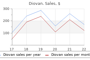
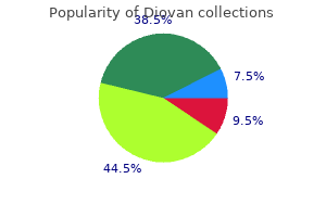
Diovan
Diovan dosages: 160 mg, 80 mg, 40 mg
Diovan packs: 30 pills, 60 pills, 90 pills, 120 pills, 180 pills, 270 pills, 360 pills

Safe diovan 40 mg
Erythropoietin deficiency happens in superior renal failure arrhythmia genetic 80 mg diovan discount with amex, ensuing within the emergence of a big normocytic anemia hypertension uncontrolled icd 9 generic diovan 160 mg with mastercard. The rising availability of recombinant erythropoietin agents, nevertheless, has all but eliminated the necessity for transfusion in dialysis patients. In either case, vitamin D undergoes numerous modifications in various organs, together with the kidneys, to turn out to be a bioactive hormone. Vitamin D synthesis begins when ultraviolet waves in sunlight cause photoisomerization of 7dehydrocholesterol to vitamin D3 (cholecalciferol), or when vitamin D2 (ergocalciferol) or D3 is ingested and absorbed. Because vitamin D is fat soluble, insufficient absorption happens in fat malabsorption states, such as pancreatic insufficiency or cystic fibrosis. Another proximal tubular enzyme, known as 24-hydroxylase, can synthesize an inactive type of vitamin D often known as 24,25-dihydroxyvitamin D. Such defects are a serious part of the phenomenon known as renal osteodystrophy, which happens in end-stage renal illness (see Plate 4-70). Most instances are acquired, reflecting publicity to substances that intervene with proximal tubular operate, similar to myeloma proteins or medicine. In rarer circumstances, generalized proximal dysfunction might end result from inherited ailments, similar to cystinosis (see Plate 4-64). Indeed, as soon as serum bicarbonate ranges decline to 15 mEq/L, reabsorption within the distal nephron can absolutely compensate for the proximal dysfunction. At this point, bicarbonate losing ceases, urine pH decreases (often turning into acidic), and serum bicarbonate concentrations stabilize. If patients are given an intravenous infusion of sodium bicarbonate, nevertheless, bicarbonate wasting resumes (with a fractional excretion of 15%) and the urine pH increases. In addition, the increased urine move through the distal tubule, which ends from proximal salt losing, stimulates K+ secretion via flowsensitive maxi-K channels. Patients with generalized proximal tubular dysfunction, for instance, exhibit salt wasting, polyuria, phosphaturia (and hypophosphatemia), glucosuria, uricosuria (and hypouricemia), aminoaciduria, microalbuminuria, and low molecular weight proteinuria. Once serum bicarbonate concentrations are low enough, nonetheless, the collecting duct can recapture all the bicarbonate wasted from the proximal tubule. In response to a load of bicarbonate salts, nonetheless, the kidneys will resume wasting bicarbonate. Extensive bicarbonate supplementation, however, often causes worsening hypokalemia, and thus potassium supplements are sometimes required as properly. Type A cells secrete protons and reabsorb bicarbonate, whereas type B cells do the reverse. Nephrolithiasis and/or nephrocalcinosis are also widespread, since calcium precipitation is favored by urine alkalinity, which results from failure of proton secretion, and hypocitraturia, which ends from the elevated citrate reabsorption that occurs in response to acidosis. Metabolic bone illness (osteomalacia or rickets) may occur due to the effects of acidosis on bone, although calcium and phosphate ranges are often normal. Acidosis displays both the absence of aldosterone-induced proton secretion and the inhibitory effects of hyperkalemia on ammoniagenesis. Fludrocortisone and/or potassium-lowering drugs, corresponding to oral resins, are additionally useful, since lowering serum potassium concentrations increases renal ammoniagenesis and ammonia excretion. Investigators have identified loss of carbonic anhydrase 2, an enzyme expressed each all through the nephron and in osteoclasts, because the biochemical defect. The illness presents in infancy, with major signs together with thickened but brittle bones, short stature, mental retardation, dental malocclusion, and visual impairment from optic nerve compression. As a end result, water is reabsorbed through a transcellular route from the tubule lumen in to the interstitium. V2 mutations account for 90% of cases and are inherited in an X-linked recessive pattern. Females may exhibit a variable stage of illness relying on their particular pattern of X-inactivation (lyonization). About 40% to 50% of patients who take lithium will experience this complication to some extent; amongst them, about half will expertise significant polyuria, beginning as early as 8 weeks after remedy begins. Lithium is freely filtered at the glomerulus and primarily reabsorbed in the proximal tubule. For reasons that are poorly understood, however which may involve a selective toxic effect on principal cells, these results can persist even after lithium is discontinued. Patients with polyuria should be questioned regarding their water consumption to assess for attainable primary polydipsia. On serum chemistries, hypernatremia is suggestive of severe dehydration secondary to polyuria, whereas hyponatremia indicates main polydipsia. Fasting glucose levels should be normal to exclude hyperglycemia as a cause of osmotic diuresis. In youngsters and adults, a water deprivation take a look at is the gold commonplace for analysis. This procedure exams the urine-concentrating capabilities of the kidneys in response to dehydration. In regular people, there shall be an applicable increase in urine osmolality because the body attempts to preserve free water. Lithium, for instance, must be discontinued, and hypokalemia or hypercalcemia must be corrected. A low-salt food plan ought to be instituted to promote solute and water reabsorption within the proximal tubule. In addition, a diuretic can be offered because it could paradoxically cut back urine output by causing a slight quantity depletion, which up-regulates salt and water reabsorption in the proximal tubule. Thiazide diuretics are preferred over loop diuretics because the latter impair creation of the solute gradient within the medulla, which interferes with urine concentration. In addition, amiloride has been proposed as a potentially preventative measure in patients taking lithium, because this agent appears to limit lithium influx in to principal cells. Such a decline is usually first evidenced as a rise in serum creatinine concentration, which may be accompanied by regular urine output, oliguria, or anuria. Second, serum creatinine concentrations is probably not in a gradual state in patients with declining renal perform. In the setting of markedly reduced move, nonetheless, these compensatory mechanisms fail, and renal filtration declines. By definition, however, the renal parenchyma remains intact, and regular operate may be restored with intravascular fluid repletion. Hyaline casts, however, could additionally be seen; these occur as a end result of low tubular flow charges improve aggregation of Tamm-Horsfall mucoproteins, that are secreted by the distal tubular epithelium. In addition, glomerular and interstitial ailments are often associated with proteinuria, in contrast to prerenal or postrenal illness. Finally, a number of glomerulonephritides cause abnormal complement levels, that are usually not seen in prerenal or postrenal disease except the patient has other comorbidities. The obstructions should due to this fact have an effect on the urethra, bladder neck, or both ureters. Such obstructions are sometimes associated with intense flank and groin pain, which results from stretching of the proximal collecting system. Irrespective of the cause, however, clinicians must be cognizant of the widespread sequelae of severely impaired renal perform, corresponding to fluid retention (with subsequent hypertension and edema), hyperkalemia, and metabolic acidosis. Any of these situations, if not correctable with medications, may require renal replacement therapy.
40 mg diovan amex
The sign of Leser-Tr�lat is the fast onset of a quantity of seborrheic keratoses associated with an underlying inside malignancy blood pressure 60 over 90 generic diovan 40 mg on line. The horn cyst develops inside the dermis and is made from a keratin-filled cystic area with a surrounding granular cell layer heart attack playing with fire purchase diovan 40 mg free shipping. A pseudo-horn cyst is fashioned by an invagination of the stratum corneum in to the underlying epidermis. A definitive inheritance pattern has not been discovered, however these keratoses show some genetic predisposition. Multiple seborrheic keratosis lesions Dermatosis papulosis nigrans Stucco keratosis human papillomavirus has been proposed but has yet to be proven. Cryotherapy and curettage are often used to deal with these benign skin growths, and both are extremely efficient. After cryotherapy remedy, a blister usually forms at the base of the seborrheic keratosis, and inside a day or two the keratosis falls off. Another extremely efficient methodology of removing that could be carried out within the office is cryotherapy adopted by a light-weight curettage; this additionally permits for histological analysis. Occasionally, dark brown or black seborrheic keratoses can mimic melanoma, and in other instances a melanoma could arise adjoining to a seborrheic keratosis and mislead the clinician. In the past, they had been additionally referred to as "benign juvenile melanoma," but that name ought to be prevented, as a result of the time period melanoma ought to be used to describe malignant tumors only. For this purpose, the phrases atypical Spitzoid melanocytic lesion, atypical Spitz nevus, and Spitzoid tumor of undetermined potential have made their means in to the dermatology lexicon to describe these difficult-toclassify cases. Clinical Findings: the basic Spitz nevus happens in childhood and has a attribute reddish-brown color. It happens equally in boys and girls and is extra generally discovered within the Caucasian population. Spitz nevi are nearly always solitary, however multiple Spitz nevi in an agminated pattern have been described. The medical differential prognosis of a Spitz nevus contains the frequent acquired nevus, pilomatricoma, dermatofibroma, adnexal tumors, and juvenile xanthogranuloma. Pathogenesis: the Spitz nevus is a melanocytic lesion derived from spindle-shaped or epithelioid melanocytes. The initiating issue or elements that trigger this melanocytic proliferation to arise are unknown. They are distinctive melanocytic lesions, and their pathogenesis is prone to be entirely different from that of congenital melanocytic or frequent acquired melanocytic nevi. It reveals the right benign maturation of melanocytes from prime to backside of the lesion. As alluded to earlier, the traditional Spitz nevus is often an easy prognosis. However, many difficult-to-classify melanocytic lesions have overlapping options of Spitz nevus and melanoma and may be exceedingly challenging diagnostically. Treatment: Complete excision for a traditional Spitz nevus is curative and allows for a whole histological evaluation. Spitz nevi in adults should all be excised to allow for full histopathological examination. Unclassifiable or difficult to classify melanocytic tumors with options of both Spitz nevus and melanoma are finest treated as in the occasion that they had been melanoma. These tumors are extremely rare and comprise nicely lower than 1% of all skin cancers recognized annually. These tumors are believed to be derived from hair follicle, sebaceous gland, apocrine gland, or eccrine gland epithelium. They are thought to come up de novo and can even arise from a preexisting benign precursor. Clinical Findings: these tumors are very uncommon, and one is unlikely to think about them in the differential diagnosis when evaluating an individual with an undiagnosed pores and skin growth. There are few clues to their origin, which makes prognosis of these cancerous tumors almost unimaginable based mostly on clinical findings alone. A punch or excisional biopsy is the most effective methodology to biopsy these lesions, as a end result of it allows the pathologist to get a large sufficient piece of tissue to consider. A punch biopsy is very essential to help differentiate microcystic adnexal carcinoma from a benign syringoma. There seems to be no genetic inheritance to these malignant tumors, with the lone exception of the sebaceous carcinoma. Sebaceous carcinoma can be seen within the Muir-Torre syndrome, which is inherited in an autosomal dominant sample. The tumors show varying amounts of cellular atypia and an invasive growth sample. They are often poorly circumscribed with various amounts of mitotic figures, necrosis, and abnormal-appearing cells. Various glandlike buildings could be seen in some tumors, which can be helpful in making the diagnosis. Often, special immunohistochemical stains are used to help differentiate the subtypes of these tumors. Treatment: these tumors ought to all be surgically excised with clear surgical margins. The Mohs surgical method has been used efficiently to deal with these tumors, as has a regular extensive native excision. Adnexal tumors are uncommon, and a biopsy for histological analysis is required for diagnosis. Slow-growing tumor that may turn out to be fairly large by the time of analysis Sebaceous carcinoma. For the same causes, the ultimate prognosis and the recurrence price of those tumors are unknown. After diagnosis and removal of those tumors, the affected person ought to have long-term follow-up to evaluate for recurrence. Adnexal tumors which have metastasized are handled with chemotherapy with or without radiotherapy. The continual lymphedema is secondary to prior mastectomy and axillary lymph node dissection. These tumors could be seen as a solitary discovering or secondary to long-standing lymphedema, such as after radiation therapy or an axillary or inguinal lymph node dissection. Soft tissue sarcomas are very uncommon and make up a small share of all malignancies reported. The tumors most commonly arise in the head and neck area and can manifest in plenty of fashions. The tumor continues to increase, varieties satellite foci of involvement, and eventually ulcerates and bleeds. The tumors sometimes present an aggressive development pattern and tend to metastasize early in the course of illness.
Diovan 160 mg order without a prescription
Conotruncal malformations heart attack 70 blockage diovan 40 mg purchase, together with interrupted aortic arch and isolated anomalies of the aortic arch arteria spanish 40 mg diovan generic with mastercard, are included within the constellation of findings in sufferers with the 22q11. Vascular ring is the broad term used to describe an aortic arch malformation during which the trachea and esophagus are compressed. The first scientific description of tracheal and esophageal compression by a double aortic arch is credited to Wolman in 1939. There are two major sixty three kinds of proper aortic arch: with no retroesophageal element (mirror-image branching) and with a retroesophageal component. The first vessel is a left innominate artery, the second and third are the right frequent carotid and right subclavian arteries. The majority of sufferers have a left ductus arteriosus arising from the left pulmonary artery and inserting in to the left subclavian artery. Bilateral patent ductus is related to congenital cardiac anomalies, usually tetralogy of Fallot or truncus arteriosus. Right Aortic Arch with Retroesophageal Component A vascular ring is usually current in the proper aortic arch with retroesophageal component. The right aortic arch extends to the left, behind the esophagus, in the form of a diverticulum. The vascular ring is formed from the proper arch and a left-sided ductus arteriosus arising from the left pulmonary artery, with extension in to the upper descending thoracic aorta. This group of abnormalities also contains anomalous retroesophageal left subclavian artery. Right aortic arch with mirror-image pattern is related to congenital coronary heart anomalies, and the commonest are tetralogy of Fallot and truncus arteriosus. Aberrant Right Subclavian Artery the most typical left arch abnormality is aberrant right subclavian artery. The proper subclavian artery arises because the fourth branch of the aortic arch, distal to the left subclavian artery. This anomaly is of significance due to its frequent affiliation with tetralogy of Fallot and the difficulty of using this vessel for the BlalockTaussig anastomosis. Interrupted aortic arch is a uncommon congenital malformation occurring in three per million reside births. It is defined as a lack of continuity between the ascending and descending thoracic aorta and has poor prognosis with out surgical remedy. For example, disruption of the forkhead transcription factor Mfh1 causes hypoplasia of the fourth aortic arch artery in mice, resulting in absence of the transverse aortic arch, resembling the interruption of the aortic arch in man. The ascending aorta arises normally, and as it leaves the pericardium it divides in to two branches, a left and a right aortic arch that be a part of posteriorly to kind the descending aorta. The left arch passes anteriorly and to the left of the trachea within the ordinary place and then becomes the descending aorta by the ligamentum arteriosum or the ductus arteriosus. The proper aortic arch passes to the right and then posterior to the esophagus to be part of the left-sided descending aorta, thereby finishing the vascular ring. The arches are usually not equal in size, the proper arch generally the bigger of the two. One arch could additionally be represented by a single atretic segment; in that case, the best arch usually persists. It is theoretically attainable, utilizing the double aortic arch mannequin, that the ductus arteriosus might be bilateral or on the proper or left aspect only. No case of functional double arch with bilateral ductus arteriosus has been reported. The descending aorta could also be on the right, on the left, or sometimes in the midline. Wheezing is the most common, followed by stridor, pneumonia, higher respiratory tract an infection, respiratory distress, cough, and respiratory cyanosis. Ascending aorta bifurcates in to an anterior left branch, supplying left widespread carotid artery and left subclavian artery, and a posterior right branch, supplying proper widespread carotid and right subclavian arteries. Continuation of aorta seen from behind demonstrates anterior left department wrapping round trachea and esophagus, in addition to right posterior department emerging from underneath esophagus. Synopsis of pathology, embryology, and pure history, Baltimore, 1977, Urban & Schwarzenberg, pp 159�166. The indicators and symptoms of tracheal and esophageal compression differ with the severity of compression. Anomalies of the Pulmonary Trunk and Arteries Isolated pulmonary artery abnormalities are uncommon and could be divided in to (1) those with anomalous arterial supply to one lung within the presence of separate aortic and pulmonary valves (and with out inherent interposition of ductal tissue) and (2) these with lungs receiving usually linked pulmonary arteries. Origin of Right or Left Pulmonary Artery from Ascending Aorta When the proper pulmonary artery arises from the aorta, it often arises from the right or posterior facet of the ascending aorta. Part of one lung may obtain anomalous vascular provide, referred to as sequestration of the lung. Pulmonary arteries might arise from the pulmonary trunk, however the left artery connects to the right lung and vice versa. In the case of the facial port-wine stain or the facial nevus flammeus, histological sections might present solely uncommon dilated capillary-like vessels in younger children, or collections of haphazardly organized dilated vessels within the papillary and reticular dermis in older patients92. However, the time period hemangioma has typically been used, particularly clinically, for these lesions. Most types of telangiectasias are current within the pores and skin, but inside organs together with the mind can be affected. There are typically few or no signs attributable to the telangiectasia itself, aside from beauty problems when it includes the pores and skin, or hemorrhagic complications of gastrointestinal hemangiomas. Incidental telangiectasias of the mind are found predominantly within the pons, and have solely hardly ever been reported to trigger signs by bleeding. The most typical congenital cutaneous telangiectasia is the nevus flammeus, or strange birthmark. Nevus flammeus appears as mottled macular lesion on the head and neck, and often regresses. The nevus vinosus, or port-wine stain, is a specialized type of nevus flammeus that demonstrates no tendency to fade and often turns into elevated, reminiscent of a true hemangioma. Unlike true hemangiomas, telangiectasias appear histologically as congested regular vessels that are separated by intervening tissue. The vascular manifestations are heralded by appearance in childhood of telangiectasias of the bulbar conjunctivae and skin of the face and extremities. The patients typically succumb to an underlying immunological abnormality that results in recurrent infections and the development of lymphoproliferative issues. Usually the defect is single, giant, and oval; occasionally (<10% of cases) the defect is small. Vascular malformations, however, are considered slow-growing congenital anomalies associated with arteriovenous shunting, and histologically are characterised by a proliferation of heterogeneous and sometimes dysplastic vascular elements, together with arteries, dysplastic arteries, veins, and arterialized veins. Fibrosis and follicular dilation and keratin plugging are present (H&E stain � 40). Watermelon abdomen has been more and more recognized as an necessary reason for occult gastrointestinal blood loss and anemia.
Buy diovan 160 mg line
Routine ophthalmological examinations ought to be beneficial Depigmented skin area to evaluate for the potential for retinal astrocytic hamartomas (phakomas) blood pressure medication and juice diovan 160 mg cheap with amex. Laser vaporization and extra conventional surgical methods have been used to remove or lessen the appearance of these tumors heart attack 02 50 heart attack enrique iglesias s and love safe 80 mg diovan. No remedy is required for the hypopigmented macules or the connective tissue nevi. All sufferers ought to be monitored routinely by their pediatrician for evaluation of developmental milestones and bodily examinations. Basis for the barrier abnormality in atopic dermatitis: Outside-inside-outside pathogenic mechanisms. Epithelial barrier function: meeting and structural features of the cornified cell envelope. The epidermal vitamin D system and innate immunity: some more light shed on this distinctive photoendocrine system Glomuvenous malformation (glomangioma) and venous malformation: distinct clinicopathologic and genetic entities. Management of benign skin lesions generally affecting the face: actinic keratosis, seborrheic keratosis, and rosacea. Palisaded encapsulated neuroma (solitary circumscribed neuroma of the skin) of the eyelid: report of two instances and review of the literature. Seborrhoeic keratoses in patients with inside malignancies: a case-control study with potential accrual of sufferers. Proliferating trichilemmal tumors: clinicopathologic evaluation as a guide to biological behavior. Palisaded encapsulated neuroma-a basic presentation of a commonly misdiagnosed neural tumor. Review of linear epidermal nevus with oral mucosal involvement-series of five new cases. The man behind the eponym: Hans Biberstein and follicular hyperplasia overlying dermatofibroma. Median raphe cyst in the scrotum, mimicking a serous borderline tumor, related to cryptorchidism after orchiopexy. Nevus lipomatosus cutaneous superficialis: a uncommon giant variant in an unusual location. Preliminary examine among truck drivers in Turkey: effects of ultraviolet gentle in some pores and skin entities. Nevus comedonicus with hidradenoma papilliferum and syringocystadenoma papilliferum within the female genital area. Mucoepidermoid (adenosquamous) carcinoma, trichoblastoma, trichilemmoma, sebaceous adenoma, tumor of the follicular infundibulum and syringocystadenoma papilliferum arising inside 2 persistent lesions of nevus sebaceous: report of a case. Reticulohistiocytoma (solitary epitheloid histiocytoma): a clinicopathologic and immunohistochemical study of forty four instances. Fibrofolliculoma/trichodiscoma and fibrous papule (perifollicular fibroma/angiofibroma): a reevaluation of the histopathological and immunohistochemical options. Lichenoid keratosis-a clinicopathological entity with lupus erythematosus-like options Blue nevi and associated lesions: a evaluate highlighting atypical and newly described variants, distinguishing options and diagnostic pitfalls. Benign pores and skin lesions: lipomas, epidermal inclusion cysts, muscle and nerve biopsies. Epidermal nevus syndrome with hypophosphatemic renal rickets with hypercalciuria: a bone marrow diagnosis. Screening of glucose/insulin metabolic alterations in males with multiple skin tags on the neck. Dermatofibroma: a possible model of local fibrosis with epithelial/mesenchymal cell interplay. Dermoscopic pattern of intermediate stage in seborrhoeic keratosis regressing to lichenoid keratosis: report of 24 instances. A twin idea of nevogenesis: theoretical issues based on dermoscopic features of melanocytic nevi. Smooth muscle hamartoma related to a congenital pattern of melanocytic nevus: a case report and review of the literature. A case of Ferguson-Smith kind multiple keratoacanthomas associated with keratoacanthoma centrifugum marginatum: response to oral acitretin. Sebaceous carcinoma: the good masquerader: rising concepts in diagnosis and treatment. Skin angiosarcoma arising in an irradiated breast: case-report and literature evaluate. Actinic keratosis: pure historical past and risk of malignant transformation in the Veterans Affairs Topical Tretinoin Chemoprevention Trial. Total pores and skin electron beam and non-myeloablative allogeneic hematopoietic stem-cell transplantation in advanced mycosis fungoides and Sezary syndrome J Clin Oncol. Cutaneous melanoma: estimating survival and recurrence danger primarily based on histopathologic features. Identification and useful validation of therapeutic targets for malignant melanoma. What is the role of adjuvant radiotherapy in the remedy of cutaneous squamous cell carcinoma with perineural invasion The usefulness of p63 detection for differentiating major from metastatic skin adenocarcinomas. Pegylated interferons: prospects for the use in the adjuvant and palliative remedy of metastatic melanoma. Bowenoid papulosis of the vulva-immunotherapeutical method with topical imiquimod. Rare skin most cancers: a population-based cancer registry descriptive examine of 151 consecutive instances recognized between 1980 and 2004. Sebaceous neoplasia and the Muir-Torre syndrome: essential connections with medical implications. Management of Merkel cell carcinoma with emphasis on small primary tumors: a case collection and evaluate of the present literature. Keratoacanthomas: overview and comparability between Houston and Minneapolis experiences. Hepatocellular carcinoma metastatic to pores and skin: diagnostic utility of antihuman hepatocyte antibody in combination with albumin in situ hybridization. Surgical remedy of dermatofibrosarcoma protuberans: the Dartmouth experience and literature evaluate. Erythema multiforme during antitumor necrosis issue remedy for plaque psoriasis. Devastating ochronotic arthropathy with successful bilateral hip and knee arthroplasties. Pathogenesis and therapeutic approaches for improved topical therapy in localized scleroderma and systemic sclerosis. State-of-the-art remedy of coccidioidomycosis: skin and soft-tissue infections. Recent insights in to atopic dermatitis and implications for administration of infectious complications. Cardiac manifestations of neonatal lupus erythematosus: pointers to management, integrating clues from the bench and bedside. Clinical and hematological presentation of youngsters and adolescents with polycythemia vera.

Order 40 mg diovan with visa
Akatsu T blood pressure in dogs buy diovan 40 mg with visa, Hayashi S arrhythmia light headed diovan 40 mg cheap line, Yamane T, et al: Emergency embolization of a ruptured pancreaticoduodenal artery aneurysm associated with the median arcuate ligament syndrome, J Gastroenterol Hepatol 19:482�483, 2004. Ogino H, Sa to Y, Banno T, et al: Embolization in a patient with ruptured anterior inferior pancreaticoduodenal arterial aneurysm with median arcuate ligament syndrome, Cardiovasc Interv Radiol 25:318�319, 2002. Baccari P, Civilini E, Dordoni L, et al: Celiac artery compression syndrome managed by laparoscopy, J Vasc Surg 50:134�139, 2009. Baldassarre E, Torino G, Siani A, et al: the laparoscopic method in the median arcuate ligament syndrome: report of a case, Swiss Med Wkly 137:353�354, 2007. Jaik N, Stawicki S, Weger N, et al: Celiac artery compression syndrome: successful utilization of robotic-assisted laparoscopic strategy, J Gastrointest Liver Dis 16:93�96, 2007. Grotemeyer D, Duran M, Iskandar F, et al: Median arcuate ligament syndrome: vascular surgical therapy and follow-up of 18 sufferers, Langenbecks Arch Surg 394:1085�1092, 2009. Wang X, Impeduglia T, Dubin Z, et al: Celiac revascularization as a requisite for treating the median arcuate ligament syndrome, Ann Vasc Surg 22:571�574, 2008. In cases with out pulmonary venous obstruction, the resistance at delivery is equal; due to this fact, the distribution of blood is equal between the pulmonary and systemic circuits. However, inside a couple of weeks of start, the pulmonary resistance decreases, and a bigger proportion of the blended venous blood returns to the pulmonary circuit, leading to a virtually three to five instances greater pulmonary-to-systemic move ratio of 3:1 to 5:1, and an equalization of oxygen saturation between the best and left coronary heart. The indicators and symptoms, therefore, depend upon the underlying hemodynamics-that is,presence or absence of pulmonary venous obstruction and the extent of mixing of blood between the best and left atrium. If interatrial mixing is inadequate, symptoms happen at delivery or shortly thereafter. The connections arise from failed growth of normal embryological venous communications or persistence, lack of regression of normal embryological venous communications, or both. Anomalous Pulmonary Venous Connections Anomalous pulmonary venous connection refers to the absence of a number of pulmonary venous connections to the left atrium, without reference to the next drainage of the anomalously disconnected pulmonary vein(s). The pulmonary veins connect instantly in to the right atrium or in to considered one of its tributaries. With development, a brand new pulmonary venous channel, the primary pulmonary vein, grows as a bulging of the left atrium. At the identical time, the intrapulmonary veins lose their connections with the splanchnic plexus and fuse with the first pulmonary vein. The intrapulmonary veins finally form the 4 pulmonary veins and are absorbed in to the left atrium. Failure of severance of the connection between the intrapulmonary and splanchnic veins leads to anomalous pulmonary venous connections that may be whole, partial, bilateral, or unilateral. The drainage site of pulmonary veins may be supradiaphragmatic or infradiaphragmatic. The main coronary heart tube is depicted in brown and the myocardium derived from the second heart subject (and included later within the heart) in yellow. A, Primary heart tube is shaped after fusion of bilateral plates of splanchnic mesoderm within the primitive plate. B, Lateral view of embryo (23 days in human), showing major coronary heart tube surrounded by cardiac jelly (blue), and second heart area located dorsally to coronary heart. The right pulmonary veins are six instances extra generally concerned than the left pulmonary veins, and the higher lobes of the lung are more generally concerned than the decrease. The left-sided pulmonary veins often connect to the derivatives of the left cardinal system. The right pulmonary veins hook up with the derivatives of the proper cardinal system. The coronary heart normally exhibits gentle dilation and hypertrophy of the right atrium and ventricle, with dilation of the pulmonary artery. The vein of the proper decrease lobe usually enters the left atrium however might connect to the proper atrium. This malformation known as scimitar syndrome, as initially described by Chassinat in 1836,eleven,12 and derives its name from the form of the anomalous vein on chest radiographs. In uncommon instances, veins of both lungs drain anomalously, however a small section of the pulmonary venous system drains usually. That condition has been termed by Edwards subtotal anomalous pulmonary venous connection. B, Anomalous connections of proper pulmonary veins to inferior vena cava in the presence of an intact atrial septum. C, Anomalous connection of left pulmonary veins to left innominate by means of a vertical vein. Accessory atrial chamber that receives all pulmonary veins and communicates with the left atrium; no other connections (classic cor triatriatum). A stenotic opening between the accessory atrium and the left atrium, nonetheless, results in features of extreme pulmonary obstruction. Patients usually have historical past of breathlessness and frequent respiratory infections. Right heart failure is normally present together with indicators of pulmonary hypertension. Surgery has been advocated as early because the neonatal interval,18 and balloon dilation may be profitable in older youngsters. Accessory atrial chamber that receives part of the pulmonary veins and connects to the left atrium. Accessory atrial chamber that receives part of the pulmonary veins and connects to the proper atrium. One is localized stenosis of the pulmonary veins on the junction of the left atrium, which may contain one or more pulmonary veins; the other is characterized by narrowing of the lumen of the pulmonary vein, both intra- or extrapulmonary in location, and is known as hypoplasia of the pulmonary veins. The latter type is occasionally current in patients 775 with pulmonary artery atresia or hypoplastic left coronary heart syndrome. Clinically patients have a historical past of respiratory signs, which can, progress to right coronary heart failure. Because of poor surgical results, balloon angioplasty and stenting currently are being evaluated as noninvasive therapies for pulmonary artery stenosis. Large outgrowth of the valve of the sinus venosus might end in full or partial subdivision of the proper atrium. As with anomalous pulmonary veins, anomalous systemic venous return is a trademark of the heterotaxy syndromes, especially asplenia. In the majority of instances (67%), the anomalous vessel programs between the aorta and pulmonary trunk, with the remainder often coursing posterior to the aorta. The ostium supplying the best coronary artery could have features much like anomalous left ostia situated in the best sinus. Namely, there may be upward displacement, location near the commissure, and slitlike ostia with ostial ridges. The proximal anomalous right coronary generally also programs between the aorta and pulmonary trunk. Awareness of this anomaly is essential throughout cardiac surgical procedure to avoid issues with myocardial safety or during prosthetic valve alternative. Origin from Pulmonary Trunk the left major coronary artery arises from the pulmonary trunk in 1/50,000 to 1/300,000 autopsies.

Common Groundsel (Groundsel). Diovan.
- Are there any interactions with medications?
- What is Groundsel?
- Colic, worms, epilepsy, irregular or painful menstrual periods (dysmenorrhea), stopping dental bleeding, and other conditions.
- Dosing considerations for Groundsel.
- Are there safety concerns?
Source: http://www.rxlist.com/script/main/art.asp?articlekey=96323
80 mg diovan cheap otc
Moudgill N arterial insufficiency diovan 40 mg purchase fast delivery, Hager E pulse pressure heart rate generic diovan 160 mg with mastercard, Gonsalves C, et al: May-Thurner syndrome: case report and review of the literature involving modern endovascular remedy, Vascular 17:330�335, 2009. Mullens W, De Keyser J, Van Dorpe A, et al: Migration of two venous stents in to the best ventricle in a affected person with May-Thurner syndrome, Int J Cardiol one hundred ten:114�115, 2006. Hartung O, Barthelemy P, Arnoux D, et al: Management of being pregnant in ladies with previous left ilio-caval stenting, J Vasc Surg 50:355�359, 2009. Ahmed K, Sampath R, Khan M: Current developments in the prognosis and management of renal nutcracker syndrome: a evaluation, Eur J Vasc Endovasc Surg 31:410�416, 2006. Shin J, Park J, Shin Y, et al: Superimposition of nutcracker syndrome in a haematuric youngster with Henoch Sch�nlein purpura, Int J Clin Pract 59:1472�1475, 2005. Fitoz S, Yalcinkaya F: Compression of left inferior vena cava: a form of nutcracker syndrome, J Clin Ultrasound 36:101�104, 2008. Mohamadi A, Ghasemi-Rad M, Mladkova N, et al: Varicocele and nutcracker syndrome: sonographic findings, J Ultrasound Med 29:1153�1160, 2010. Kim J, Joh J, Choi H, et al: Transposition of the left renal vein in nutcracker syndrome, Eur J Vasc Endovasc Surg 31:80�82, 2006. Salehipour M, Rasekhi A, Shirazi M, et al: the function of renal autotransplantation in therapy of nutcracker syndrome, Saudi J Kidney Dis Transpl 21:237�241, 2010. Xu D, Liu Y, Gao Y, et al: Management of renal nutcracker syndrome by retroperitoneal laparoscopic nephrectomy with ex vivo autograft repair and autotransplantation: a case report and evaluate of the literature, J Med Case Reports 3:82, 2009. Zhang Q, Zhang Y, Lou S, et al: Laparoscopic extravascular renal vein stent placement for nutcracker syndrome, J Endourol 354�357, 2010. Hartung O, Azghari A, Barthelemy P, et al: Laparoscopic transposition of the left renal vein in to the inferior vena cava for nutcracker syndrome, J Vasc Surg fifty two:738�741, 2010. Rogers A, Beech A, Braithwaite B: Transperitoneal laparoscopic left gonadal vein ligation may be the right remedy possibility for pelvic congestion signs secondary to nutcracker syndrome, Vascular 15:238�240, 2007. Shaper K, Jackson J, Williams G: the nutcracker syndrome: an unusual explanation for haematuria, Br J Urol seventy four:144�146, 1994. Basile A, Tsetis D, Calcara G, et al: Percutaneous nitinol stent implantation within the remedy of nutcracker syndrome in young adults, J Vasc Interv Radiol 18:1042�1046, 2007. Chen W, Chu J, Yang J, et al: Endovascular stent placement for the remedy of nutcracker phenomenon in three pediatric sufferers, J Vasc Interv Radiol 16:1529�1533, 2005. Cohen F, Amabile P, Varoquaux A, et al: Endovascular therapy of circumaortic nutcracker syndrome, J Vasc Interv Radiol 20:1255�1257, 2009. Hartung O, Grisoli D, Boufi M, et al: Endovascular stenting in the therapy of pelvic vein congestion brought on by nutcracker syndrome: lessons realized from the primary 5 circumstances, J Vasc Surg forty two:275�280, 2005. Wang L, Yi L, Yang L, et al: Diagnosis and surgical treatment of nutcracker syndrome: a single-center expertise, Urology seventy three:871�876, 2009. Zhang H, Li M, Jin W, et al: the left renal entrapment syndrome: diagnosis and remedy, Ann Vasc Surg 21:198�203, 2007. Haidar S, Thomas K, Miller S: Popliteal artery entrapment syndrome in a younger girl, Pediatr Radiol 35:440�443, 2005. Ferreira J, Canedo A, Gra�a S, et al: Thrombosed popliteal aneurysm-first manifestation of bilateral popliteal entrapment syndrome, Int Angiol 29:83�86, 2010. Tercan F, Oguzkurt L, Kizilkilic O, et al: Popliteal artery entrapment syndrome, Diagn Interv Radiol eleven:222�224, 2005. Misselbeck T, Dangleben D, Celani V: Isolated popliteal vein entrapment by the popliteus muscle: a case report, Vasc Med 13:37�39, 2008. Pillai J: A present interpretation of popliteal vascular entrapment, J Vasc Surg 48: 61s�65s, 2008. Holden A, Merrilees S, Mitchell N, et al: Magnetic resonance imaging of popliteal artery pathologies, Eur J Radiol 67:159�168, 2008. Kukreja K, Scagnelli T, Narayanan G, et al: Role of angiography in popliteal artery entrapment syndrome, Diagn Interv Radiol 15:57�60, 2009. Anil G, Tay K, Howe T, et al: Dynamic computed tomography angiography: function in the analysis of popliteal artery entrapment syndrome, Cardiovasc Interv Radiol 34:259�270, 2011. Zhong H, Liu C, Shao G: Computed tomographic angiography and digital subtraction angiography findings in popliteal artery entrapment syndrome, J Comput Assist Tomogr 34:254�259, 2010. Meier T, Schneider E, Amann-Vesti B: Long-term follow-up of sufferers with popliteal artery entrapment syndrome treated by endoluminal revascularization, Vasa 39: 189�195, 2010. Paraskevas N, Castier Y, Fukui S, et al: Superficial femoral artery autograft reconstruction for complicated popliteal artery entrapment syndrome, Vasc Endovasc Surg forty three:165�169, 2009. Gagnon J, Doyle D: Adventitial cystic illness of common femoral artery, Ann Vasc Surg 21:84�86, 2007. Jindal R, Majed A, Hamady M, et al: Cystic adventitial disease of the iliofemoral artery: case reports and a short review, Vascular 14:169�172, 2006. Morizumi S, Suematsu Y, Gon S, et al: Adventitial cystic disease of the femoral vein, Ann Vasc Surg 24:1135e5�1135e7, 2010. Fran�a M, Pin to J, Machado R, et al: Case 157: Bilateral adventitial cystic disease of the popliteal artery, Radiology 255:655�660, 2010. Ortmann J, Widmer M, Gretener S, et al: Cystic adventitial degeneration: ectopic ganglia from adjoining joint capsules, Vasa 38:374�377, 2009. Asciut to G, Mumme A, Marpe B, et al: Different approaches in the treatment of cystic adventitial illness of the popliteal artery, Chir Ital fifty nine:467�473, 2007. Setacci F, Sirignano P, de Dona to G, et al: Adventitial cystic illness of the popliteal artery: expertise of a single vascular and endovascular heart, J Cardiovasc Surg forty nine:235�239, 2008. Tsilimparis N, Hanack U, Yousefi S, et al: Cystic adventitial disease of the popliteal artery: an argument for the developmental concept, J Vasc Surg forty five:1249�1252, 2007. Taurino M, Rizzo L, Stella N, et al: Doppler ultrasonography and exercise testing in diagnosing a popliteal artery adventitial cyst, Cardiovasc Ultrasound 7:23, 2009. Nano G, Dalainas I, Casana R, et al: Case report of adventitial cystic illness of the popliteal artery offered with the "dog-leg" sign, Int Angiol 26:75�78, 2007. Khoury M: Failed angioplasty of a popliteal artery stenosis secondary to cystic adventitial disease, Vasc Endovasc Surg 38:277�280, 2004. Foertsch T, Koch A, Singer H, et al: Celiac trunk compression syndrome requiring surgery in three adolescent sufferers, J Pediatr Surg forty two:709�713, 2007. Desmond C, Roberts S: Exercise-related abdominal pain as a manifestation of the median arcuate ligament syndrome, Scand J Gastroenterol 39:1310�1313, 2004. Loukas M, Pinyard J, Vaid S, et al: Clinical anatomy of celiac artery compression syndrome: a evaluate, Clin Anat 20:612�617, 2007. Most instances are recognized in the first year of life, and sudden death occurs in approximately 40% of cases. In a minority of instances (20%), adequate myocardial collaterals develop from the usually structured proper coronary artery, enabling potential survival in to maturity. Sudden death usually happens at rest, but could happen after strenuous activity in older children.
Syndromes
- Irritability
- Blood tests to rule out other conditions similar to MS
- Medications that increase blood pressure
- Infection of the lining of the heart chambers and heart valves (infectious endocarditis). This can be caused by bacteria, fungi, or other infectious substances
- Do a complete physical exam
- Lymphangiography
- Idiopathic aplastic anemia
- Chest retracts unless the child is breathing through mouth or crying
Diovan 160 mg cheap line
They are believed to be triggered not by vasculitis however by irritation of the small cutaneous capillaries heart attack during sex discount 40 mg diovan overnight delivery, which produces a capillaritis blood pressure pills joint pain purchase diovan 80 mg without a prescription. The rash is typically of no scientific significance to the patient, however it can trigger important beauty concern, and the purpuras have to be differentiated from different situations that may cause similar rashes. They are believed by some to be slightly different manifestations of the identical disease state, and the histopathology of all of the variants is strikingly similar. Over time, a brownish-red hyperpigmented background types secondary to the extravasation of purple blood cells and their subsequent breakdown inside the pores and skin to launch hemosiderin. The rash might unfold proximally up the decrease extremity but not often affects other areas of the body. Most patients are referred to the dermatologist to rule out vasculitis, which is easily carried out by not finding any evidence of palpable purpura. The rash is almost all the time completely asymptomatic, and patients frequently complain only of the looks. If one sees widespread petechia, a platelet rely should be performed to search for thrombocytopenia. If the platelet rely is normal, a skin biopsy of the higher extremity or truncal space of involvement should be performed to consider for the very rare form of pigmented purpuric mycosis fungoides. Eczematoid pigmented purpura of Doucas and Kapetanakis is a uncommon variant that manifests with petechiae and hyperpigmentation however can be related to an overlying eczematous eruption. Pigmented purpura of Gougerot and Blum is also called lichenoid pigmented purpura. Lichen aureus may be seen at any age and manifests with the presence of a quantity of tiny, golden-colored macules that coalesce in to a large macule or patch. Extravasated erythrocytes and a lymphocytic vasculitis are the necessary thing histological options. This is a rare type of pigmented purpura that sometimes begins on the legs and spreads slowly over time. Pathogenesis: the pigmented purpuric dermatoses are believed to be caused by capillaritis. Oral vitamin C and bioflavonoids have been reported to be successful, again mostly in anecdotal stories. The major objective in treatment is to differentiate pityriasis rosea from different rashes that may have an analogous clinical picture. Clinical Findings: Pityriasis rosea is a common rash of younger adults and youngsters. A small but important subset of sufferers have had a preceding higher respiratory tract infection. This has led some to search for a viral reason for the rash, although none have been found. The rash of pityriasis rosea can have a varying morphology, nevertheless it most commonly begins with a herald patch. The herald patch is a 2- to 4-cm, pink-red patch with fine adherent scale that commonly occurs on the trunk. The primary differential analysis includes guttate psoriasis and, in cases that affect the palms and soles, secondary syphilis. Tinea corporis is nearly always within the differential diagnosis of any rash that has a patchtype morphology and fine floor scale. Tinea corporis can be simply recognized with a microscopic analysis of a small scraping of the pores and skin. Any affected person who has pityriasis rosea that affects the palms and or soles should be examined for syphilis. On healing, postinflammatory hyperpigmentation or hypopigmentation may result and will persist for a quantity of months. Generalized skinny oval patches the palms and soles are sometimes unaffected in Secondary syphillis are distributed on the trunk pityriasis rosea. Varying amounts of extravasated red blood cells are appreciated throughout the higher dermis. Pathogenesis: Many makes an attempt to isolate a viral or a bacterial factor in patients with pityriasis rosea have been met with frustration. The use of oral erythromycin, twice a day for two weeks, was shown to decrease the period of the rash. Patients with pityriasis rubra pilaris typically have erythroderma with a few islands of sparing. These islands of completely normal pores and skin are normally small, a few centimeters in diameter, but they can be much larger. Fissuring is fairly common inside the keratoderma and could be a supply of pain and a site for secondary infection. They sometimes run a chronic medical course, with most instances spontaneously resolving a number of years after onset. Skin biopsy and clinical pathological correlation help the clinician make a agency prognosis. Carnauba wax�like thickening of the palms and soles is a common medical finding in pityriasis rubra pilaris. Topical corticosteroid wet wraps, oral retinoids, and ultraviolet therapy have long been used as first-line agents. The retinoids are considered first-line remedy, and each isotretinoin and acitretin have been used. Other immunosuppressants have been used, including methotrexate, azathioprine, and the newer anti�tumor necrosis factor inhibitors. Cut part reveals organizing infarcts and thrombosed aneuysms in corticomedullary area. Polyarteritis nodosa has been found in some circumstances to be a continual, non�life-threatening illness that affects only the pores and skin. Many other organ system may be involved, and the pores and skin options will be the presenting sign of the illness. Excisional pores and skin biopsies of cutaneous lesions of polyarteritis nodosa show the attribute necrotizing vasculitis of medium vessels throughout the deep reticular dermis. The cutaneous diagnosis of polyarteritis nodosa should alert the clinician to the possibility of systemic illness, and appropriate testing ought to be undertaken to consider for widespread illness. Most instances of polyarteritis nodosa are idiopathic, but this condition could be seen in association with viral infections, malignancy, or autoimmune disease. Coinfection with the hepatitis B virus is essentially the most classic and most frequent association with polyarteritis nodosa. Clinical Findings: the primary cutaneous manifestation of polyarteritis nodosa is palpable purpura. The affected person could develop livedo reticularis of the extremities, and secondary ulcerations may form as the vasculitis progresses and causes necrosis of the overlying skin. The analysis of the kind of vasculitis is difficult to make from medical examination alone. Tissue sampling is required to decide the sort of vessel affected by the inflammatory vasculitis. Polyarteritis nodosa has additionally been shown to have nonspecific findings, corresponding to red macules and papules, that mimic drug eruptions or viral infections.

Cheap 80 mg diovan otc
This bacterial disease was as quickly as limited to tropical areas pulmonary hypertension 70 mmhg diovan 160 mg buy generic online, however with the benefit of worldwide travel blood pressure medication and weight gain generic diovan 80 mg free shipping, it may possibly now be seen globally. Trachoma, which often starts as conjunctivitis, ends in chronic intense irritation of the bulbar and eyelid conjunctiva that causes scarring and eventually blindness if left untreated. The disease is seen more regularly in sufferers with a low socioeconomic status and in those with a number of sexual partners. After a short incubation interval (a few days to a few weeks), a painless papule forms and ultimately ulcerates. This ulceration is commonly described as painless, nevertheless it causes the affected person irritation and discomfort with strain and manipulation. Initial involvement occurs within 2 to three weeks after therapeutic of the ulcer and typically leads to discrete, painful lymph nodes on each side of the inguinal crease. The lymph nodes coalesce over time and mat together in to a big mass of tissue called buboes. The huge adenopathy may turn into necrotic, and suppurative lymph nodes are incessantly seen. If the first and secondary ailments have affected the rectum, rectal fissures and strictures could additionally be current, leading to persistent pain. This incapability to create a steady supply of power forces the bacterium to reside inside a bunch cell. The infectious form of the bacterium, called the elementary body, positive aspects entry in to a host cell. The finding of iodine-staining, glycogen-containing inclusion bodies is sensitive and specific for the presence of C. Treatment: the routine software of erythromycin to the eyes of newborns has dramatically decreased the danger of trachoma. The bacteria, Neisseria meningitidis, is able to inflicting septicemia, pneumonia, and meningitis. These are all relentless diseases that are universally deadly if not promptly treated. The latter syndrome, also referred to as acute adrenocortical insufficiency, is immediately attributable to hemorrhagic destruction of each adrenal glands. This syndrome can result from a broad range of circumstances, together with infections, and N. Clinical Findings: Children youthful than 1 yr of age are these most probably to develop illness from N. Other threat factors embrace a deficiency of the complement parts C5, C6, C7, and C8. Chronic immunosuppression increases the chance, as does dwelling in crowded circumstances. This is why military barracks and faculty dormitories are sometimes sources of outbreaks. Cutaneous findings embrace palpable purpura, ecchymosis, widespread macular purpura, and necrosis of the skin with secondary vesiculopustules. Fulminant meningococcal septicemia might result in hemorrhagic necrosis of the adrenal glands; this is termed the Waterhouse-Friderichsen syndrome. This Meningococci from blood, spinal fluid, and/or throat Extensive purpura, shock, prostration, cyanosis Hemorrhagic destruction of adrenal gland Characteristic fever chart a hundred and five Temperature (�F) a hundred 1 Days 2 syndrome is seen in fewer than 5% of patients with N. They have signs and signs of hemodynamic collapse, hypotension, acute renal failure, and a biphasic fever. The pores and skin findings are caused by small-vessel embolization or endothelial destruction from the septicemia. Blood extravasates by way of the broken endothelial partitions and produces large purpura. The extra extensive the cutaneous purpura in meningococcal septicemia, the upper the incidence of Waterhouse-Friderichsen syndrome. The gram-negative diplococcal micro organism grows on the chocolate agar plate and seems as small, spherical, moist, gray colonies. Pathogenesis: Meningococcal infections, including septicemia and meningitis, are brought on by the gramnegative micro organism, N. The meningococcus micro organism may be discovered as a transient colonizer within the oropharynx of as a lot as 10% of sampled individuals. These carriers specific no sequelae however function a potential reservoir for meningococcal illness. If the bacteria is ready to reproduce to such an extent as to trigger bacteremia, it then turns into a possible pathogen. The micro organism exhibit a neurotrophic behavior and assault the lining of the central nervous system. Currently, a vaccine is out there that protects towards the serotypes that most incessantly cause disease: serotypes A, C, Y, and W-135. The remaining five serotypes can affect any individual regardless of vaccination standing. The bacteria expresses a toxin (lipooligosaccharide) on its floor that causes lots of the systemic signs of illness. Histology: Most pores and skin biopsy specimens present proof of vasculitis with neutrophils, fibrinoid necrosis, and extravasated purple blood cells. Embolism of capillaries and small venules is commonly seen, and necrosis and ulceration can be secondary findings. Treatment: Treatment requires prompt recognition of symptoms and immediate intravenous antibiotic therapy. Any shut contacts of the affected person ought to be screened for proof of illness and given prophylactic oral therapy to decrease the potential of an epidemic. Skin (furuncles) Inflammation and suppurative process on floor of leptomeninges of brain and spinal wire Thrombophlebitis of superior sagittal sinus and suppurative ependymitis, with beginning hydrocephalus ceftriaxone, followed by penicillin or by chloramphenicol in penicillin-allergic sufferers. Patients with Waterhouse-Friderichsen syndrome need adrenal gland alternative therapy. This prophylactic remedy, in addition to intravenous remedy, ought to be started instantly if medical suspicion is excessive sufficient; delaying remedy for even a couple of hours to anticipate laboratory confirmation could be the distinction between life and death. Immunization is helping to keep the disease incidence low, and guidelines have been established for which high-risk teams should get the vaccine and when. The diagnosis is made on scientific grounds after inspection of the attribute pores and skin findings. When seen in the genital region of adults, molluscum contagiosum is considered to be a sexually transmitted disease. This infection rarely happens in immunocompetent adults outdoors sexual transmission.
Diovan 80 mg order fast delivery
Topical corticosteroids help relieve the itching and reduce a number of the redness arteria omerale diovan 80 mg buy cheap. It is believed to have a greater prognosis arrhythmia diagnosis code diovan 160 mg generic online, as a outcome of few cases are related to an underlying most cancers. It is assumed that early remedy of juvenile dermatomyositis decreases the chance of developing severe calcinosis cutis in the course of the course of the disease. Livedo reticularis with pores and skin necrosis Large quantities of phospholipoprotein membranes coming into the circulation activate extrinsic pathway excessively. It has a grave prognosis except caught and treated early in the midst of disease. Skin manifestations happen early and continue to progress until the patient recovers. The skin lesions could result in gangrene and secondary infection, additional worsening the prognosis. The preliminary cutaneous scientific look is that of small petechiae that enlarge and coalesce in to giant macules and plaques of erythema. Ulceration, necrosis, and blister formation are generally seen within the areas of involvement. As the illness progresses, gangrene could develop within the affected areas as the blood move to the skin is considerably decreased due to clotting of assorted parts of the vascular system. An inciting event corresponding to trauma or an infection initiates the clotting cascade in which the clotting components are used up (or misplaced, in instances of extreme bleeding) faster than they are often replaced. This units off a cascade of events within the clotting system that ends in consumption of all the components utilized in clotting, leading to thrombosis and hemorrhage. Histology: Examination of pores and skin biopsies reveals necrosis of the overlying epidermis and parts of the dermis. Thrombosis of the small veins and arterioles is seen, as Tumor necrosis Giant hemangioma Abortion Extensive exposure of subendothelium activates intrinsic pathway excessively. Treatment: Treatment requires prompt recognition of the condition and instant supportive care. Many agents are used to assist lower thrombosis and exchange misplaced clotting elements. This uncommon cutaneous eruption is believed to be attributable to an irregular expulsion of fragmented elastic fibers from the dermis. The elastic fibers penetrate the surface of the dermis and manifest as an uncommon serpiginous eruption. It has been seen as an isolated discovering but also can be seen in affiliation with many underlying situations, together with Down syndrome, Ehlers-Danlos syndrome, and Marfan syndrome. Clinical Findings: Elastosis perforans serpiginosa is a rare cutaneous perforating pores and skin illness. It is far more commonly seen in the young adult population, and it has a significant male predominance, with a ratio of four: 1 to 5: 1. The eruption usually begins as small purple papules with an excoriated or barely ulcerated floor. The rash runs a waxing and waning course, but most cases resolve spontaneously with or with out therapy. Resolution on average happens inside 6 months, however cases lasting as much as 5 years have been reported within the literature. Patients with underlying Down syndrome could have just one lesion or widespread cutaneous involvement. It has been estimated that up to 1% of patients with Down syndrome will develop evidence of this rash over the course of their lifetime. Approximately 33% of cases of elastosis perforans serpiginosa are related to an underlying disorder (see field to right). An autosomal dominant sample of inheritance has been described in a small number of instances, impartial of any of the listed underlying circumstances. The medicine penicillamine has long been identified to trigger abnormalities of elastic fibers, and use of this medicine has been shown to induce an eruption resembling elastosis perforans serpiginosa. As the lesions progress, the dermis ulcerates in pinpoint areas and the underlying fragmentized and abnormal elastic tissue extrudes. Histology: Abnormally fragmented eosinophilic elastic tissue can be appreciated on routine hematoxylin and eosin staining. Special elastic tissue stains can be utilized to better isolate and recognize the elastic tissue. Examination of biopsy specimens exhibits an isolated area of acanthotic dermis by which a passageway has shaped. The passage begins in the superficial dermis and leads to the floor of the epidermis. This is crammed with the abnormal elastic tissue, a couple of histiocytes, and an occasional big cell. This unusual pores and skin finding is usually related to Down syndrome, osteogenesis imperfecta, and Marfan syndrome. Associations with Elastosis Perforans Serpiginosa Acrogeria Chronic renal failure Down syndrome Ehlers-Danlos syndrome Marfan syndrome Medications-penicillamine Osteogenesis imperfecta Pseudoxanthoma elasticum Rothmund-Thomson syndrome Scleroderma Dense connective tissue Longitudinal bundles of collagen and elastic fibers Fibroblast nuclei Transverse fibers of unfastened connective tissue elastic fibers. The cause for the abnormality within the elastic fibers has but to be decided, besides in these circumstances induced by penicillamine. Treatment: Many therapies have been attempted, and their use is anecdotal at greatest. There have been no randomized, prospective, placebo-controlled trials for the remedy of this eruption. Cryotherapy has the most information to help its use, however ablative carbon dioxide lasers have additionally been used with good results. No therapy is required, as a result of these eruptions virtually at all times spontaneously remit. The xanthomatous ailments are a diverse group of circumstances with distinctive medical, laboratory, and systemic findings. An abnormality in lipid and ldl cholesterol metabolism is what hyperlinks these circumstances collectively. Proteins and carbohydrates, when current in extra, can be converted to triglycerides to be saved as a future power supply. This process makes up the remaining supply of free fatty acids and triglycerides equipped to the physique. Triglycerides are transformed in to free fatty acids, which are damaged down in to acetyl-coenzyme A (acetyl-CoA). Ingested triglycerides are damaged down in to free fatty acids in the lumen of the gut by bile acids.