
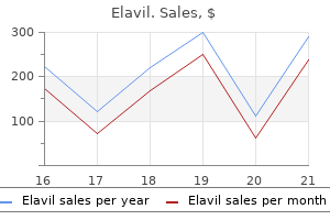
Elavil
Elavil dosages: 75 mg, 50 mg, 25 mg, 10 mg
Elavil packs: 90 pills, 180 pills, 270 pills, 360 pills, 30 pills, 60 pills, 120 pills

Cheap 50 mg elavil amex
Possible signals embrace the halting of urine circulate advanced pain treatment center mason ohio 25 mg elavil discount otc, increased hydrostatic stress on tubular epithelial cells midwest pain treatment center fremont ohio 75 mg elavil buy visa, adjustments in blood flow to the tubules or in interstitial strain, and technology of natriuretic substances within the kidney that result in longterm inhibition of transporter operate. These findings suggest that obstruction induces acute molecular changes within the renal cytoskeleton, partly mediated by increased stretch of the renal tubular cells during obstruction. Consequently, sodium delivery to every tubular section is decreased, and apical membrane Na+ entry slows dramatically as a result of the electrochemical gradients for Na+ entry between the stationary apical fluid and the cell interior become increasingly unfavorable for continued sodium transport. Reduced Na+ entry might then instantly stimulate downregulation of transporter exercise and expression. When apical Na+ entry was blocked either by substituting one other cation for sodium in the apical answer, or by adding amiloride to the apical solution, apical sodium entry was markedly decreased for some hours after the blockade was eliminated. There is a strong labeling at the base of the internal medulla in obstructed kidneys positioned completely in the interstitial cells (B). In addition to the direct results of halting urine move, modifications in intrarenal mediators and subcellular pathways likely play a crucial position in the discount of salt transport observed with obstruction. As discussed earlier, obstruction brings on a monocellular infiltrate within the kidney194; and this infiltrate tends to observe a peritubular distribution. When both ureters are obstructed, extrarenal components markedly enhance the sodium-wasting tendency already present within the obstructed kidney. Following launch of 24 hours of unilateral obstruction, removing or obstruction of the contralateral kidney markedly enhances salt wasting by the obstructed kidney. Indeed, interstitial osmolality has been proven to be decreased in obstructed kidneys. As was the case with sodium transport, the effects had been related in unilateral and bilateral obstruction. In addition to faulty amassing duct H+ transport, decreased era of the principle buffer that carries acid equivalents within the urine, ammonia, has also been noticed in kidneys launched from obstruction. Cortical slices of obstructed kidneys exhibit decreased glutamine uptake and oxidation, decreased gluconeogenesis, and reduced complete oxygen consumption, all including as a lot as a decreased ability to generate ammonia from glutamine. It is assumed that persistent obstruction damages tubular epithelial cells by rising hydrostatic strain, lowering blood flow (due to the renal vasoconstriction that occurs in obstruction, see earlier), and growing oxidative stress. All these factors accelerate the development of interstitial fibrosis by increased extracellular matrix, cell infiltration, apoptosis, and accumulation of activated myofibroblasts. It has been hypothesized that changes in the intratubular dynamic forces-so-called tubular stretch-in urinary tract obstruction also are an essential determinant for development of tubulointerstitial fibrosis within the kidney. Along this line, data suggest that mast cells even have the capacity to release chymase, a protease, which may restrict development of tubulointerstitial fibrosis by decreasing infiltration of inflammatory cells and launch of proinflammatory and profibrotic chemokines and cytokines. Thus the process resulting in kidney fibrosis is complex, and numerous processes contribute to regulating the mobile adjustments which are answerable for these pathophysiologic adjustments. However, if the research are relevant to human obstructive nephropathy, they counsel that sufferers present process release of obstruction may benefit from therapies that block proapoptotic, proinflammatory, or profibrotic mediators or from remedies that stimulate epithelial cell progress and differentiation. These outcomes support the view that irritation is a vital determinant for the onset of renal deterioration in urinary tract obstruction. However, in experimental models, obstruction in utero may cause pulmonary hyperplasia and renal impairment instantly or indirectly, resulting in vital morbidity and mortality. The sort of intervention depends on the location of the obstruction, its diploma, and its cause, in addition to the presence or absence of concomitant ailments and complications, and the overall condition of the patient. Calculi, the commonest form of acute unilateral urinary obstruction, can usually be managed conservatively with analgesics for control of pain and intravenous fluids to increase urine circulate. Ninety percent of stones smaller than 5 mm move spontaneously, however as stones get bigger, spontaneous stone passage turns into progressively much less possible. Active efforts to fragment or remove the stone are indicated for persistent obstruction, uncontrollable pain, or urinary tract an infection. Current possibilities for remedy embrace extracorporeal shock wave lithotripsy (which could require ureteral stent placement if the affected person is symptomatic),308 ureteroscopy with stone fragmentation (usually with laser lithotripsy), and, in uncommon circumstances, open excision of the stone. In the previous, complex stones excessive up within the ureter or in the renal pelvis have been difficult to remove with out open surgical procedure. Compared with adult obstructive nephropathy, fetal obstructive nephropathy is particularly devastating because renal growth and continued nephron development are impaired by the progression of fibrosis. Several research have examined aspects of obstructive nephropathy in the newborn utilizing a neonatal rat model of unilateral obstruction, and the pathophysiology involved in fetal urinary tract obstruction shall be mentioned in Chapter 73. Internal urethrotomy with direct visualization could additionally be efficient in the treatment of urethral strictures, as a result of dilation often has only a brief lived impact. Suprapubic cystostomy may be needed in sufferers with impassable urethral strictures, adopted by open urethroplasty to restore urinary tract continuity, when possible. The actual course chosen depends on the likelihood that renal function will improve with the reduction of obstruction. Factors that help decide whether to function and what type of surgical intervention to use embody the age and common situation of the patient, the appearance and function of the obstructed kidney and the contralateral one, the purpose for the obstruction, and the absence or presence of infection. Robotic surgical procedure has advanced from simple extirpative surgical procedure to advanced reconstructions, including hydronephrosis, which is possible and safe. In addition, imaging research that reveal both anatomy and performance of the obstructed kidney predict the extent of functional restoration poorly (see earlier), as a end result of the extent of anatomic distortion during obstruction correlates poorly with the extent of recovery as quickly as the obstruction is relieved. However, there are presently no methods out there to predict reliably the practical potential restoration of an obstructed kidney. In instances of prenatal urinary tract obstruction, medical choice making is complicated because the risks of not intervening may be very high, as can the dangers of prenatal surgery. Because fetal intervention may be associated with frequent issues and a excessive price of fetal wastage, patients for the intervention ought to be fastidiously chosen. Fetal renal biopsy, which demonstrated a 50% to 60% success rate, correlates properly with consequence and has few maternal problems. Studies demonstrate that antenatal intervention may assist fetuses with probably the most severe forms of obstructive uropathy, otherwise often associated with a fatal neonatal course. However, different elements, such because the presence of other diseases and the presence or absence of urinary tract an infection, play an necessary function as properly. However, recovery of renal perform in people has been documented following release of obstruction of sixty nine days or longer. As noted earlier, studies in experimental animals have implicated quite a lot of factors in continual kidney illness as a outcome of prolonged obstruction, including excessive production of renal vasoconstrictors similar to renin and angiotensin, growth components that may improve fibrosis. Decramer S, Wittke S, Mischak H, et al: Predicting the medical end result of congenital unilateral ureteropelvic junction obstruction in newborn by urinary proteome evaluation. It is notable that clinically vital postobstructive diuresis often happens only within the setting of prior bilateral obstruction, or unilateral obstruction of a solitary functioning kidney. The mechanisms involved have been described in detail earlier and contain the mixture of intrinsic damage to tubular salt, solute, and water reabsorption, in addition to the results of volume growth, solute. Management of the patient with postobstructive diuresis focuses on avoiding severe volume depletion because of salt losing, and different electrolyte imbalances, such as hypokalemia, hyponatremia, hypernatremia, and hypomagnesemia. It normally lasts for several days to per week however may, in rare circumstances, persist for months. In many instances, extreme quantity or fluid substitute prolongs the diuresis and natriuresis.
Buy 25 mg elavil with mastercard
Chanmugam P myofascial pain treatment center virginia buy generic elavil 25 mg line, Feng L pain treatment for postherpetic neuralgia elavil 25 mg order otc, Liou S, et al: Radicicol, a protein tyrosine kinase inhibitor, suppresses the expression of mitogen-inducible cyclooxygenase in macrophages stimulated with lipopolysaccharide and in experimental glomerulonephritis. Hirose S, Yamamoto T, Feng L, et al: Expression and localization of cyclooxygenase isoforms and cytosolic phospholipase A2 in anti-Thy-1 glomerulonephritis. Tomasoni S, Noris M, Zappella S, et al: Upregulation of renal and systemic cyclooxygenase-2 in patients with lively lupus nephritis. Zoja C, Benigni A, Noris M, et al: Mycophenolate mofetil mixed with a cyclooxygenase-2 inhibitor ameliorates murine lupus nephritis. Kitahara M, Eitner F, Ostendorf T, et al: Selective cyclooxygenase-2 inhibition impairs glomerular capillary therapeutic in experimental glomerulonephritis. Schneider A, Harendza S, Zahner G, et al: Cyclooxygenase metabolites mediate glomerular monocyte chemoattractant protein-1 formation and monocyte recruitment in experimental glomerulonephritis. Miyajima A, Ito K, Asano T, et al: Does cyclooxygenase-2 inhibitor stop renal tissue injury in unilateral ureteral obstruction Ozturk H, Ozdemir E, Otcu S, et al: Renal effects on a solitary kidney of specific inhibition of cyclooxygenease-2 after 24 h of full ureteric obstruction in rats. Tonshoff B, Busch C, Schweer H, et al: In vivo prostanoid formation during acute renal allograft rejection. Hocherl K, Dreher F, Vitzthum H, et al: Cyclosporine A suppresses cyclooxygenase-2 expression within the rat kidney. Laffi G, La Villa G, Pinzani M, et al: Arachidonic acid derivatives and renal operate in liver cirrhosis. Lopez-Parra M, Claria J, Planaguma A, et al: Cyclooxygenase-1 derived prostaglandins are concerned in the maintenance of renal perform in rats with cirrhosis and ascites. Bosch-Marce M, Claria J, Titos E, et al: Selective inhibition of cyclooxygenase 2 spares renal operate and prostaglandin synthesis in cirrhotic rats with ascites. Epstein M, Lifschitz M: Renal eicosanoids as determinants of renal function in liver disease. Komers R, Mar D, Denisenko O, et al: Epigenetic adjustments in renal genes dysregulated in mouse and rat models of sort 1 diabetes. Asakura J, Hasegawa H, Takayanagi K, et al: Renoprotective impact of pioglitazone by the prevention of glomerular hyperfiltration through the possible restoration of altered macula densa signaling in rats with type 2 diabetic nephropathy. Yao B, Xu J, Qi Z, et al: Role of renal cortical cyclooxygenase-2 expression in hyperfiltration in rats with high-protein consumption. Okawara M, Seki H, Matsuoka K, et al: Examination of the expression of cyclooxygenase-2 in placenta villi from victims of pregnancy-induced hypertension. Klockenbusch W, Rath W: Prevention of pre-eclampsia by lowdose acetylsalicylic acid-a crucial appraisal. Virdis A, Colucci R, Fornai M, et al: Cyclooxygenase-2 inhibition improves vascular endothelial dysfunction in a rat model of endotoxic shock: function of inducible nitric-oxide synthase and oxidative stress. Kiritoshi S, Nishikawa T, Sonoda K, et al: Reactive oxygen species from mitochondria induce cyclooxygenase-2 gene expression in human mesangial cells: potential position in diabetic nephropathy. Nie D, Tang K, Diglio C, et al: Eicosanoid regulation of angiogenesis: position of endothelial arachidonate 12-lipoxygenase. Ma J, Natarajan R, LaPage J, et al: 12/15-lipoxygenase inhibitors in diabetic nephropathy in the rat. Stern N, Nozawa K, Kisch E, et al: Tonic inhibition of renin secretion by the 12 lipoxygenase pathway: augmentation by excessive salt consumption. Role of antiglomerular basement membrane antibody dose, complement, and neutrophiles. Noiri E, Yokomizo T, Nakao A, et al: An in vivo method displaying the chemotactic activity of leukotriene B(4) in acute renal ischemic-reperfusion harm. Makita K, Takahashi K, Karara A, et al: Experimental and/or genetically controlled alterations of the renal microsomal cytochrome P450 epoxygenase induce hypertension in rats fed a high salt food regimen. Yared A, Albrightson-Winslow C, Griswold D, et al: Functional significance of leukotriene B4 in normal and glomerulonephritic kidneys. Yokose T, Doy M, Taniguchi T, et al: Immunohistochemical research of cytochrome P450 2C and 3A in human non-neoplastic and neoplastic tissues. Makita K, Falck J, Capdevila J: Cytochrome P450, the arachidonic acid cascade, and hypertension: new vistas for an old enzyme system. Katoh T, Takahashi K, Capdevila J, et al: Glomerular stereospecific synthesis and hemodynamic actions of 8,9-epoxyeicosatrienoic acid in rat kidney. Vazquez B, Rios A, Escalante B: Arachidonic acid metabolism modulates vasopressin-induced renal vasoconstriction. Chen G, Xu R, Wang Y, et al: Genetic disruption of soluble epoxide hydrolase is protecting towards streptozotocin-induced diabetic nephropathy. Iwai N, Inagami T: Identification of a candidate gene answerable for the high blood pressure of spontaneously hypertensive rats. Disturbances of both or each of these elements have severe medical penalties, are comparatively frequent, and are among the commonest situations encountered in hospital clinical practice. In fact, abnormalities of Na+ and water steadiness are responsible for, or associated with, a wide spectrum of medical and surgical admissions or issues. The principal problems of Na+ balance are manifested clinically as hypovolemia or hypervolemia, whereas disruption in water balance can be identified solely in the laboratory as hyponatremia or hypernatremia. Although issues of Na+ and water steadiness are often interrelated, the latter are thought-about in a separate chapter. In healthy people in regular state, dietary consumption is carefully matched by urinary output of Na+. Conversely, on a high-Na+ diet (200 mmol/day, or 12 g/day), approximately 200 mmol of Na+ is excreted within the urine. The balance between transcapillary hydraulic and colloid osmotic (oncotic) gradients (Starling forces) favors the net transudation of fluid from the intravascular to interstitial compartment. Relative volumes of each compartment are proven as fractions; approximate absolute volumes of the compartments (in liters)ina70-kgadultareshowninparentheses. The traditional two-compartment model of volume regulation, according to which the intravascular and interstitial spaces are in equilibrium, has been lately challenged. It now seems that Na+ can be bound to and saved on proteoglycans in interstitial sites, the place it becomes osmotically inactive; accordingly, a novel mechanism of quantity regulation has been elucidated. Finally, a novel human examine involving astronauts on the Mars expedition, who received diets with fastened salt intake that varied between 6 and 12 g daily, each for 35 days, was recently reported (reviewed in Reference 14). However, changes in total body Na+ solely occurred after 7 days, and blood stress reached a new regular state after 3 weeks. From these data, it appears that intrinsic rhythms with a periodicity of 30 days or more exist for aldosterone and Na+ retention, impartial of salt consumption. Any change in perfusion stress (or stretch) at these websites evokes applicable compensatory responses.
Elavil 50 mg effective
Quigley R allied pain treatment center news discount elavil 50 mg on line, Baum M: Developmental adjustments in rabbit proximal straight tubule paracellular permeability pain treatment center hartford hospital 50 mg elavil generic visa. Capasso G, Unwin R, Agulian S, et al: Bicarbonate transport alongside the loop of Henle. Capasso G, Unwin R, Rizzo M, et al: Bicarbonate transport along the loop of Henle: molecular mechanisms and regulation. Capasso G, Unwin R, Ciani F, et al: Bicarbonate transport alongside the loop of henle. Unwin R, Capasso G, Giebisch G: Bicarbonate transport alongside the loop of Henle results of adrenal steroids. Bastani B, Purcell H, Hemken P, et al: Expression and distribution of renal vacuolar proton-translocating adenosine triphosphatase in response to chronic acid and alkali hundreds within the rat. Melo Z, Cruz-Rangel S, Bautista R, et al: Molecular evidence for a job for K(+)-Cl(-) cotransporters in the kidney. Bastani B, McEnaney S, Yang L, et al: Adaptation of inner medullary accumulating duct vacuolar H-adenosine triphosphatase to persistent acid or alkali masses within the rat. Shiraishi N, Kitamura K, Kohda Y, et al: Increased endothelin-1 expression in the kidney in hypercalcemic rats. Tashima Y, Kohda Y, Nonoguchi H, et al: Intranephron localization and regulation of the V1a vasopressin receptor during persistent metabolic acidosis and dehydration in rats. Izumi Y, Hori K, Nakayama Y, et al: Aldosterone requires vasopressin V1a receptors on intercalated cells to mediate acid-base homeostasis. Manucha W, Valles P: Effect of glandular kallikrein on distal bicarbonate transport. Siga E, Houillier P, Mandon B, et al: Calcitonin stimulates H+ secretion in rat kidney intercalated cells. Ferrier B, Martin M, Baverel G: Reabsorption and secretion of alpha-ketoglutarate along the rat nephron: a micropuncture examine. Tokonami N, Morla L, Centeno G, et al: -Ketoglutarate regulates acid-base steadiness via an intrarenal paracrine mechanism. Bagnis C, Marshansky V, Breton S, et al: Remodeling the mobile profile of accumulating ducts by continual carbonic anhydrase inhibition. Trepiccione F, Capasso G, Nielsen S, et al: Evaluation of cellular plasticity within the accumulating duct through the restoration of lithiuminduced nephrogenic diabetes insipidus. Welsh-Bacic D, Nowik M, Kaissling B, et al: Proliferation of acidsecretory cells within the kidney during adaptive remodelling of the collecting duct. Takito J, Hikita C, Al-Awqati Q: Hensin, a model new amassing duct protein involved within the in vitro plasticity of intercalated cell polarity. Silbernagl S: Tubular reabsorption of L-glutamine studied by freeflow micropuncture and microperfusion of rat kidney. Hoffmann N, Thees M, Kinne R: Phosphate transport by isolated renal brush border vesicles. Nowik M, Picard N, Stange G, et al: Renal phosphaturia during metabolic acidosis revisited: molecular mechanisms for decreased renal phosphate reabsorption. Adler S, Zett B, Anderson B: Renal citrate within the potassiumdeficient rat: role of potassium and chloride ions. Conjard A, Komaty O, Delage H, et al: Inhibition of glutamine synthetase in the mouse kidney: a novel mechanism of adaptation to metabolic acidosis. The absence of alterations in the activity of renal ammonia-producing enzymes within the dog. Attmane-Elakeb A, Amlal H, Bichara M: Ammonium carriers in medullary thick ascending limb. Simon E, Martin D, Buerkert J: Contribution of individual superficial nephron segments to ammonium dealing with in chronic metabolic acidosis within the rat. Biver S, Belge H, Bourgeois S, et al: A function for Rhesus issue Rhcg in renal ammonium excretion and male fertility. Li S, Sato S, Yang X, et al: Pyk2 activation is integral to acid stimulation of sodium/hydrogen exchanger 3. Yang B, Zhao D, Solenov E, et al: Evidence from knockout mice against physiologically vital aquaporin 8-facilitated ammonia transport. For example, body fluid tonicity is tightly managed by regulation of renal water excretion; extracellular fluid volume is controlled by regulation of NaCl excretion; systemic acid-base steadiness is managed by regulation of net acid excretion; systemic K+ stability is managed by regulation of K+ excretion; and the body maintains nitrogen stability through regulation of urea excretion. The impartial regulation of water and solute excretion is important for the homeostatic capabilities of the kidney to be performed concurrently. This implies that within the absence of modifications in solute intake or metabolic manufacturing of waste solutes, the kidney is prepared to excrete completely different volumes of water upon modifications in water consumption. Under normal circumstances, the circulating vasopressin level is decided by osmoreceptors within the hypothalamus that trigger will increase in vasopressin secretion (by the posterior pituitary gland) when the osmolality of the blood rises above a threshold worth, approximately 292 mOsm/kg H2O. Short-looped nephrons generally have glomeruli which are situated more superficially within the cortex and have loops that bend in the outer medulla. Long-looped nephrons usually have glomeruli which may be situated more deeply within the cortex and have loops that bend at varied ranges of the inside medulla. The inner-outer medullary border is defined by the transition from skinny to thick ascending limbs. Thus the outer medulla contains solely thick ascending limbs, no matter the sort of loop. The long-looped nephrons bend at numerous levels of the internal medulla from the inner-outer medullary border to the papillary tip. Thus progressively fewer loops of Henle prolong to deeper ranges of the internal medulla. The descending portion of the loop of Henle consists of the S2 proximal straight tubule within the medullary ray, the S3 proximal straight tubule (or pars recta) within the outer stripe of the outer medulla, and the thin descending limb in the inner stripe of the outer medulla and the inner medulla. Althoughtheurineflowratewas markedly decreased at higher vasopressin infusion rates, the osmolar clearance (solute excretion, Cosm) changed little. For instance, throughout excessive antidiuresis (high vasopressin levels), water excretion is 100- or more-fold decrease than during extensive water diuresis (low vasopressin levels). These main adjustments in water excretion are obtained without substantial adjustments in steady-state solute excretion. During low circulating vasopressin ranges, urine osmolality is less than that of plasma (290 mOsm/kg H2O)-the diluting function of the kidney. In distinction, when the circulating vasopressin levels are high, urine osmolality is much higher than that of plasma-the concentrating function of the kidney. Themajorportionsofthenephron are proximal tubules (medium blue), thin limbs of loops of Henle (single line), thick ascending limbs of loops of Henle (green), distal convolutedtubules(lavender),andthecollectingductsystem(yellow). ClC-K1 expression, similar to skinny descending limb prebend phase, begins, on average, 165 �m before the loop bend (arrows). It is first detected in the ultimate approximately165 �m of the thin descending limb.
25 mg elavil discount fast delivery
Moderate hypercalcemia (12 to 14 mg/dL) midsouth pain treatment center oxford ms cheap 25 mg elavil overnight delivery, especially if acute and symptomatic pain treatment for gout elavil 25 mg buy visa, requires extra aggressive therapy. Patients with severe hypercalcemia (>14 mg/dL), even without signs, should be handled intensively. Volume Repletion and Loop Diuretics Immobilization, especially in high bone turnover states. Increased sclerostin manufacturing by osteocytes during mechanical unloading and disuse of the bone is implicated within the pathogenesis of hypercalcemia. Granulomatous Disease A number of granulomatous illnesses are associated with hypercalcemia. The most common is sarcoidosis (prevalence of hypercalcemia and hypercalciuria of 10% and 20%, respectively), but tuberculosis, berylliosis, histoplasmosis, coccidioidomycosis, pneumocystosis, leprosy, histiocytosis X, eosinophilic granulomatosis, and inflammatory bowel illness may current with hypercalcemia. It could be achieved with a traditional isotonic saline infusion at 200 to 500 mL/hour, adjusted to acquire a urine output of one hundred fifty to 200 ml/hour and with appropriate hemodynamic monitoring. Ibandronate and clodronate can be found in continental Europe, United Kingdom, and elsewhere. Pamidronate is usually used at a dose of ninety mg, but the 60-mg dose could also be used to treat patients of small stature or these with renal impairment or mild hypercalcemia. Inhibition of Bone Resorption the rise in bone resorption, as the most common pathology leading to hypercalcemia, should be addressed concurrently with volume enlargement and hydration. Bisphosphonates are at present the agents of alternative in the remedy of gentle to severe hypercalcemia, especially that associated with most cancers and vitamin D toxicity. Zoledronate was barely more efficacious than pamidronate in a randomized medical trial. Fever is noticed in about 20% of patients taking bisphosphonates; rare unwanted effects embody acute renal failure, collapsing glomerulopathy, and osteonecrosis of the jaw. The dose of bisphosphonates ought to be adjusted in sufferers with preexisting kidney disease. It has a rapid onset (within 12 hours), its impact is transient, and it has minimal toxicity. Gallium nitrate inhibits bone resorption by increasing the solubility of hydroxyapatite crystals. It has also been used for the treatment of hypercalcemia of malignancy not corrected by bisphosphonates in an open label examine. Glucocorticoids are helpful therapy for hypercalcemia in a selected subset of causes. Thus, ionized calcium should be instantly measured earlier than a major workup for the causes of hypocalcemia is undertaken. Hypocalcemia is very prevalent in hospitalized sufferers (10% to 18%) and is particularly frequent in the intensive care unit (70% to 80%). Their presentation displays absolutely the calcium focus and the rapidity of its fall. The classic signs of hypocalcemia embody neuromuscular excitability within the type of numbness, circumoral tingling, feeling of pins and needles within the toes and arms, muscle cramps, carpopedal spasms, laryngeal stridor, and frank tetany. Bilateral cataracts affecting the anterior and posterior subcapsular areas of the cortical portions of the lens could develop after 1 yr of hypocalcemia. In sufferers with idiopathic hypoparathyroidism, the skin could be dry and scaly, eczema and psoriasis could worsen, and candidiasis can happen. Because some forms of this illness have an autoimmune trigger, manifestations of other autoimmune illnesses, corresponding to adrenal, thyroid, and gonadal insufficiency, diabetes mellitus, pernicious anemia, vitiligo, and alopecia areata may be current and ought to be sought. Long-lasting hypocalcemia in kids and adults may cause congestive heart failure caused by cardiomyopathy, which is reversible with correction of the calcium. X-ray examination of the skull or computed axial tomography scanning of the brain might reveal intracranial calcifications, particularly of the basal ganglia. Such calcifications are additionally encountered in patients with pseudohypoparathyroidism. Bone illness may be noticed, but its findings differ within the varied causes of hypocalcemia (see later). These entities should be considered early within the analysis of hypocalcemic individuals. All these conditions current during the neonatal interval with severe hypocalcemia without any other organ involvement and respond well to therapy with vitamin D analogues. Renal calcium excretion requires monitoring as a outcome of these patients may develop frank hypercalciuria and nephrocalcinosis. This condition, reported in 1942 by Albright, was the primary described instance of a hormone resistance disease. Congenital defects leading to hypomagnesemia and hypocalcemia are mentioned later (see "Magnesium Disorders") and in Chapters 43 and seventy five. Transient hypocalcemia after thyroid surgical procedure was observed in 2% to 23% of cases, whereas permanent hypocalcemia occurred in approximately 1% to 2%. Hypoparathyroidism might outcome from inadvertent elimination of the parathyroids, harm from bleeding, or devascularization. The combination of calcium deficiency and vitamin D deficiency accelerates skeletal abnormalities and the development of hypocalcemia. Inherited and purchased disorders of vitamin D and its metabolites may be associated with hypocalcemia. Vitamin D is present naturally in a few meals, is artificially added to others, and is out there as a food supplement or drug. Vitamin D deficiency with hypocalcemia is usually seen in patients with renal insufficiency (see Chapter 55). Prolonged vitamin D deficiency causes rickets in kids (a disorder of Medications. Medication-induced hypocalcemia is a relatively widespread reason for hypocalcemia, notably in hospitalized sufferers. Calcium readings could be as little as 6 mg/dL, however with no symptoms or signs of hypocalcemia. Foscarnet can cause hypocalcemia by way of the chelation of extracellular calcium ions, so normal total calcium measurements could not mirror ionized hypocalcemia. Patients treated with foscarnet should endure whole calcium and ionized calcium measurements. Oral sodium phosphate�induced hyperphosphatemia may cause hypocalcemia, notably in patients with renal failure. It might be because of calcium chelation by free fatty acids generated by the motion of pancreatic lipase, although some animal studies have challenged this speculation. The treatment depends on pace of onset and the severity of scientific and laboratory options. The most popular calcium salt is calcium gluconate (10 mL of 10% calcium gluconate incorporates ninety three mg of elemental calcium). Oral calcium supplementation, starting with 500 to 1000 mg of elemental calcium every day and increasing up to a maximum of 2000 mg daily, is a good strategy.
Buy 75 mg elavil with amex
These cytokines recruit neutrophils and different immunocompetent cells to the kidney and bladder texas pain treatment center frisco generic elavil 10 mg overnight delivery. A vigorous native and systemic humoral immune response happens in patients with pyelonephritis pain treatment for lyme disease buy elavil 75 mg mastercard. Bacteria typically persist in the renal parenchyma despite very excessive levels of particular antibodies. However, acute cystitis is related to a decreased or undetectable serologic response, presumably reflecting the superficial nature of the an infection. The local immune response is of quick period and is reactivated for each infection. However, animal research have reported some safety towards same-strain reinfection by systemic and native antibodies. Recruitment of B and T lymphocytes to the bladder wall is observed with secondary infections. Urine specimens for culture should all the time be obtained before antimicrobial remedy is initiated because urinary excretion of antimicrobial brokers quickly sterilizes urine. If the specimen is delayed in reaching the laboratory, it must be refrigerated at 4� C until transported. For both women and men, a clean-catch voided specimen with out further periurethral cleansing is normally acceptable. For men, a specimen could additionally be obtained in an external condom catheter after utility of a clear condom catheter and amassing bag. Specimens obtained from patients with short-term indwelling catheters ought to be collected by puncture of the catheter port. Women often have low numbers of contaminating organisms from vaginal or periurethral flora isolated from voided specimens, and this quantitative criterion distinguishes bacteriuria from contamination. Application of this quantitative normal is always acceptable for the diagnosis of asymptomatic bacteriuria, but for symptomatic circumstances, the quantitative urine culture results must be interpreted in the context of the clinical presentation and with consideration of the tactic of specimen assortment (Table 37. A long-term catheter ought to be replaced and the specimen collected via a new catheter. Quantitative counts may also be lower when an infection is caused by some fastidious organisms or if the patient is receiving a urinary antiseptic. Other relevant considerations in interpreting a urine tradition outcome include the quantity and sort of organisms isolated. In young healthy ladies, group B streptococci and Entero coccus species isolated in any quantitative depend are additionally often contaminants. Antimicrobial ranges in renal tissue, that are correlated with serum ranges, decide outcome for pyelonephritis. The urine focus is determined by the interplay of glomerular filtration, energetic tubular secretion, and tubular reabsorption, all influenced by pH, protein binding, and the molecular structure of the drug. The "intermediate" susceptibility designation reported by the clinical microbiology laboratory implies medical efficacy in body websites the place antimicrobial agents are physiologically concentrated, such because the urine, and is relevant to therapy of urinary tract infection. Thus, when an organism isolated from the urine is reported to have intermediate susceptibility to an antimicrobial agent, the drug is usually appropriate for treatment of urinary tract infection with that organism. The urine bactericidal activity of some antimicrobial brokers is modified by the urine pH. Penicillins, tetracyclines, and nitrofurantoin are extra active in acidic urine, and aminoglycosides, fluoroquinolones, and erythromycin are extra active in alkaline urine. Drug entry and exercise rely upon concentration gradient, protein binding, lipid solubility, molecular dimension, local pH, and pKa of the antimicrobial agent. Alkaline medication corresponding to trimethoprim diffuse into the prostate and are trapped, and high concentrations are thus achieved, but the drug remains in an inactive, ionized type. Acute uncomplicated urinary tract an infection is rare in wholesome young males, with an estimated incidence of lower than 0. Other potential urovirulence characteristics embody adhesins, iron sequestration methods, and toxins. Salmonella species and bacteria related to sexually transmitted infections, such as Ureaplasma urealyticum, Gard nerella vaginalis, and Mycoplasma hominis, are often isolated. In as many as 30% of early reinfections-those occurring within 1 month of therapy of an episode of acute cystitis-an E. This discovering is assumed to be a consequence of failure of the antimicrobial remedy to remove virulent strains from the gut or vaginal flora reservoirs. Host Factors the most important behavioral affiliation of urinary tract infection in premenopausal women is sexual activity. Spermicide use for contraception is another unbiased behavioral danger issue for acute cystitis in premenopausal women. Case-control research have persistently demonstrated that behavioral variables popularly recognized as dangers for cystitis-such as sort of underwear, bathing quite than showering, postcoital voiding, frequency of voiding, perineal hygiene practices, vaginal douching, and tampon use-are not related to an elevated danger of an infection. However, potential cohort research and case-control studies uniformly show no association of oral or topical estrogen use with recurrent urinary tract an infection, regardless of restoration of vaginal lactobacilli and acid pH. The differential prognosis contains sexually transmitted infections, vulvovaginal candidiasis, and noninfectious syndromes corresponding to interstitial cystitis. The mixture of new-onset frequency, dysuria, and urgency, together with the absence of vaginal discharge and ache, has a constructive predictive worth for acute cystitis of 90%. Nonsecretors specific cell-surface glycosphingolipids on the vaginal epithelium and, presumably, urethral mucosa that differ from these expressed by secretors and that bind uropathogenic E. Thus, the limited dwell time of urine within the bladder appears the doubtless clarification for the remark of a excessive frequency of urine cultures with decrease quantitative counts. Failure to reply to applicable empirical antimicrobial therapy or an early (<1 month) symptomatic recurrence after remedy is suggestive of an infection with a resistant organism. In these situations, a urine culture should be obtained to affirm whether or not antimicrobial resistance is present and to facilitate selection of an efficient various routine. The presence of pyuria, identified by routine urinalysis or leukocyte esterase dipstick testing, is a consistent accompaniment of acute cystitis. Nitrite exams uncommonly have falsepositive outcomes, however these might happen when blood, urobilinogen, or some dyes are present within the urine. The rates of medical remedy were 77% with nitrofurantoin, compared with 54% with placebo at three days, and 88% and 52%, respectively, at 7 days. The anticipated remedy fee for really helpful first-line empirical regimens is 80% to 95%. The fluoroquinolones-norfloxacin, ciprofloxacin, and levofloxacin-are not typically beneficial as first-line therapy due to concerns that their widespread use will lead to the emergence of resistance. Fosfomycin is given as a single dose; multiple doses of this antimicrobial are related to rapid emergence of resistance. Nitrofurantoin, fosfomycin trometanol, and pivmecillinam presently stay effective for a lot of of those strains. Effective control can be achieved with low-dose prophylactic antimicrobial therapy given either daily or each other day at bedtime or after intercourse (see Table 37. This technique is recommended for women who expertise more than two episodes in 6 months.
Brazilian Ipecac (Ipecac). Elavil.
- Causing vomiting (emetic).
- Thinning mucous to make coughing easier, bronchitis associated with croup, hepatitis, amoebic dysentery, loss of appetite, cancer, and other conditions.
- What is Ipecac?
- Are there any interactions with medications?
- Are there safety concerns?
- How does Ipecac work?
Source: http://www.rxlist.com/script/main/art.asp?articlekey=96194
50 mg elavil discount with mastercard
Pain ayurvedic treatment for shingles pain order elavil 75 mg line, diploma of renal dysfunction pain treatment satisfaction scale (ptss) elavil 10 mg buy mastercard, and the presence of infection dictate the speed and nature of the evaluation. Numerous imaging techniques are available; every has benefits and downsides, including the ability to establish the positioning and reason for the obstruction and to separate practical obstruction from mere dilation of the urinary tract. Patientspecific components, similar to the danger of radiocontrast within the setting of renal insufficiency or the chance of exposure to radiation in pregnant women, must also be weighed. Finally, ultrasonography could detect perinephric abscesses, which may complicate some forms of obstructive nephropathy. Unexplained kidney failure with benign urinary sediment should counsel urinary tract obstruction. Pyuria and bacteriuria could point out pyelonephritis; bacteriuria alone might suggest stasis. Hematologic evaluation includes the hemoglobin degree, hematocrit, and mean corpuscular volume (to determine anemia of persistent kidney disease), and white blood cell rely (to establish possible hematopoietic system neoplasm or infection). Every skilled nephrologist has seen cases of obstruction with negative ultrasonographic study outcomes. Therefore the prognosis of obstruction should nonetheless be considered in patients with worsening renal operate, persistent azotemia, or acute changes in renal function or urine output, even within the absence of hydronephrosis on ultrasonography. However, in some cases of acute urinary obstruction, ultrasonography could fail to detect pathologic processes. Renal ultrasound elastography supplies measurement of kidney elasticity by the Shearwave technique. This discovering is essential within the prenatal counseling and remedy of boys with bilateral hydronephrosis and marked bladder dilation. Persistent postnatal renal abnormalities seem probably when the anteroposterior diameter of the fetal renal pelvis measures more than 6 mm at less than 20 weeks, more than eight mm at 20 to 30 weeks, and greater than 10 mm at greater than 30 weeks of gestation. The long-term morbidity of delicate hydronephrosis (pelviectasis with out calyceal dilation) is low. Overall, because only roughly 5% to 25% of sufferers with antenatal hydronephrosis will finally require surgical intervention,102,137 careful long-term follow-up of these sufferers is required all through childhood and into adulthood. However, in the absence of bilateral hydronephrosis, a solitary kidney, or suspected posterior urethral valve, useful imaging could be deferred till the primary 4 to 6 weeks of life. However, an infection within the setting of ureteral obstruction can cause important morbidity, leading to an toddler with sepsis, and renal injury is a possible comorbid condition. In urinary tract obstruction, pathophysiologic adjustments affecting the strain within the collecting system and kidney perfusion are well imaged and type the premise for the correct interpretation of real-time ultrasonography and shade duplex Doppler ultrasonography. A, Left hydronephrosis: dilated renal pelvis (arrows), with regular kidney on right. Isotopic renography is often used to estimate the fractional contribution of each kidney to total renal perform. The noninvasive character of this examination with its high reproducibility makes it wonderful for monitoring patients, and it helps the urologist to decide whether or not to carry out surgical intervention or watchful ready. Diuretic renography was introduced into medical follow in 1978152 and could additionally be used to distinguish between hydronephrosis or pelvic dilation with obstruction and dilation without obstruction. Following administration of radioisotope, when the isotope seems within the renal pelvis, a loop diuretic such as furosemide is given intravenously. Magnetic resonance urographic image exhibits giant dilation of the left pelvicalyceal system and narrowing of the left ureteropelvic junction phase. Renal excretion of the tracer with a t 12 between 15 and 20 minutes is taken into account equivocal. An absent or blunted diuretic response ensuing from decreased renal perform or grossly dilated pelvis makes interpretation of the test troublesome and limits its usefulness and may require assist instruments to increase the diagnostic performance. Estimating the drainage as residual exercise somewhat than any parameter on the slope may be more enough, particularly if the time of furosemide administration is modified. The urinary tract is taken into account nonobstructed if renal pelvic strain is less than 15 cm H2O, equivocal at a stress between 15 and 22 cm H2O, and obstructed if strain exceeds 22 cm H2O. When retrograde pyelography not often is performed, this takes place throughout cystoscopy, by cannulating the ureteral orifice and injecting contrast. Because the procedure passes by way of the bladder to attain the higher urinary tract, the chance for introducing an infection proximal to the obstruction have to be saved in thoughts, and the obstruction must be relieved instantly after retrograde pyelography. Antegrade pyelography is carried out by percutaneous cannulation of the renal pelvis and injection of the contrast material into the kidney and ureter. B, Stones (arrowheads) as filling defects within the distal ureter (not seen on plain film). Complete obstruction of short period strikingly alters renal blood move, glomerular filtration, and tubular perform, while producing minimal anatomic changes in blood vessels, glomeruli, and tubules. Similarly, as a end result of obstruction reduces urine circulate previous the macula densa, this construction induces afferent vasodilation. Indomethacin blocks the hyperemic response, which indicates that vasodilator prostaglandins are critical to afferent vasodilation. Increased efferent nerve exercise to the right kidney was accompanied by decreased blood move to that kidney. This vasoconstrictor response was ablated by denervation of either the left or proper kidney before induction of left ureteral obstruction, which suggests that increased afferent renal nerve visitors triggers vasoconstrictive renorenal reflex exercise that counteracts the early intrinsic renal vasodilator results of obstruction in bilateral ureteral obstruction. In bilateral obstruction, renal blood flow is reduced to ranges 30% to 60% beneath normal179,one hundred eighty,182 (Table 38. First, release of obstruction strikingly augments the circulate of tubule fluid past the macula densa. Although the absolute price of flow continues to be far below regular, the macula densa doubtless senses the dramatic change in the price of circulate, and this will lead to intense vasoconstriction. Ureteral obstruction quickly increases renal vein renin ranges at a time when renal blood flow is normal or elevated, but at later time factors, renal vein renin levels return to normal. In micropuncture research, some nephrons by no means regain filtration function, whereas others reveal striking hyperfiltration. This demonstrates that early launch of neonatal obstruction offers dramatically higher safety of renal operate than launch of obstruction after the maturation course of is completed. In obstruction, increased proportions of filtered salt and water are delivered to the loop of Henle in juxtamedullary nephrons J1 and J2, which indicates decreased reabsorption. In bilateral obstruction, there was web addition or secretion of salt and water into the lumen of the internal medullary accumulating duct, which means that in this setting the internal medullary amassing duct secretes salt and water. Pathologically, prolonged obstruction results in profound tubular atrophy and chronic interstitial irritation and fibrosis (see later), whereas at early time points following the onset of obstruction, corresponding to at 24 hours, there are solely slight structural and ultrastructural modifications, including mitochondrial swelling, modest blunting of basolateral interdigitations in the thick ascending limb and proximal tubule epithelial cells, as properly as flattening of the epithelium and a few widening of the intercellular spaces within the collecting ducts. As discussed later, regulation of tubular transport is complicated and is due to each direct injury of epithelial cells and the motion of extratubular mediators, arising from each the kidney and extrarenal sources. Data from Buerkert J, Martin D, Head M, et al: Deep nephron perform after launch of acute unilateral ureteral obstruction within the younger rat.
Syndromes
- Bloody stools
- Make sure the blood-acid balance is normal
- Sleep with your head raised about 6 inches. Sleeping with the head higher than the stomach helps prevent digested food from backing up into the esophagus. Place books, bricks, or blocks under the legs at the head of your bed. You can also use a wedge-shaped pillow under your mattress. Sleeping on extra pillows does NOT work well for relieving heartburn because you can slip off the pillows during the night.
- Over-the-counter pain medicines may be helpful for mild pain (neuralgia).
- X-rays of the abdomen
- Increased heart rate
Discount 10 mg elavil mastercard
Kallistratos G pain treatment quotes elavil 25 mg purchase, Timmermann A best treatment for pain from shingles buy elavil 10 mg cheap, Fenner O: Influence of the salting-out impact on the formation of calcium oxalate crystals in human urine. �stberg O: Studien �ber die Zitronens�ureausscheidung der Menschenniere in normalen und pathologischen Zust�nden. Chutipongtanate S, Chaiyarit S, Thongboonkerd V: Citrate, not phosphate, can dissolve calcium oxalate monohydrate crystals and detach these crystals from renal tubular cells. Mateos Anton F, Garcia Puig J, Gaspar G, et al: Renal tubular acidosis in recurrent renal stone formers. Sakhaee K, Nigam S, Snell P, et al: Assessment of the pathogenetic position of physical exercise in renal stone formation. Baggio B, Gambaro G, Favaro S, et al: Prevalence of hyperoxaluria in idiopathic calcium oxalate kidney stone disease. Sidhu H, Enatska L, Ogden S, et al: Evaluating children in the Ukraine for colonization with the intestinal bacterium Oxalobacter formigenes, using a polymerase chain reaction-based detection system. Osswald H, Hautmann R: Renal elimination kinetics and plasma half-life of oxalate in man. Cochat P, Deloraine A, Rotily M, et al: Epidemiology of primary hyperoxaluria type 1. Kamoun A, Lakhoua R: End-stage renal illness of the Tunisian youngster: epidemiology, etiologies, and consequence. Hoppe B, Beck B, Gatter N, et al: Oxalobacter formigenes: a potential tool for the treatment of major hyperoxaluria sort 1. Marangella M, Fruttero B, Bruno M, et al: Hyperoxaluria in idiopathic calcium stone illness: further proof of intestinal hyperabsorption of oxalate. Chen Z, Ye Z, Zeng L, et al: Clinical investigation on gastric oxalate absorption. Durrani O, Morrisroe S, Jackman S, et al: Analysis of stone disease in morbidly overweight patients undergoing gastric bypass surgery. Daudon M, Bouzidi H, Bazin D: Composition and morphology of phosphate stones and their relation with etiology. Heimbach D, Jacobs D, Hesse A, et al: How to enhance lithotripsy and chemolitholysis of brushite-stones: an in vitro study. Yu T, Weinreb N, Wittman R, et al: Secondary gout related to continual myeloproliferative disorders. In Kelley W, Weiner I, editors: Uric acid, vol 51, Berlin-Heidelberg, 1978, Springer, pp 325�336. Knudsen L, Marcussen H, Fleckenstein P, et al: Urolithiasis in persistent inflammatory bowel disease. Vinay P, Lemieux G, Cartier P, et al: Effect of fatty acids on renal ammoniagenesis in in vivo and in vitro research. Brodehl J, Gellissen K, Kowalewski S: Isolated cystinuria (without lysin-, ornithinand argininuria) in a household with hypocalcemic tetany. Font-Llitjos M, Jimenez-Vidal M, Bisceglia L, et al: New insights into cystinuria: 40 new mutations, genotype-phenotype correlation, and digenic inheritance causing partial phenotype. Fukushima T, Yamazaki Y, Sugita A, et al: Prophylaxis of uric acid stone in patients with inflammatory bowel disease following in depth colonic resection. Daudon M, Lacour B, Jungers P: Influence of body size on urinary stone composition in men and women. Lee Y, Hirose H, Ohneda M, et al: Beta-cell lipotoxicity in the pathogenesis of non-insulin-dependent diabetes mellitus of overweight rats: impairment in adipocyte-beta-cell relationships. Bollee G, Harambat J, Bensman A, et al: Adenine phosphoribosyltransferase deficiency. Ichida K, Amaya Y, Okamoto K, et al: Mutations related to functional disorder of xanthine oxidoreductase and hereditary xanthinuria in people. Buyukgebiz B, Arslan N, Ozturk Y, et al: Complication of brief bowel syndrome: an infant with quick bowel syndrome creating ammonium acid urate urolithiasis. Pichette V, Bonnardeaux A, Cardinal J, et al: Ammonium acid urate crystal formation in grownup North American stone-formers. Taiwan Bureau of Health, Taipei City Government: Results of investigation in dairy products with melamine, 2008. Gambaro G, Vezzoli G, Casari G, et al: Genetics of hypercalciuria and calcium nephrolithiasis: from the rare monogenic to the widespread polygenic forms. Trinchieri A, Mandressi A, Luongo P, et al: Familial aggregation of renal calcium stone illness. Monga M, Macias B, Groppo E, et al: Genetic heritability of urinary stone danger in similar twins. Nishijima S, Sugaya K, Naito A, et al: Association of vitamin D receptor gene polymorphism with urolithiasis. Lerolle N, Coulet F, Lantz B, et al: No proof for level mutations of the calcium-sensing receptor in familial idiopathic hypercalciuria. Vezzoli G, Tanini A, Ferrucci L, et al: Influence of calcium-sensing receptor gene on urinary calcium excretion in stone-forming sufferers. Kohjimoto Y, Sasaki Y, Iguchi M, et al: Association of metabolic syndrome traits and severity of kidney stones: outcomes from a nationwide survey on urolithiasis in Japan. Pigna F, Sakhaee K, Adams-Huet B, et al: Body fats content and distribution and urinary danger components for nephrolithiasis. Borghi L, Meschi T, Guerra A, et al: Essential arterial hypertension and stone disease. Elmfeldt D, Wilhelmsen L, Tibblin G, et al: Registration of myocardial infarction in the city of Goteborg, Sweden. Domingos F, Serra A: Nephrolithiasis is associated with an elevated prevalence of cardiovascular disease. Tibblin G: High blood strain in males aged 50-a inhabitants research of men born in 1913. Cirillo M, Laurenzi M: Elevated blood pressure and constructive history of kidney stones: results from a population-based examine. Strazzullo P, Nunziata V, Cirillo M, et al: Abnormalities of calcium metabolism in essential hypertension. Hess B: Tamm-Horsfall glycoprotein-inhibitor or promoter of calcium oxalate monohydrate crystallization processes Rendina D, Mossetti G, De Filippo G, et al: Association between metabolic syndrome and nephrolithiasis in an inpatient population in southern Italy: function of gender, hypertension and belly obesity. Tiwari R, Campfield T, Wittcopp C, et al: Metabolic syndrome in obese adolescents is associated with risk for nephrolithiasis. Kobayashi S, Ohmori M, Akaeda T, et al: Primary adenocarcinoma of the renal pelvis. Maclure M, Willett W: A case-control research of food plan and risk of renal adenocarcinoma. Schlehofer B, Pommer W, Mellemgaard A, et al: International renal-cell-cancer research. Lawoyin S, Sismilich S, Browne R, et al: Bone mineral content in sufferers with calcium urolithiasis. Fuss M, Gillet C, Simon J, et al: Bone mineral content material in idiopathic renal stone illness and in main hyperparathyroidism.
25 mg elavil discount otc
Gerard Y eastern ct pain treatment center norwich ct elavil 75 mg generic without prescription, Maulin L neuropathic pain treatment guidelines 2010 25 mg elavil buy visa, Yazdanpanah Y, et al: Symptomatic hyperlactataemia: an emerging complication of antiretroviral remedy. Pessler F, Emery H, Dai L, et al: the spectrum of renal tubular acidosis in paediatric Sjogren syndrome. Antonipillai I, Wang Y, Horton R: Tumor necrosis issue and interleukin-1 could regulate renin secretion. Bjerneroth G: Alkaline buffers for correction of metabolic acidosis during cardiopulmonary resuscitation with focus on Tribonat- a evaluation. Uchida H, Yamamoto H, Kisaki Y, et al: D-lactic acidosis in shortbowel syndrome managed with antibiotics and probiotics. Zar T, Yusufzai I, Sullivan A, et al: Acute kidney damage, hyperosmolality and metabolic acidosis related to lorazepam. An understanding of the underlying physiology is important to the diagnostic and management method to hyperkalemic and hypokalemic sufferers. The increasing listing of medication with a potential to have an result on plasma potassium concentration (K+) has sophisticated medical administration and provided new insights. These advances could be integrated into an increasingly mechanistic, molecular understanding of potassium disorders. In a healthy individual at regular state, the complete day by day intake of potassium is excreted, roughly 90% in the urine and 10% in the stool. The enzyme advanced is made up of a tissue-specific mixture of a number of -, -, and -subunits, that are additional subject to tissue-specific patterns of regulation. Notably, the digoxinouabain binding website of -subunits is extremely conserved, suggesting a potential position in the physiologic response to endogenous ouabain and digoxin-like compounds. Novel knock-in mice have been generated that express 2-subunits with engineered resistance to ouabain. This provocative knowledge have given extra credence to the controversial position of such ouabain-like molecules in hypertension and heart problems. Exercise is thus a well-described explanation for transient hyperkalemia; interstitial K+ in human muscle can reach levels as high as 10 mmol/L after fatiguing train. Some of these elements are of specific scientific relevance and are subsequently reviewed here intimately. Again, as is the case for insulin, the impact of catecholamines on plasma K+ has been identified for the rationale that 1930s47; nonetheless, a complicating problem is the differential effect of stimulating - and -adrenergic receptors (Table 18. Uptake of K+ by liver and muscle, with resultant hypokalemia, is stimulated by way of 2-receptors. It is assumed that -adrenergic stimulation increases K+ uptake during train to avoid hyperkalemia, whereas -adrenergic mechanisms help blunt the ensuing postexercise nadir. In addition to apical K+ channels, considerable evidence implicates apical K+-Cl- cotransport in distal K+ secretion. However, an more and more dominant theme is that aldosterone plays a permissive and synergistic function in K+ homeostasis. The mechanisms that underlie this aldosterone paradox, the independent regulation of Na+ and K+ dealing with by the aldosterone-sensitive distal nephron, have been delineated. Although a quantity of tissues are able to renin secretion, renin of renal origin has a dominant physiologic impact. The adrenal launch of aldosterone as a outcome of increased K+ relies on an intact adrenal renin angiotensin system,166 significantly throughout Na+ restriction. The utility of the K+/creatinine ratio was evaluated in a examine of forty three sufferers with severe hypokalemia (range, 1. The K+/creatinine ratio was thus significantly decrease in the patients with periodic paralysis (11 versus 36 mEq/g creatinine; 1. In accordance with the Nernst equation, the resting membrane potential is said to the ratio of the intracellular to extracellular potassium focus. Hypokalemia causes K2P1 channels, which are usually selective for potassium, to transport sodium into cells suddenly, inflicting the paradoxic depolarization. Weakness and paralysis is therefore a not infrequent consequence of hypokalemia of diverse causes. Hyperkalemia depolarizes cardiac myocytes, reducing the membrane potential from -90 mV to roughly -80 mV. This brings the membrane potential closer to the threshold for generation of an motion potential; delicate and/or rapid-onset hyperkalemia will initially enhance cardiac excitability, since a lesser depolarizing stimulus is required to generate an motion potential. Cardiac arrhythmias related to hyperkalemia embrace sinus bradycardia, sinus arrest, slow idioventricular rhythms, ventricular tachycardia, ventricular fibrillation, and asystole. However, these adjustments are notoriously insensitive, such that only 55% of sufferers with serum K+ more than 6. Relevant variables embody the rapidity of the onset of hyperkalemia218,219 and the presence or absence of concomitant hypocalcemia, acidemia, and/or hyponatremia. Care must also be taken to adequately distinguish the symmetrically peaked, church steeple T waves induced by hyperkalemia from T wave changes of different causes. This presentation of hyperkalemia can mimic Guillain-Barr� syndrome and may embrace diaphragmatic paralysis and respiratory failure. A, this dysfunction is particularly widespread in thoroughbred quarter horses; an affected horse is proven during a paralytic attack, triggered by rest afterheavyexercise. These agents are chiefly encountered within the remedy of bronchial asthma, but tocolytics similar to ritodrine can induce hypokalemia and arrhythmias throughout maternal labor. Barium salts are broadly used in industry, and poisoning has been described by varied mechanisms in industrial accidents. Treatment of barium poisoning with K+ serves to enhance plasma K+ and displace barium from affected K+ channels263; hemodialysis can be an efficient treatment. Paralysis is related to multiple other causes of hypokalemia, acquired and genetic. Polyethylene glycol�based bowel preparation regimens for colonoscopy can also lead to hypokalemia in older patients. For a given degree of natriuresis, thiazides usually trigger more profound levels of hypokalemia21,210,244 than loop diuretics, regardless of their decrease natriuretic efficacy. Whereas thiazides and loss-of-function mutations in the Na+Cl- cotransporter lower Ca2+ excretion,304 loop diuretics trigger a major calciuresis. The traditional explanation is that K+ excretion increases in order to stability the adverse cost of these nonreabsorbable anions. Hypertension and hypokalemia, usually attributed to will increase in circulating 11-deoxycorticosterone,323 are seen in patients with congenital adrenal hyperplasia due to defects in steroid 11-hydroxylase323 or steroid 17hydroxylase324; poor 11-hydroxylase leads to virilization and other signs of androgen excess,323 whereas lowered intercourse steroids in 17-hydroxylase deficiency lead to hypogonadism. The diagnosis can be biochemically confirmed by a dexamethasone suppression take a look at, with a suppression of aldosterone to lower than 4 ng/dL in preserving with the diagnosis. These knockout mice have significant nephromegaly, as a result of an enormous hypertrophy and hyperplasia of distal convoluted tubules. They might have a rise in urinary calcium (Ca2+) excretion, and 20% are hypomagnesemic. Prostaglandin synthesis and excretion is significantly elevated and should account for most of the systemic signs. Regardless, paying homage to the scientific effect of thiazides on bone, there are clear differences in bone density between affected and unaffected members of particular Gitelman kindreds.
Buy discount elavil 50 mg line
Dynamic contrast media�enhanced T1-weighted sequences with fats suppression are also routinely obtained knee pain treatment running elavil 25 mg effective. Coronal(A)andaxial (B) T2-weighted images show decreased signal intensity of the renal cortex in relation to the medullary pyramids back pain treatment natural discount elavil 25 mg on-line. By taking benefit of some of these factors, diagnostic angiography and venography may be performed without using intravenous contrast; these sequences are typically referred to as "bright-blood" sequences. The most robust sequences are based mostly on inversion recovery, balanced steady-state free precession strategies. Although a great morphologic examination, T2-weighted urography is ultimately limited by a lack of practical information. In patients with nondilated methods, each methods require hydration and furosemide for sufficient distension of the renal amassing system. This speedy breath-hold sequence takes less than 5 seconds to obtain and is presented as a urogram-like image. The T2-weighted sequence is used as an preliminary survey of fluid within the accumulating system. What prevents widespread clinical use, however, is the shortage of consensus on optimal imaging method and methods of knowledge evaluation. Also, hepatobiliary excretion is approximately 3%, which will increase with renal insufficiency. Depth photon attenuation is corrected in accordance with a formula relating body weight and height. The relative tubular function could additionally be obtained by drawing renal regions of interest, corrected for background activity. Planar images are obtained in the anterior, posterior, left anterior oblique/right anterior oblique, and right posterior oblique/ left posterior oblique projections. Normal variations embrace dromedary hump (splenic impression on the left kidney), fetal lobulation, horseshoe kidney, crossed fused ectopy, and hypertrophied column of Bertin. The renal images additionally permit correct evaluation of the relative renal size, position, and axis. Prerenal and renal causes include hypotension or dehydration resulting in hypoperfusion of the kidneys and nephrotoxic drugs138 and account for greater than 90% of all cases. Hydronephrosis is usually graded in accordance with the extent of calyceal dilation and the degree of cortical thinning. The calyces seem as giant, ballooned, fluid-filled constructions with a dilated renal pelvis of variable dimension. Cortical loss is obvious, with the dilated calyces approaching or reaching the renal capsule. The central echo complicated is separated by the mildly distended calycesandrenalpelvis. In hydronephrosis the dilated calyces have a visibly direct communication with the renal pelvis, which can be dilated. These findings, nonetheless, are nonspecific, and kidney biopsy could additionally be required for diagnosis. The regular corticomedullary differentiation is lost with growing cortical echogenicity. Sequential studies over time could additionally be used to assess the progression of illness by monitoring the renal dimension and cortical echogenicity. The course of the dilated ureters may be followed distally to set up the site of obstruction. The cause of obstruction is incessantly visible and should embody pelvic tumors, distal ureteral stones, and retroperitoneal adenopathy or mass. In general, the general measurement and thickness of the renal parenchyma seem to decrease with age. The density of the inner contents of the cysts can also differ on account of hemorrhage or proteinaceous particles. However, this imaging pattern is nonspecific and must be interpreted in the scientific context. Obstruction can be ruled out if the clearance half-time of the renal pelvic emptying is lower than 10 minutes. A gradual downward slope after furosemide administration may be indicative of partial obstruction. A 17-year scientific experience at one institution proved that this protocol is beneficial for sufferers of all ages and for all indications. Nephrocalcinosis refers to diffuse or punctate renal parenchymal calcification occurring in both the medulla or cortex, normally bilaterally. Calcifications additionally happen in vascular structures, significantly in sufferers with diabetes and advanced atherosclerotic disease. Theleftkidneyhasnocontrast materials in the pelvicalyceal system and incorporates only nonopacifiedurine. The stippled calcifications of hyperoxaluria could additionally be found in both the cortex and the medulla, in addition to in different organs, such as the guts. Nephrocalcinosis happens in other illnesses in which hypercalcemia or hypercalciuria happen, similar to hyperthyroidism, sarcoidosis, hypervitaminosis D, immobilization, multiple myeloma, and metastatic neoplasms. The calcifications are inclined to be uniform and distributed throughout the renal pyramids bilaterally. With medullary sponge kidney and renal tubular ectasia, small calculi type in the distal collecting tubules, most likely because of stasis. The calcifications are small, round, and inside the peak of the pyramid adjoining to the calyx. Medullary sponge kidney is also associated with nephrolithiasis, as a result of the small calculi in the distal accumulating tubules might cross into the collecting systems and ureters, leading to renal colic. Medullary calcifications are also seen in sufferers with renal papillary necrosis. Retained tissue fragments could calcify and have the looks of medullary nephrocalcinosis. Distal ureteral stones near the ureterovesical junction could additionally be visualized by way of the urine-filled bladder transabdominally. The research are carried out with 3-mm collimation or less, and the slices are reconstructed to be contiguous or barely overlapping. At the point of obstruction the stone could also be visible throughout the ureter, with delicate tissue thickening of the ureteral wall at that level. This thickening might be caused by edema and irritation associated with the passage of the stone. Axial pictures of the kidneys show perinephric and peripelvic stranding and fluid on the proper (A) attributable to forniceal rupture and leakage of urine on account of the distal obstructing stone at the right ureterovesicaljunction(B). Images reconstructed within the coronal airplane along the course of the ureters right down to the level of the stone may be useful. Coronal T2-weightedsequence(A)demonstrateshigh�signalintensityblood contained by left renal capsule (arrowheads). Axial T1-weighted image (B) and gadolinium-enhanced T1-weighted picture (C) show mass effect on left kidney (arrowheads) brought on by a subcapsular hematoma.