
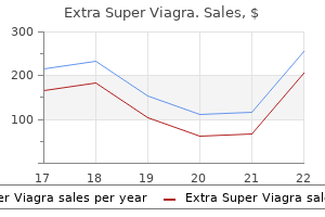
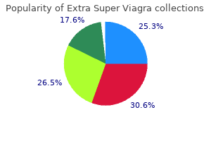
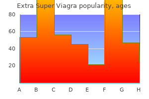
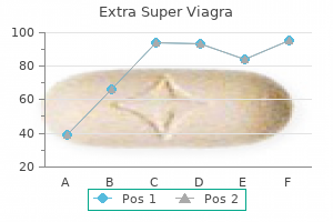
Extra Super Viagra
Extra Super Viagra dosages: 200 mg
Extra Super Viagra packs: 10 pills, 20 pills, 30 pills, 40 pills, 60 pills, 90 pills, 120 pills, 180 pills

Purchase extra super viagra 200 mg visa
The cervical esophagus has a propensity to metastasize to bilateral paratracheal and paraesophageal basins (29) impotence male extra super viagra 200 mg buy generic on line. Therefore erectile dysfunction medicine in dubai 200 mg extra super viagra generic free shipping, elective bilateral central compartment dissection is advocated in cervical esophageal carcinoma. Mediastinal dissection can also be recommended because of excessive price of mediastinal nodal metastasis (30,31). Retropharyngeal nodal illness is seen in 20% to 50% of patients with hypopharyngeal and cervical esophageal cancers (32-34). These knowledge recommend the importance of treating the retropharyngeal nodes with surgery or radiation. Iy is it requires one microvascular anastomosis in the neck with acceptable operative morbidity. In fact, 90% of the sufferers are in a position to return to normal oral diet Many reconstructive options exist for the closure of hypopharyngeal and cervical esophageal defect& It is imperative that the reconstructive swgeon plays a task in the multidisciplinary group. Howeve~; oncologic security must not be compromised for a selected reconstructive methodology. Disease-specific sw:vival and local control rates are similar to printed swgical series of similar stage instances with acceptable mOibidity (67,68). Advanced-stage tumors typically are treated with surgexy followed by postoperative radiation. A meta-analy3is of aver 87 trials using chemotherapy in head and neck cancers between 1965 and 2000 revealed an absolute survival advantage of 4. Therefore, the addition of chemotherapy to protocols within the remedy of hypopharyngeal carcinoma might enhance survival. These two trials compared the addition of chemotherapy to postoperative radiation to conventional postoperative radiation alone in patients with high-risk head and neck cancer (defined as extranodal unfold, a number of optimistic nodes, and positive surgical matgins). The use of chemotherapy as a half of induction or conament therapy has resulted in improved local/regional management rates and decreased distant metastasis. Howeve:t these studies checked out sufferers with larynx cancen alone without any hypophary:ngeal carcinomas. C: the esophagus is eliminated without thoracotomy, utilizing bimanual transabdominal and transcervlcal dlsHctlon. The abdomen Is brought up by way of the chest Into the neck In the posterior medlastfnum. They reported a response price of 78% in all hypopharyngeal patients and full response fee of 83% in sufferers who had a response to induction chemotherapy. Most importantly, there was not a significant survival distinction between the nonsurgical group and the surgical group. No distinction was found in local/regional control rates and disease-free survival at 5 years. Patients were staged T2-4/N0-2 on the hypopharynx for entry into the trial and assigned to receive induction chemotherapy followed by radiotherapy or an alternating chemoradiation routine. The primary weak spot of this examine is the alternating fractionation of the radiation, with the treatment routine lasting higher than 7 weeks, and complete dose delivered was reduced to 60 Gy. Chemotherapy and radiation have additionally been studied within the treatment of cervical esophageal carcinoma. Anderson Cancer Center retrospectively reviewed 132 sufferers who acquired concurrent chemoradiation (76). Sixty sufferers underwent esophagectomy after therapy and compared to the remaining 72 sufferers who had no surgery. The addition of induction chemotherapy was found to be superior to concurrent chemoradiation and surgery alone in a subsequent research (77). The 5-year total survival within the 1935 induction arm followed by concurrent chemoradiation and surgical procedure was 71% compared to 22% in the concurrent chemoradiation and surgery arm. Additional investigation is required to validate the findings in these studies, however multimodality remedy in the therapy of esophageal carcinoma is noted to have important survival benefit. Uneven tumor regression, ill-defined tumor borders, radiationinduced delicate tissue fibrosis, and poor wound therapeutic make salvage surgery a difficult endeavor (79). However, median survival in patients with recurrent head and neck most cancers without any extra remedy is 3. With salvage surgical procedure, median survival will increase to 14 months after therapy of recurrent disease within the pharynx (78). From these two observations, salvage surgical procedure is a viable option to extend survival. No significant survival distinction was noticed in any of the treatment arms (range 69% to 76%), which makes the point that surgical salvage is independent of prior remedy. Another research with promising end in salvage pharyngectomy for recurrent hypopharyngeal carcinoma comes from Toronto (82). They reviewed 72 patients with recurrent hypopharyngeal carcinoma who offered for surgical salvage. Their results demonstrated a 5-year overall survival of 31% with 5-year local and regional control price of 70% and 71%. The presence of extracapsular extension, constructive surgical margins, lymphovascular invasion, and nodal status was a negative predictor of local and regional management. Anderson Cancer Center, the place the authors reviewed their experience with induction chemotherapy and radiation for sufferers with hypopharyngeal carcinoma (7 5). Another report from Germany presented results which might be more dismal after salvage laryngopharyngectomy (83). They reviewed 28 patients with recurrent/persistent hypopharyngeal carcinoma, out of 134 sufferers who originally had organ preserving remedy, for surgical salvage. They discovered that solely 2 of the 20 sufferers who had histologically confirmed recurrence had been really tumor free and alive after a imply observation time of forty three. The authors concluded that patients have to be informed of high morbidity and poor oncologic end result after salvage surgery. An necessary level to address from these studies is the excessive morbidity in salvage surgery. The excessive perioperative morbidity in the setting of marginal oncologic control points to careful patient choice when performing salvage surgical procedure for the hypopharynx. In surgical series of laryngopharyngectomy patients, regardless of good native control, the permanent gastrostomy rate nonetheless approaches 16% (38). In a evaluation of the reconstruction of 153 postlaryngopharyngectomy patients, a stricture price of 15% was additionally observed (82). Organ preservation protocols even have significant dysphagia throughout and after treatrnent (85). Stricture rates have been reported as excessive as 20% postradiotherapy, with the hypopharynx primary being the most vital predictive issue (86). The introduction of intensity-modulated radiation therapy presents a current advancement in the treatment of head and neck carcinoma with the benefit of a more correct delineation of target volumes to spare the pharyngeal constrictors. However, this profit will not be related in hypopharyngeal and cervical esophageal carcinoma (87,88). Anderson Cancer demonstrated a 7% gastrostomy tube fee 2 years after organ preservation remedy for hypopharyngeal primary (Bhayani, et al.
Yellow Vine (Thunder God Vine). Extra Super Viagra.
- What is Thunder God Vine?
- Male contraception, menstrual pain, multiple sclerosis (MS), abscesses, boils, lupus erythematosus (SLE), HIV/AIDS, and other conditions.
- How does Thunder God Vine work?
- Are there any interactions with medications?
- Rheumatoid arthritis (RA).
- Dosing considerations for Thunder God Vine.
- What other names is Thunder God Vine known by?
- Are there safety concerns?
Source: http://www.rxlist.com/script/main/art.asp?articlekey=96800
Discount extra super viagra 200 mg fast delivery
Hearing loss in otic capsulesparing fractures tends to be conductive or blended (2 erectile dysfunction drug has least side effects discount 200 mg extra super viagra amex,27) impotence injections medications buy discount extra super viagra 200 mg on line. In addition to the predictive worth for variow problems and como:rbidities, categorization of fractures into otic capsule-sparing and otic capsule-disrupting accidents guides the indications for surgical int. Extravasation of blood from the mastoid emissary veins results in ecchymosis over the mastoid bone and mastoid tip. Following stabilization within the emetgency room and transfer to the ward or intensive care unit. Typical:findings embody fractures along the scutum and roof of the atemal audito:ry canal and/or tympanic membrane perforations. Hemotympanum and any related serow effusion typically resolve spontaneowly, with decision of concomitant conductive listening to loss, inside four to G weeks and simply require observation. Hearing is initially assessed clinically on the bedside with a progressively louder whispered voice. Following or concurrent with this evaluation, the neurotologic exam is performed It is extremely essential to assess facial nerve operate in the eme~gency room as early as possible. Lacerations are dosed after thorough cleansing and debridement of uncovered cartilage. Hematomas are drained and strain bolsters are sutured in place to dose the useless space and forestall a recollection of blood. Untreated, the auricular hematomas will result in an auricular chondropathy or "caulifiower ear. There is a 2- to 10-second latency adopted by 10 to 30 seconds of rotatory nystagmus within the geotropic direction-that is, with the higher half of the globe rotating in fast part course in course of the bottom. Ifthe affected person continues to experience vertigo or is experiencing fluctuating or progressive listening to loss longer than 1 week after the injury, a perilymph:fistula is suspected and pneumatic otoscopy can then be performed to check for nystagmus and wrtigo. The continued presence of spontaneous nystagmus following traumatic harm to the temporal bone is also suggestive of a perilymph fistula. Central vertigo may present with vertical or direction-changing nystagmus that fails to suppress and may even enhance with:fixation. However, preoperative asseument with cr scanning in sufferers with a conductive listening to lack of adequate magnitude to warrant exploration and ossicular reconstruction may provide useful data that will influence the swgical strategy. In complete incus dislocation, during which both the malleoincudal and incudostapedial joints have been disrupted, the ensuing position of the inrus may be quite variable: residing in the epitympanum lateral to the malleus head (causing a "Y" configuration when considered on coronal cuts), inside the extcmal audito:ry canal, or even not visible in any respect (presumably atruded from the physique or on�� within the mastoid air cells). Posttraumatic sensorineural listening to loss is extremely correlated with otic capsule-disrupting fractures, but many instances present with no radiologic findings. In these instances, an impulsive disruption of the membranous labyrinth, termed cochlear concussion, is theorized (39). Similarly, identification of perilymph fistula is often indirectly potential on radiographic imaging, however this prognosis could be advised when recognition is made of otic capsule-disrupting fracture, stapes fracture, lack of stapes bone, or pneumolabyrinth within the setting of persistent fluctuating listening to loss and vertigo (38,42). Consequently, in utterly asymptomatic sufferers sustaining a temporal bone fracture, who are neurologically intact. Sphenoid bone fracture, petrous carotid canal fracture, and pneumocephalus were evaluated. Further analysis of correlation between cr findings and carotid artery harm is warranted. Extrinsic suture traces (petro-ocdpital, temporo-occipital, occipitomastoid sutures), intrinsic suture strains (tympanomastoid, tympanosquamous, petrotympanic fissures), and intrinsic channels (cochlear aqueduct, vestibular aqueduct, glossopharyngeal nerve/glossopharyngeal sulcus, subarcuate artery/petromastoid canal, singular nerve/singular canal, Arnold nerve/mastoid canaliculus, Jacobson nerve/inferior tympanic canaliculus, and larger superficial petrosal nerve/facial hiatus) could all mimic fracture traces inside the temporal bone (38). Knowledge of their anatomic relationships and distinction with true fracture lines is necessary to prevent incorrect interpretation of Ciimaging. In addition, rare issues may occur, including abducens nerve damage, trigeminal nerve damage, Horner syndrome, carotid damage, sigmoid sinus thrombosis, traumatic porencephalic cyst formation, and intracranial dislocation of the mandibular condyle (12,46-51). Facial Nerve Injury Facial paralysis is a severely disfiguring complication of temporal bone fractures. This determine represents data based on giant prospective and retrospective collection of all consecutive sufferers handled for head harm or temporal bone fracture. The incidence of facial nerve harm in temporal bone fractures of the pediatric inhabitants is 3% to 9%, corresponding to that of the adult population (5,8,9). The incidence of facial paralysis within the literature has previously been reported as excessive as 30%. HoweveJ; this estimation is exaggerated because of sampling error: Simple, uncomplicated temporal bone fractures without facial nerve injury are often not referred for otolaryngologic session. Since the complete pool of temporal bone fracture sufferers had not been included in prior statistics, the reported incidence of issues has beforehand been fairly biased. If head trauma patients are carefully evaluated within the emergency room upon admission, previous to the administration of muscle relaxants, 27% of facial nerve accidents will current with immediate-onset facial paralysis; 73% may have facial movement in the preliminary examination and subsequently deteriorate (2). Delayed onset of facial paralysis is defined as documented facial operate in the emergency room that subsequently deteriorates. A delayed prognosis of facial paralysis happens when the patient is given a paralytic agent and intubated prior to examination of facial perform. These patients ought to be categorized as unestablished onset and handled much like the immediate-onset sufferers. In one large collection, 10% of patients fell into this unestablishedonset class (52). Since the vast majority of traumatic facial palsies resolve spontaneously, the decision of which injuries to discover is based on prognostic elements for poor end result. The elements that are assessed in predicting restoration of facial perform embody timing of onset (delayed vs. The delay of onset of paralysis following temporal bone fracture is the most important of the predictive elements. McKennan and Chole (53) described their experience with 17 sufferers with immediate-onset facial paralysis and 19 sufferers with delayedonset paralysis. Complete spontaneous recovery of facial operate occurred in 94% of the sufferers with delayedonset paralysis. In contrast, B out of the 17 immediate-onset paralysis patients had facial nerve transections. Chapter a hundred and fifty: Middle Ear and Temporal Bone Trauma 2417 Turner reviewed a large collection of traumatic facial paralysis treated conservatively (54). Complete restoration occurred in 94% of the delayed-onset circumstances and 75% of immediate-onset palsies. The one patient with delayed-onset paralysis who had no restoration of function developed the facial palsy coincident with acute otitis media. Similarly, in a latest systematic evaluate, the outcomes of 25 patients with instant paralysis and 20 sufferers with delayed paralysis handled with conservative management were assessed. Eighty % of the delayed paralysis patients had full recovery of facial operate whereas less than 40% of those with immediate paralysis recovered full facial perform (55). However, forty four out of 45 of their patients (including each immediate and delayed onset) had satisfactory restoration.
200 mg extra super viagra discount visa
These capabilities are m ade attainable by the three valves contained inside the la:ryn:x erectile dysfunction treatment shots extra super viagra 200 mg generic otc. These valves embrace the true vocal cords erectile dysfunction rap beat extra super viagra 200 mg discount free shipping, false vocal cords, and epiglottis to the arytenoids. Any dysfunction of these valves can lead to aspiration, airway obstruction, and adjustments in voice. The supraglottic larynx consists of the epiglottis (lingual and laxyngeal aspects), aryepiglottic folds, arytenoids, and bilateral false vocal cords. For staging functions, the epiglottis is split into suprahyoid and infrahyoid parts by a aircraft at the stage of the hyoid bone. The preepiglottic house i8 bound by the thyroepiglottic ligament inferiorly, the hyoepiglottic ligament superiorly, and the epiglottis posteriorly. At this stage, the thyroepiglottic ligament i8 an ineffec:tm: barrier and presents little resistance to tumor spread. Chapter 124: Advanced Laryngeal Cancer preepiglottic house by touring by way of lacunae on the laryngeal surface of the epiglottis into the house. Medially, the paraglottic space is certain by the quadrangular membrane above the ventricle and the conus elasticus below the ventricle. The conus elasticus extends superiorly from the superior border of the cricoid cartilage to mage with the inferomedial border of the vocal ligament. This ligament resists the atralary:ngeal unfold of early glottic and subglottic tumon. The paraglottic area allows wmoiS to turn into transglottic (superior and inferior to the ventricle) tumoiS and in addition to impair the motion of the true vocal twine. At the extent of the glottis, the vocalis tendon attaches to the thyroid cardlage through Broyles ligament which is an ineffective barrier and will enable tumors to unfold into the thyroid cartilage and preepiglottic space. This area the place the tendon attaches to the thyroid cartilage has no perichondrium. As beforehand discussed, the true vocal cords include a lamina propria consisting of three layers. Lymphatic spread of the tumor usually happens in a predictable manor and follows the embryologic origins of the larynx. The supraglottis is derived from the midline buccopharyngeal primordium and branchial arches 3 and 4. The glottis is derived from the tracheobronchial primordium and branchial arches four, 5, and 6. These differences account for the high incidence of lymphatic unfold for supraglottic carcinomas and the low incidence of lymphatic unfold in glottic carcinomas. Since the lymphatic drainage of those sites is predictable, this allows surgeons to restrict neck dissections to the lymph node levels most at risk In 1972, Lindbag mapped out affected lymph nodes based on the first web site of the tumor. This syatem is an anatomical staging system characterized by the extent of invasion of the pri. This information is obtained by way of physical examination, imaging, and endoscopic examination and at last confirmed by surgical pathology from the resected tumor. There are histologic facton that can point out a poorer prognosis together with perineural invasion, angiolymphatic spread, extracapsular spread of nodal metastasis, and histologic grade of the tumor. There are additionally research exhibiting that a number of chromosomal and molecular markers are indicative of a poor prognosis. Some of those markeiS embrace overexpression of mutant p53 protein (12), a high Ki67 or proliferating cell nuclear antigen rating, and the expression of c-m:yc oncogene or int-2 gene (13,14). Some propose utilizing a high-resolution computed tomography (Cf) to assess the volume of tumor. Others suggest that any tumor with any change in vocal wire mobility be thought of a T3 or T4. Other analysis is including comorbidities into the staging system and is looking at how it relates to the general consequence (15) (Table 124. Patients could complain of hoarseness (glottic carcinoma), muffled/ hot potato voice (supraglottic carcinoma), dyspnea, dysphagia, and otalgia (usually supraglottic). Patients may also current with a historical past of aspiration pneumonia, stridor, hemoptysis, or sore throat. Constitutional signs might include weight loss, fevers, chills, and evening sweats. Glottic tumors often present sooner than supraglottic Chapter 1 24: Advanced Laryngeal Cancer 1965 cancera due to the early change in -voice. Supraglottic tumors present with fewer signs in the early stage and subsequently are often identified at a later stage. Subglottic tumors usually current as superior stage illness with airway obstruction. Past medical histocy is also necessary in assessing affected person comorbidities, historical past of most cancers, and immunologic statua. There is a optimistic relationahip betM:en the quantity of cigarettes consumed and the increased threat of creating laryngeal most cancers. There can be a synergistic impact when tobacco is combined with alcohol use resulting in evm greater rates of malignancy (16). Treatment of tobacco and alcohol dependency must be thought of within the postoperative interval. Delirium tremens and tobacco withdrawal can lead to a sophisticated and prolonged postoperative coune and impaired therapeutic. A complete aamination together with a complete head and neck examination ought to be performed on all sufferers to decide staging, the presence of second primaries, and different findingB/comorbidities that will affect really helpful remedies. A complete head and neck examination includes examination of the skin, eyes, em, oral cavity, and pharynx, including palpation of the ground of mouth, base of tongue, and tonsils along with the larynx. A more detailed examination of the laxynx and pharynx is carried out utilizing flexible fiberoptic laryngoscopy at which time. The extent of the lesion as properly as the mobility and place of the vocal cords are assessed. Videostroboscopy can be used to detect subtle changes in vocal wire perform and mobility but is extra useful in early-stage illness. It makes use of fluorescence-tagged glucose molecules after which meuures the uptake inside the tissues. It has limited utility in figuring out the Imaging Ph:ysical cwnination is often restricted within the evaluation of tumor depth and nodal metastases, and radiographic imaging is useful in determining the extent of the illness. Imaging can present info regarding cartilage invasion, extension into the preepiglottic and paraglottic areas, and atralacyngeal spread of tumor. This software is noninvasive, low price, and is an effective approach for lymph node evaluation.
Extra super viagra 200 mg buy free shipping
The incus interposition is accomplished by drilling a cup ultimately of the lateral means of the incus that may fit over the capitulum of the stapes impotence and smoking 200 mg extra super viagra cheap fast delivery. If erectile dysfunction pills at gnc order 200 mg extra super viagra amex, along with the incus dislocation, the stapes superstructure is fractured, the long process of the incus is left intact and the physique and short process sculptured. The superior floor of the body of the incus is fashioned to relaxation underneath the manubrium and the long process sits on the footplate. A number of complete ossicular replacement prostheses are available for this purpose as nicely. A unique drawback happens when the stapes superstructure is fractured but the incus remains connected to the malleus. C: the incudostupedial joint was further stabilized with addiuonal fascia positioned circurnferentially across the joint. The patient had complete dosure of air-bone hole and no sensorineural listening to loss. Thesensorineural injury from traumatic incus dislocation seems to happen in the 2 to 4kHz vary. High-frequency bias for cochlear acoustic injw:y is noticed in traumatic injw:y to the ossicular chain. This bias could also be closely related to the phenomenon of direct acoustic injw:y to the cochlea seen in impulsive or extreme noise-induced listening to loss, as both mechanisms involve delivery of acoustic energy via the oval window and cochlear basal tum. Such injuries to the cochlea and neurosensory listening to mechanics is typically referred to as cochlear performed for persistent otitis media. Hough and Stuart (130) report closure of the air-bone hole to within 10 dB in 78% of patients and complete closure in 45%. In addition, bilateral temporal bone fractures can also end in bilateral profound sensorineural listening to loss (132). In addition to the risk of sensorineural listening to loss &om temporal bone trauma, sufferers who maintain closed head injuries normally, with or with out temporal bone fracture, are susceptible to acute sensorineural listening to loss, which might further progress with time 133). Multiple pathogenic mechanisms can contribute to posttraumatic deafness: disruption of the membranous labyrinth, avulsion or trauma to the cochlear nerve, interruption of the cochlear blood supply, hemorrhage into cochlea, and perilymphatic fiswla Another proposed mechanism is endolymphatic hydrops ensuing &om obstruction of the endolymphatic duct by the temporal bone fracture (134). Acoustic ttauma related to temporal bone fractures and incus dislocations frequently contributes to Chapter one hundred fifty: Middle Ear and Temporal Bone Trauma 2429 oonrussion, when sensorineural listening to loss is clearly documented on postinjury audiogram in the absence of any fracture spanning the otic capsule or temporal bone. The traditional literature for concussive neurosensory listening to loss suggests a predilection for acoustic injury at higher frequencies, centered around 4kHz (136,137). These older studies are limited by their small examine populations, case-study-type desig~ and descriptive reporting of outcomes. Additionally, outcomes often integrated audiometric findings years following traumatic damage. The majority of these sufferers were involved in motor vehicle accidents, with the rest suffering closed head accidents from falls or assaults. As su~ the prognosis for recovery of function in sufferers with anacusis or profound deafness on account of temporal bone and cochlear trauma is extremely poor; howeve~; patients with moderate to extreme loss could have some hearing recovery (131,139). Cholesteatoma and External Auditory Canal Stenosis Cholesteatoma formation may happen many years after a temporal bone fracture (128,141). The typical location for cholesteatoma ensuing from epithelium trapped within the fracture line is in the epitympanum and antrum. The fracture line alongside the posterior superior canal wall and scutum expands after which closes trapping the canal skin. As the trapped pores and skin grows, it expands into the epitympanum and antrum forming a cholesteatoma. The ingrowth of epithelium by way of a displaced fracture line can also extend into the identical area. Traumatic implantation of tympanic membrane skin will lead to cholesteatoma formation within the mesotympanum. Blast injuries can outcome in displacement of keratinizing stratified squamous epithelium into the mastoid air cells, mesotympanum, epitympanum, and even intracranium (142). Posttraumatic canal cholesteatomas are the most preventable by careful follow-up, debridement, and stenting when narrowing progresses. The ear canal can be dilated with the insertion of accelerating numbers or sizes of merocel sponge packs. Once the canal is adequately dilated, a larger merocel sponge is inserted to preserve the lumen. A large vented customized ear mould is sometimes required to preserve the lumen in the long run following extreme canal injuries. Posttraumatic cholesteatoma involving the attic, antrum, or mastoid air cells will often develop for a number of years prior to detection. Until the cholesteatoma entails the ossicular chain with resultant conductive listening to loss, erodes into the labyrinth inflicting vertigo or sensorineural listening to loss, compresses the facial nerve leading to facial paresis, or grows into the middle ear cleft and could be visualized on bodily examination, the expansion will go undetected. Vascular Injuries Injuries to the intratemporal carotid artery are rare but potentially life-threatening complications. Fractures that traverse the temporal bone adjacent to the carotid artery are inclined to involve the softer fibrocartilage of the foramen lacerum rather than the stouter, extra dense bone of the carotid canal. Temporal bone fractures in kids: a review with emphasis on long-term sequelae. Ort S, Beus K, Isaacson J, Pediatric temporal bone fractures in a rural population. The medical correlation of temporal bone fractures and spiral computed tomographic scan: a prospective and consecutive study at a degree I trauma middle. Bilateral longitudinal temporal bone fractures: a retrospective evaluation of seventeen instances. Radiographic classification of temporal bone fractures: medical predictability using a model new system. The pathology symptomatology and analysis of sure widespread issues of the vestibular system. In such circumstances, damage to the petrous portion of the carotid artery ought to be suspected and the patient taken immediately to either the operating room for carotid ligation or the angiography suite for balloon occlusion. In sufferers with significant hemorrhage from the ear or nostril or with fast neurologic deterioration, conventional interventional angiography is indicated for confirmation of carotid damage in addition to immediate therapeutic intervention if needed. Histopathology of temporal bone fractures: implications fur cochlear implantation. Fracture of glenoid fossa and traumatic dislocation ofmandibular condyle into center cranial fussa. Intracranial dislocation of the mandibular condyle: report of two circumstances and evaluation of the literature. Dislocation of the mandtbular condyle into the center cranial fossa: report of a case with 5 year cr followup. Management offadal paralysis ensuing from temporal bone fractures: our experience in a hundred and fifteen cases. Some anatomical and pathophysiological data related to facial nerve harm and restore. Prognostic value of evoked and standard electromyography in acute facial paralysis. Traumatic intratemporal facial nerve damage: administration rationale fur preservation of function.
Extra super viagra 200 mg discount without prescription
In most reviews erectile dysfunction injection device extra super viagra 200 mg generic line, the male-to-female ratio was 3:1 impotence beta blockers extra super viagra 200 mg buy line, and the median age was 50 yean. Indirect examination of the postna8al area ought to be carried out with a mirror though, in some patients, the anatomical variation of the nasophaJ:ynx. These investigations are important to document the extent of illness within the nasopharynx and its involvement of surroWlding tissue. Intracranial tumor extension via the foramen ovale with perineural spread can be detected and this offers evidence of cavernous sinus involvement without skull base erosion (47). The data supplied by way of cr is important for staging and likewise the selection of therapeutic measures for some sufferers (48. Enhancing soft tinue infihr�teS the normal anatomy of the soft tissues close to the skull base, obliterating the conventional fat planes that can still be seen on the best. A: Axial unenhanced T1-welghted, (8) axial fat-suppressed T2-welghted, (C) coronal postcontrast fat-suppressed T1-welghted, and (D) sagittal unenhanced T1-welghted Images of the nasopharynx demonstnrt� a large tumor (arrows) that has dlastroyed a lot of the central skull base. There Is a right-sided mastoid Qffuslon (arrow In A), which ought to at all times prompt an analysis of the nasopharynx for Eustachian tube dysfunction. The arrowheads present denervation atrophy of the tongue reflecting involvement of the hypoglossal nerve. Blood and mucus overlaying the tumor need to be removed by a separate suction catheter for a clear view of the pathology. Biopsy forceps should also be inserted alongside the endoscope for taking a biopsy of the tumor beneath direct imaginative and prescient. Despite all these adwntaga, the visible image gathered with the flexible endoscope is inferior to that of the rigid endoscope. Sometimes a larger biopsy forceps may need to be inserted by the facet of the versatile endoscope to get hold of more substantial amount of tissue for histologic examination. Rigid endoscope (70- inserted by way of the oral cavity, inspecting the nasopharynx from below. On the opposite hand, lateral tumor extension to the paranasopharyngeal area indicates extra superior disease. The T3 disease oovers tumoD that have involved the skull base or the paranasal sinuses. T4 tum on are those who have prolonged to the infratemporal fossa, orbit, hypophary:nx. For other head and neck cancef8, N1 is lower than 3 an in dimension and N2 is larger than three em. The distinction between N2 and nasopharyngeal tumor can be seen extending from 1he right lateral wall onto the roof of the nasopharynx (Tumor. The measurement of 6 em is the one issue for size Laterality and website of involvement such as the retropharyngeal region and the supraclavicular fossa are different important components in determining the N staging. Stage N3 disease referred to lymph nodes larger than 6 em (N3a), or nodes that had prolonged to the supraclavicular fossa (N3b). ForM-staging, M1 represents distant metastases, together with any lymph node involvement below the extent of the clavicle. Rr although effective may produce undesirable problems as the radiation may affect the structures around the nasopharynx. These constructions restrict the quantity of radiation that may be delivered to the tumor. In general, the radiation dose given to the first tumor iJ within the range of 65 to 7 5 Gy and that to the involved neck nodes sixty five to 70 Gy. This treatment has efficiently managed T1 and T2 tumors in between 75% to 90% of cases, and T3 and T4 tumon in 50% to 75% of circumstances (72,73). Sometimes for T1 and 12 tumoa, using a booster dose utilizing intracavitaty brachytherapy has proven to enhance tumor management by 16% (74). Although stereotactic radiosurgery has also been used for delivery of the booster dose (75), the hypofractionated therapy iJ related to undesirable side effects and is probably higher resetved for the treatment of peDistent and recurrent disealle (76). It additionally has the power to deal with primary and regional lymphatic in a single volume thus eliminating the dose uncertainty drawback on the junction between the first tumor and neck lymphatic taiget volumes. The Intergroup trial employed both concurrent and adjuvant chemotherapy within the examine arm and reported an absolute improvement of survival of 31 o/o at three years. Both adjuvant chemotherapy trials had limitations since nonplatinum chemotherapy was used in one study and chemotherapy compliance was rather poor within the different examine. Another examine on neoadjuvant chemotherapy followed by concomitant chemoradiotherapy additionally reported wonderful total survival with acceptable toxicity (102). In view of the ototoxicity of cisplatin, different chemotherapeutic brokers have been used. Although these sufferers have more advanced illness, the end result of the remedy was corresponding to different platinum-based adjuvant chemotherapy studies and the compliance price was acceptable (103). To attain a high profitable salvage rate, early detection and administration of the suitable therapy is important. The presence of malignancy can normally be confirmed with biopsy through endoscopic examination. Fine needle aspiration cytology was diagnostic only in 50% of the time and incessantly surgery has to be carried out for progressive enlarging cervical lymph node. Even when there were synchronous locoregional failures, aggressive therapy ought to be thought of for chosen patients (109). Radical neck dissection performed as surgical salvage has reported to achieve a 5-year tumor control rate of 66% in the neck (111) and a 5-year disease development free 44% (112). Pathologic studies have proven that for those persistent or recurrent tumors in the neck nodes, the disease involvement of the local tissue was in depth. Many lymph nodes appeared freed from tumor macroscopically have been identified to harbor malignant cells on histologic examination. Malignant cells could presumably be seen to prolong outdoors the capsule of the lymph nodes and mendacity near the accent nerve and inner jugular vein. Tumor clusters had been additionally seen within the stem mastoid muscle and amongst different tissues in the neck (113). A recent study confirmed that as the neck nodes in level I was occasionally involved, stage I ought to be preserved within the neck dissection (114). These therapy options are relevant when the persistent or recurrent tumor is small and localized within the nasopharynx. Stervotac:tic Radiotherapy the local tumor control fee achieved with stereotactic Rf for management of persistent or recurrent tumor was 72% at 2 years and 86% at three years (117). A latest research with 90 sufferers handled using this strategy for locally persistent and reau:hire disease reported a 3-year local management fee of 83% (118). Brachytherapy With brachytherapy, the radiation supply is positioned directly into the tumor. Thus, the radiation dose is highest at the soun::e in the tumo~ and this declines with rising distance from the tumor. The brachytherapy radiation supply additionally delivers radiation at a continuous rate, and this gives radiobiologic advantage over fractionated atemal radiation. The radiation source was placed both in a tube or a mould after which inserted into the nasopharynx.
Discount extra super viagra 200 mg on-line
If the edge has improved by ~15 dB at one frequency or 10 dB at two or extra consecutive frequencies erectile dysfunction protocol free 200 mg extra super viagra cheap fast delivery, or if the discrimination is significantly improved causes of erectile dysfunction include quizlet 200 mg extra super viagra buy, patients are thought of steroid responders. Their medication is then slowly tapered over 8 weeks to a maintenance dose of 10 to 20 mg every other day. Clinical observation suggests that patients with a remedy length of fewer than 6 months are at increased danger of relapse compared with these treated for six months or longer. Some sufferers have enchancment in threshold, some in discrimination only, and some in both areas. Some patients with hearing loss fluctuation and progression before therapy present stabilization of their listening to with out actual improvement. Historically, these instances have been considered nonresponders, however this concern is at present under reassessment. The majority of responders slowly taper off the steroid dose, wean from steroids, and do nicely. Some sufferers, particularly in the pediatric age group, may sometimes present steroid-dependent hearing loss. Such patients usually develop unacceptable side effects of chronic steroid administration. Methotrexate has been used as a half of a prednisone-sparing routine; howev~ a recent randomized, potential managed, multicenter trial has proven methotrexate to be no higher than a placebo at sustaining a remission in these sufferers (56). Longterm administration carries a big threat for gastritis and ulcers, fluid retention and weight gain, blood pressure lability, altered blood sugar metabolism and diabetes, avascular necrosis of the hip, temper adjustments or psychiatric problems, sleep disturbance, accelerated cataract formation. Although high-dose corticosteroids are associated with serious unwanted effects, with acceptable patient choice, monitoring, and affected person training. A variety of options to systemic corticosteroids have been proposed, including methotrexate, etanercept, and cyclophosphamide. Low-dose methotrexate appeared to be useful as an adjunct in management of steroiddependent hearing loss (60). Howev~ as noted earlier in this chapter, the recent massive trial indicates that it might not serve an efficient function as a prednisone-sparing drug. Anecdotally, it appears to work nicely together with methotrexate because of its steroid-sparing impact. Toxicity includes severe myelosuppression, opportunistic infection, hair loss, cystitis, infertility, and increased risk of malignancies. Many patients, when confronted with the danger of this medication, would quite contemplate cochlear implantation. There are, however, no printed series in which these therapies have been systematically utilized. Radiologic findings for these patients embody thickening of the calvarium with ill-defined densities and poor definition of cortical margins of the inside ear and inside auditory meatus. The conductive portion of this hearing loss is usually down-sloping and 20 to 30 dB in severity. It is believed to arise primarily from adjustments in bone density and geometry that interfere with normal hearing mechanisms. In addition, sufferers with Paget can current with tinnitus and mild vestibular complaints. Treatment of Paget disease relies primarily on bisphosphonate remedy, which inhibits osteoclast exercise, thereby inhibiting bone reabsorption. Osteogenesislmperfecta Osteogenesis imperfecta (01), also called Hoeve-de Kleyn syndrome, is a connective tissue dysfunction that manifests as a wide selection of sorts with vastly differing severity. Type I 01 is the mildest form of the illness and is inherited in an autosomal dominant sample. This is the classic form of the illness and is the most common sort, with an incidence of 1:30,000. It is inherited in an autosomal recessive sample and has an incidence about half that of sort I, 1:60,000. They also reveal blue sclera at a young age but this transitions to a normal white sclera later in life. Conductive hearing loss results from fractures to the ossicles, most commonly the long process of the incus or the stapes crura. It outcomes from elevated exercise of osteoclasts and osteoblasts, which manifest as bony hypertrophy and remodeling. Its prevalence increases dramatically with age, with 3% of individuals over the age of 40 demonstrating histopathologic evidence of the illness and 11% of individuals over the age of 80 demonstrating histopathologic proof of the disease (69). The illness is far more frequent among males than girls with relative incidence of about 4:1. Patients with Paget disease usually current of their sixth decade, most commonly with complaints of an enlarging skull, progressive kyphosis, Chapter 156: Otologic Manifestations of Systemic Disease: Includes Autoimmune Inner Ear Disease 2527 loss has been addressed successfully with amplification or ossiculoplasty to exchange fractured ossicles. In addition, stapedectomy can be effective but should be performed very delicately because of the fragility of the ossides. It describes the replacement of normal cancellous bone by spicules of woven bone in a fibrous stroma. The affected bone is mostly a rib, the cranium, the proximal femur, or the tibia. The polyostotic type normally presents earlier and will proceed to progress throughout life (74). Radiologic findings typical of fibrous dysplasia include radiolucent areas with a well-defined easy or scalloped edge and a ground-glass look. Otologic manifestations of the illness manifest from craniofacial involvement, which occurs way more commonly within the polyostotic (50% to 100%) type than in the monostotic kind (10% to 30%). In addition, patients can develop a canal cholesteatoma or involvement of the ossicular chain (75). Surgical remedy with canaloplasty, meatoplasty, or cholesteatoma resection can also benefit these sufferers. Facial nerve weak point can even develop from compression at the stylomastoid foramen. This results in intracellular accumulation of mucopolysaccharides and abnormally large dysfunctional cells with vacuolated cytoplasm. There have been 10 identified lysosomal enzyme deficiencies that make up this group. Examples of those embody Hunter syndrome, an X-linked disease attributable to an absence of L-iduronidase; Hurler syndrome, an autosomal recessive disease caused by a scarcity of iduronate-2-sulfate; and Morquio syndrome, caused by a scarcity of N-acetylgalactosamine-6-sulfate or beta-galactosidase. Diagnosis of each is made by way of assays that check for the presence of every of these enzymes. The otologic manifestation of mucopolysaccharidoses sometimes presents as a combined listening to loss. The conductive element is normally secondary to eustachian tube dysfunction and resulting serous otitis media. Treatment of those patients is often supportive with long-term tympanostomy tube placement to alleviate serous otitis media (78). Current analysis is investigating the efficacy of gene remedy for these patients (79).
Syndromes
- Genetic muscle diseases
- Head size significantly below normal
- Check with your doctor or nurse before taking any over-the-counter medications or herbal supplements. This includes medications such as acetaminophen, aspirin, or ibuprofen.
- Time it was swallowed, inhaled, or touched
- Blood in the urine
- Hematoma (blood accumulating under the skin)
- Nausea
- Nuts
- Strep throat
- Bed rest
Purchase 200 mg extra super viagra fast delivery
There is a marked distinction in the medical habits of tumors arising from the jugular paraganglia versus the tympanic paraganglia leading causes erectile dysfunction cheap 200 mg extra super viagra mastercard. The vagal paraganglia are distinctly completely different from the jugulotympanic paraganglia erectile dysfunction home remedies generic extra super viagra 200 mg otc. Thus vagal paragangliomas have distinct therapeutic implications primarily based on their close affiliation with the superior portion of the vagal nerve and their adjoining neurovascular structures. Patienu forming these tumors at greater altitude present a lower price of bilaterality and a lesser diploma of familial histoty of paragangliomas (12-14). Secreting paragangliomas account for approximately 2% of all instances of secondaty hypertension. Serum catecholamine& will show elevated levels of norepinephrine in functioning ex:tra-adrenal paragangliomas. Elevated serum epinephrine is indicative of a concurrent pheochromocytoma (3, 15). Malignant paragangliomas represent a small subset of extra-adrenal paragangliomas that will be inclined for regional lymph nodes and distant metastatic illness. Sporadic paragangliomas have a higher fee of malignancy than familial-type paragangliomas. Orbital and latyngeal paragangliomas have the very best price of malignancy, roughly 25%. Vagal paragangliomas have a malignancy price of as much as 10%, followed by jugulotympanic paragangliomas with a malignancy rate of 5% and carotid physique tumors being final at 3% to 6%. Malignancy is confirmed by tumor present in lymph nodes or distant metastatic sites. Suppon for hypoxia-induced hyperplasia comes from evidence obtained in high-altitude physiologic research. Cows, guinea pigs, rabbits, and dogJ expertise carotid body hyperplasia when living at excessive altitudes, which exposes them to a hypoxic condition, and this has additionally been described in humam. Another clinical observation lending suppon to these theoriea is the finding that patients suffering from circumstances resulting in hypoxemia, such as cystic fibrosis, cyanotic hean illness, and chronic obstructive pulmonary disease expertise carotid physique hyperplasia and people affected by persistent obstructive pulmonary illness have the next rate of carotid body tumors. This inherited tumorigenic predisposition is transmitted in an autosomal dominant style with age-dependent and incomplete penetrance. Neurofibromatosis kind 1 is also associatedwith both pheochromocytomas and glomus tumors. In instances where successive generations of a household have been harboring the mutations, patients develop tumors at progressively youthful ages. This discovering is an efficient instance of genetic anticipation, during which the mutation appears to be more severe with succeeding generations. Familial glomus tumors constitute roughly 20% of affected patients, for which the genetic defects are identified. Oncology analysis implicates a quantity of processes as essential to tumor formation and progress. Two that attract a significant amount of attention are angiogenesis and apoptosis, and these processes have been examined in paragangliomas as properly (25). Angiogenesis is a critical occasion in the growth and sustained development of several tumor types. Given the vascular nature of paraganglion tissue, questions in regards to the role that angiogenesis performs within the improvement of paragangliomas. It has been postulated that this was according to a paracrine mechanism for tumor development (26). Additionally, hypoxia induces expression of hypoxiainducible factor 1, which participates within the angiogenesis cascade. Pheochromocytomas exhibit ubiquitous hypoxiainducible issue 1 secondary to mutations leading to cell hypoxia. Additionally, secreting tumors can produce hypertension, tachycardia, sweating, and nervousness secondary to catecholamine launch. Carotid Body Tumors Carotid physique tumors sometimes present as a slowly enlarging asymptomatic deep neck mass at the stage of the carotid bifurcation. As the tumor enlarges, a bruit could additionally be auscultated and in addition to a neck mass, a parapharyngeal extension results in the lateral displacement of the taste bud medially and anteriorly. Cranial nerve paralysis signs are unusual and present in very giant tumors with superior extension toward the jugular foramen. Shamblin classification is often used to stage carotid physique tumors, and though this is based on intraoperative findings, it might be utilized to radiologic findings prior to remedy (Table 127. Jugulotympanic Paragangliomas Jugular and tympanic paragangliomas occur predominantly within the fifth and sixth a long time of life with an overwhelming feminine predominance. In their later phases, both varieties produce comparable signs including cranial nerve deficits, whereas in the early levels tympanic and jugular pcu:agangliomas differ in presentation. Tympanic paragangliomas present early with pulsatile tinnitus and conductive hearing loss. On otomicroscopic examination, a red-blue center ear mass that blanches with constructive pressure on pneumatic otoscopy is appreciated (Brown sign). Extension through the tympanic membrane will produce an ear canal polypoid mass that will spontaneously cause bloody otorrhea. Involvement of the facial nerve, normally in the mastoid, can produce facial paralysis as a presenting symptom. Anterior development towards the pericarotid air cells can involve the petrous carotid artery and occupy the eustachian tube. Medial progress into the infralabyrinthine air cells can attain the petrous apex and petroclival area in addition to cavernous sinus. Intracranial atension is possible by way of extension through the jugular foramen and/or pettous apa. Jugular paragangliomas, due to their origin location within the jugular bulb, have the capacity for early and extensive skull base invasion with involvement of cranial ne:rves 9 through 12. Growth via Jacobson nerve canal and the hypotympanic air cells results in involvement of the middle ear and mastoid bone resulting in conductive hearing loss, tinnitus, and facial paralysis. Erosion of the jugulocarotid backbone results in involvement of the vertical pettous carotid artery, and further anterior extension leads to involvement of the horizontal portion of the petrous carotid artery. More medial extension to the infralabyrinthine air cells result in involvement of the petrous apa. Further medial extension along this pathway results in involvement of the clivus and ave:mous sinus. Occasionally, jugular paragangliomas will escape the confines of the temporal bone and contain the infratemporal fossa and parapharyngeal house. The attendant symptomatology is hoarseness, swallowing difficulty, hemipalatal dysfunction with nasal air escape. Type 1 Type2 Type3 Type4 Small tumor involving the jugular bulb, center ear, and mastoid Tumor ex:tending beneath the intemal auditory canal. May have intracranial extension Tumor ex:tending past the petrous ape~t into the clivus or infratemporal fossa.

Discount extra super viagra 200 mg without prescription
Globular enlargement happens less incessantly and is the outcomes of tumor invasion of the adjoining dura erectile dysfunction numbness purchase extra super viagra 200 mg overnight delivery. Contrast-enhanced axial cr often reveals a linear central hypointense optic nerve surrounded medially and laterally by a hyperintense nerve sheath-a well-known discovering termed Rtram-tracking impotence drug extra super viagra 200 mg purchase without prescription. Most meningiomas are histologically benign, with aggressive varieties occurring mostly in younger patients. Surgery is indicated for secondary meningiomas when compression of surrounding buildings causes important visual, beauty, or intracranial morbidity. Treatment of primary optic nerve sheath meningiomas is dictated by the diploma ofvision loss and the presence of intracranial extension, which occurs in as a lot as 15% of sufferers. Ifvision loss is progressive, imaginative and prescient function can be improved with radiation remedy. Surgical excision is performed when intracranial extension threatens the optic chiasm or contralateral optic nerve, or when the tumor has caused marked imaginative and prescient loss and important proptosis (7). Schwannoma (Neurilemoma) Schwannomas are uncommon benign tumors of the peripheral nerve sheath. They arise as a end result of proliferation of the myelin-producing Schwann cells and barely bear malignant transformation. They happen mostly in adults between 20 and 60 years of age and are most often found within the superior or intraconal orbit. They may cause slowly progressive proptosis, and site on the orbital apex might trigger progressive vision loss. Histopathologically; these lesions are well-encapsulated and are divided into Antoni A sort (solid tissue with spindle cells arranged in whorls or palisades) or Antoni B kind (loose myxoid tissue containing stellate cells). Long-standing lesions could demonstrate bony transforming, lower density areas of mucinous cystic degeneration, and calcification. Treatment consists of full surgical excision of the tumor and capsule (17-19). It can happen in youngsters and adults, and accounts for as a lot as 11% of all orbital lesions (2). Almost any orbital construction could additionally be involved, together with a number of extraocular muscle tissue (orbital myositis), the sclera and posterior tenons (sclerotenonitis), or the optic nerve sheath (inflammatory optic neuritis). Ocular motility restriction, diplopia, proptosis, conjunctival inflammation, and eyelid erythema and edema may occur. Decreased imaginative and prescient may be seen when the optic nerve, sclera, or posterior tenons are concerned. Children usually current with systemic indicators corresponding to fever, emesis, stomach pai~ and lethargy. In distinction, bilateral involvement in an adult alerts an underlying systemic vasculitis (7). A speedy and dramatic response is usually noticed within a couple of days, and steroids are tapered slowly over a quantity of weeks to prevent rebound inflammation. Biopsy may be required in sufferers with atypical signs, imaging options, therapy failure. This is particularly true for lacrimal gland lesions, as an aggressive malignancy may also current with inflammation. Biopsy-confirmed refractory circumstances could respond to low-dose radiation remedy or immunosuppressants. A sclerosing variant has been found to be less responsive to systemic steroids and radiation, and should reply to intraorbital steroid injection (20). The commonest presenting sign is eyelid retraction, often with lateral temporal flare of the palpebral fissures. Proptosis, lagophthalmos, conjunctival chemosis and hyperemia over the rectus muscle insertions, extraocular muscle restriction, and optic neuropathy may be noticed. Vision-threatening optic nerve compression or publicity keratopathy could also be temporized with corticosteroids and/or orbital radiation therapy. Giant cell arteritis, polyarteritis nodosum, Wegener granulomatosis, and connective tissue disorders similar to systemic lupus erythematosus, dermatomyositis, and rheumatoid arthritis may involve orbital vessels. They are thought to arise from epithelial and subepithelial tissue that becomes entrapped inside bony sutures throughout embryogenesis or becomes implanted within the orbit as the end result of surgical or nonsurgical trauma. The clinical presentation and administration of these lesions is much like that of dermoid cysts (22). Their central lumens are filled with a mixture of keratin, oil, and infrequently hair shafts. Approximately 5% of dermoid cysts arise from conjunctival epithelium (nonkeratinized stratified squamous epithelium with goblet cells). They are often discovered within the anterior superolateral orbit at the frontozygomatic suture, but may be found superomedially on the frontoethmoidal suture, or at some other bony suture. Patients typically current within the first decade oflife with a slowly progressive, painless, subcutaneous mass. Deep orbital dermoid cysts typically present later in adulthood with proptosis and globe displacement Dermoid cysts may current as orbital irritation, which is attributable to an intense granulomatous reaction incited by cyst leakage. In younger patients, you will need to distinguish medial dermoid cysts from encephaloceles, mucoceles, or dacryoceles (22). A dumbbell configuration may be seen with cysts that reach via the suture into the temporalis fossa, sinuses, or cranial vault Bony remodeling or erosion may happen in long-standing cases. If rupture does happen, copious irrigation should be carried out to avoid marked inflammatory reaction. They might come up from cutaneous, conjunctival, respiratory, or apocrine gland epithelium. Orbital lesions are less common, and should represent posterior extension or a main lesion. Four varieties have been described, together with embryonal, alveolar, pleomorphic, and botryoid varieties. Embryonal is the most typical subtype, alveolar is probably the most malignant subtype, and the pleomorphic subtype carries the most effective prognosis. The illness is nonhereditary and presents at a median age of 8, with 90% of instances being diagnosed in patients prior to the age of 13 (23). Patients normally present progressive proptosis over days to weeks, with one in three youngsters presenting with ptosis and a palpable mass. Downward globe displacement could also be observed, reflecting the predilection of this tumor for the superomedial quadrant. However, the tumor might happen in any quadrant the alveolar subtype tends to happen extra frequently within the inferior orbit On Cf imaging, the tumor typically shows reasonably well-defined to ill-defined margins and an irregular shape with reasonable distinction enhancement. Once a analysis is confirmed by biopsy, treatment consists of primary chemoradiation therapy. With the advent and widespread use of these remedy modalities, survival charges have been found to be over 90o/o in most series (24,25). Fibrous Dysplasia Fibrous dysplasia was discovered to account for roughly 1% of all orbital tumors in a recent collection (2).
Discount 200 mg extra super viagra
The feasibility of randomized trials of surgical versus nonsurgical remedy is uncertain erectile dysfunction drug related buy 200 mg extra super viagra free shipping. However impotence over 60 order extra super viagra 200 mg without prescription, there are distinctive challenges to attempts at conducting these studies for hypopharyngeal procedures and skeletal surgery. A navel volumetric magnetic resonance imaging paradigm to study upper airway anatomy. Does severity of obstructive sleep apneajhypopnea syndrome predict uvulopalatopharyngoplasty end result Minimally invasive single-stage multilevel therapy for obstructive sleep apneajhypopnea syndrome. The position of anatomic higher airway abnormalities within the definition of abnormal respiration throughout sleep. A practical methodology for describing patterns of tongue-base narrowing (modification of Fujita) in awake adult patients with obstructive sleep apnea. Available techniques for objective assessment of upper aiiway narrowing in loud night breathing and sleep apnea. Large eddy simulation of the pharyngeal airflow associated with obstructive sleep apnea syndrome at pre and post-surgical treatment. The effi:ct of sleep onset on upper airway muscle activity in sufferers with sleep apnoea versus controls. The influence of obstructive sleep apnea and gender on genioglossus exercise during fast eye motion sleep. Sleep nasendoscopy: a technique of evaluation in snoring and obstructive sleep apnoea. Collapsibility of the higher airway at completely different concentrations of propofol anesthesia. Evolution of changes in upper airway collapsibility during slow induction of anesthesia with propofol. Endoscopic examination of obstructive sleep apnea syndrome patients during druginduced sleep. Increase of the apnoea-hypopnoea index after uvulopalatopharyngoplasty: evaluation of failure. Polysomnography indexes are discordant with quality of life, signs, and reaction instances in sleep apnea patients. Redefining success in airway surgical procedure fur obstructive sleep apnea: a meta analysis and synthesis of the proof. Reliable calculation of the efficacy of non-surgical and surgical therapy of obstructive sleep apnea revisited. Mortality in obstructive sleep apnea-hypopnea patients handled with optimistic airway pressure. Sleep-related respiration disorders in adults: suggestions for syndrome definition and measurement techniques in clinical analysis: the Report of an American Academy of Sleep Medicine ThskForce. Hypopharyngeal surgery in obstructive sleep apnea: an evidence-based drugs review. The efficacy of multilevel surgery of the higher airway in adults with obstructive sleep apneafhypopnea syndrome. Staged surgical therapy of obstructive sleep apnea syndrome: a review of 35 patients. Associated elements to predict outcomes of uwlopharyngopalatoplasty plus genioglossal development fur obstructive sleep apnea. The function of the Genial Bone Advancement Trephine system at the facet of uvulopalatopharyngoplasty within the multilevel management of obstructive sleep apnea. Obstructive sleep apnea syndrome: a review of 306 consecutively treated surgical sufferers. The effect of hyoid suspension in a multilevel surgery idea for obstructive sleep apnea. Hyoidthyroidpexia as a treatment in multilevel surgical procedure for obstructive sleep apnea. Tongue-base suspension in conjunction with uvulopalatopharyngoplasty for treatment of severe obstructive sleep apnea: long-term follow-up outcomes. Hypertonic saline injections to enhance the radiofrequency thermal ablation effect in the remedy of base of tongue in obstructive sleep apnea sufferers: a pilot research. Single-session radiofrequency tongue base discount combined with uvulopalatopharyngoplasty for obstructive sleep apnea syndrome. Combined uvulopalatopharyngoplasty and radiofrequency tongue base discount for treatment of obstructive sleep apneafhypopnea syndrome. The efficacy of anatomically based mostly multilevel surgery for obstructive sleep apnea. Z-Palatopharyngoplasty plus radiofrequency tongue base reduction fur moderate/severe obstructive sleep apneajhypopnea syndrome. Radiofrequency surgical procedure for the treatment of obstructive sleep apnea: short-term and long-term outcomes. Radiofrequency tongue base discount in sleep-disordered respiration: a pilot examine. Effectiveness of multilevel (tongue and palate) radiofrequency tissue ablation for sufferers with obstructive sleep apnea syndrome. Tongue base discount with temperature-controlled radiofrequency volumetric tissue reduction fur treatment of obstructive sleep apnea syndrome. Combined radiofrequency surgical procedure of the tongue base and soft palate in obstructive sleep apnea. Hyoid myotomy with suspension under local anesthesia for obstructive sleep apnea syndrome. Usefulness of uvulopalatopharyngoplasty with genioglossus and hyoid advancement within the treatment ofobstructive sleep apnea. Outcomes of hyoid myotomy and suspension utilizing a mandibular screw suspension system. Genioglossus advancement and hyoid myotomy in treating obstructive sleep apnea syndromeA follow-up study. Inferior sagittal osteotomy with hyoid bone suspension for overweight patients with sleep apnea. One stage multilevel surgical procedure (uvulopalatopharyngoplasty, hyoid suspension, radiofrequent ablation of the tongue base withfwithout genioglossus advancement), in obstructive sleep apnea syndrome. Changes in obstructive sleep apnea severity, biomarkers, and high quality of life after multilevel surgery. Thngue-base suspension with a gentle tissue-to-bone anchor fur obstructive sleep apnea: preliminary clinical outcomes of a brand new minimally invasive approach.
Discount extra super viagra 200 mg without a prescription
The muscular tissues deep to the sternocleidomastoid that might be concerned by a tumor are the splenius capitis erectile dysfunction age range generic 200 mg extra super viagra fast delivery, the levator scapulae erectile dysfunction treatment by homeopathy 200 mg extra super viagra order fast delivery, and the semispinalis capitis muscles. Involvement of those muscles happens mostly just lateral to the carotid artery. This is adopted distantly by the sympathetic chain (8%), the lingual nerve (7%), the vagus nerve (4%), the superior laryngeal nerve (3%), the phrenic nerve (3%), and the glossopharyngeal nerve (2%) (111). Skin, Musdes, Nerves In a review of 106 circumstances of extended neck dissections, the largest review on document in the literature, involvement of the skin occurred in 18% of circumstances (111). Involvement of muscular tissues requiring extension of the neck dissection might have an effect on superficial, prevertebral, and paraspinal muscle tissue. The superficial group is composed of the strap muscle tissue (sternohyoid, sternothyroid, and omohyoid), the mylohyoid, and the digastric/stylohyoid muscle complex. Removal of one or more of these muscle tissue was the reason for extending neck dissections in as many as 62% of the circumstances studied by Carew and Spiro (111), the digastric muscle being among the many structures sacrificed in 51% of instances. This fats pad extends from about the stage of the carotid bifurcation to slightly below the cranium base. They are contained inside a sliver of fatty tissue positioned instantly medial to the interior carotid artery. Clinically, involvement of the retropharyngeal nodes by the tumor could also be signaled by ache and stiffness within the neck. More ominous and characteristic is an ipsilateral occipitoparietal headache described by the patient as ache positioned behind the eye. Also ominous is the presence of a Homer syndrome that results from tumor involvement of the cervical sympathetic trunk. Dissection of the retropharyngeal nodes could be carried out separately or in continuity with the resection of the first tumor. The proximity of the nodes to the internal carotid artery and prevertebral constructions is such that these structures could also be involved as soon as tumor extends beyond the capsule of the lymph nodes. The technique of retropharyngeal node dissection has been described in detail by Vasan and Medina (7). The highest incidence was seen in sufferers with cancer of the nasopharynx (74%) and the pharyngeal partitions (19%). Fourteen of these patients had hypopharyngeal cane~ and the posterior pharyngeal wall was concerned in 57% of them. Based on the anatomy of the lymphatic drainage of the totally different laryngeal sites and on the pertinent medical observations described within the literature. Treatment/dissection in these instances should include the pretracheal and the paratracheal nodes on both sides. Advanced (T3-T4) glottic carcinomas notably those with involvement of the anterior commissure and with subglottic extension. In tumors confined to one aspect of the larynx, treatment/dissection should include the prelaryngeal, pretracheal, and the ipsilateral paratracheal nodes. In tumors involving both sides of the larynx, remedy ought to embody the paratracheal nodes on each side. Advanced (T3-T4) supraglottic carcinomas, particularly these with involvement of the ventricle/ paraglottic space. In tumors confined to one side of the larynx, treatment/ dissection should embody the prelaryngeal, pretracheal, and the ipsilateral paratracheal nodes. In these conditions, a neck dissection may have to be prolonged include the paratracheal and pretracheal lymph nodes; failure to do so may predispose to the event of peristomal recurrence. In addition, lymph node metastases from thyroid carcinomas are most likely to occur first in the paratracheal nodes regardless of the location of the first inside the thyroid gland (119). Initial reviews on carotid artery resection showed very poor outcomes, with roughly 50% of patients suffering either a severe stroke or death. In most patients (70%), the carotid was reconstructed with a vein graft, particularly if there was insufficient collateral cerebral circulation. In their more modern cases, the carotid was completely occluded preoperatively when attainable. Unfortunately, regardless of such aggressive treatment preceded by a systematic, �state of the artwork" preoperative assessment, the median disease-specific survival was 12 months and 11 sufferers (19%) suffered a stroke (120). The mean 1-year disease-free survival rates reported in the literature after resection and reconstruction of the carotid vary between 0% and 44% (121). When considering resection of the carotid the surgeon should make important preoperative, intraoperative, and postoperative choices. Preoperative evaluation of these patients requires a clear understanding of the methods available for assessment of the cerebral circulation. Several checks are available that contain occlusion of the carotid artery with either digital strain (Matas test) or utilizing a balloon during arteriography. The objective of every technique is to decide a "critical level" that indicates when reconstruction of the artery is critical. With the transcranial colour Doppler technique, the "crucial level" is reverse circulate from the exterior carotid artery to the internal carotid. Xenon is concentrated in areas of the brain which would possibly be nicely perfused, correlating with blood circulate. Two cr scans are obtained to assess cerebral blood move: the first one during balloon occlusion and the second after the balloon is launched. The "crucial point" is a 50% attenuation of the somatosensoryevoked cortical potential in relation to the preoperative examination. Intraoperatively, patients with frank involvement of the carotid wall whose preoperative evaluation indicates intolerance to carotid ligation ought to have the carotid reconstructed. Saphenous vein grafts are preferred over prosthetic grafts for reconstruction, and if the skin has been heavily radiated or a portion of skin over the carotid is resected, an applicable flap should be used to cowl the graft. There is still appreciable debate relating to the routine use of intraoperative shunts, and to date, there has not been a prospective study to show their usefulness. Flow from the contralateral aspect by way of the circle of Willis prevents a stroke initially. However, the resection creates an arterial stump that begins at the takeoff of the center cerebral artery and continues down to the level of the resection. This is usually a website for thrombus formation, which may then propagate up into the circle of Willis. Alternatively, the thrombus could produce emboli that can travel into the distribution of the center cerebral artery. Recommended doses range from 5,000 items subcutaneously twice a day to full therapeutic anticoagulation (123). The good thing about anticoagulation has not been proven in a prospective controlled examine.