
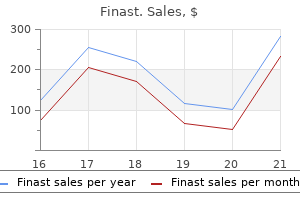
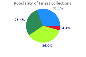
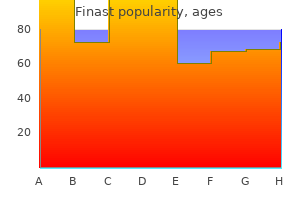
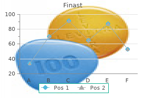
Finast
Finast dosages: 5 mg
Finast packs: 30 pills, 60 pills, 90 pills, 120 pills, 180 pills, 270 pills

Finast 5 mg free shipping
Systemic vasc:ulitis during the course of systemic sclerosis: report of 12 circumstances and evaluate of the literature hair loss cure 32 finast 5 mg effective. Association between environmental exposures and granulomatosis with polyangiitis in Canterbury hair loss nyc finast 5 mg purchase free shipping, New Zealand. Systemic vasculitis with asthma and eosinophilia: a scientific approach to Churg-Strauss syndrome. Cutaneous manifestations of Churg-Strauss syndrome: a clinicopathologic correlation. Prevalence and scientific significance of antineutrophil cytoplasmic antibodies in Churg-Strauss syndrome. Epidemiological patterns of perniosis, and its association with systemic disorder. Cold-associated perniosis ofthe thighs ("equcstriantype� chilblain): a reappraisal based mostly on a clinicopathologic and immunohistochemical research of6 cases. Progression of pigmented purpura-like eruptions to mycosis fungoides: report of3 instances. Fink-Puches R, Wolf P, Kerl H, et al Lichen aureus: clinicopathologic options, natural historical past, and relationship to mycosis fungoides. Study on dermatoses and their prevalence in groups of confirmed alcoholic people in comparison to a non-alcoholic group of individuals. Lichen aureus: an unusual histopathological presentation: a case report and a review of literature. Granulomatous pigmented purpuric dermatosis: report of a case with atypical scientific presentation including dermoscopic findings. Mutliple rheumatoid papules attribute of the early stage of rheumatoid vasculitis. A case of main immunodeficiency as a result of a defect ofthe major histocompatibilitygene complex class I processing and presentation pathway. Stimulator of interferon genes-associated vasculopathy with onset in infancy: a mimic of childhood granulomatosis with polyangi. Antineutrophil cytoplasmic antibodies, abnormal angiograms, and pathological findings in polyarteritits nodosa and Churg-Strauss syndrome: Indicatiomi for the classification of vasculitides of the polyarteritits nodosa group. Consensus statement on surgical pathology of the aorta from the Society for Cardiovascular Pathology and the Association for European Cardiovascular Pathology: I. Ueber eine bisher nicht beschriebene eigentuemliche Arterien veraenderung (periarteritis nodosa), die mit Morbus Briggti und rapide fortschreitender all gemeiner Muskellaehmung einhergeht. Veber Polylarteritis acuta nodosa (sogerannte Periarteritis nodosa) undihre Beziehurgen zur polylmyositis and polyneuritis acuta. Panarteritis cutanea benigna-an entity restricted to the pores and skin or cutaneous presentation of a systemic necrotizing vasculitis Nerve conduction study of decrease extremities in cutaneous arteritis patients with neurological manifestations. Treatment for cutaneous arteritis sufferers with mononeuritis multiplex and elevated C-reactive protein. Polyarteritis nodosa-like vasculitis in affiliation with minocycline use: a single-center case series. Significance of two skin biopsy performances with consecutive deeper sections within the differential diagnosis between cutaneous polyarteritis nodosa and livedo vasculopathy. Penile Mondor illness and its impact on erectile operate: outcomes of 30 sufferers. Re-examination of features to distinguish polyarteritis nodosa from superficial thrombophlebitis. Diagnosis, therapy, and long-term management of Kawasaki disease: a scientific assertion for well being professionals from the American Heart Association. Varicella zoster virus in temporal arteries of patients with large cell arteritis. Infections and the chance of incident giant cell arteritis: a population-based, cue-control examine. In search of a candidate pathogen for giant cell arteritis: sequencing-based characterization of the giant cell arteritis microbiome. Scalp necrosis in big cell arteritis: case report and evaluation of the relevance ofthis cutaneous signal oflarge-vessel vasculitis. Association between specimen size and diagnostic yield of temporal artery biopsy. Incidence of discordant temporal artery biopsy in the diagnosis ofgiant cell arteritis. Histological parameters helpful in recognising steroid-treated temporal arteritis: an analysis of 35 instances. Calciphyww of the temporal artery masquerading as temporal arteritis: a case presentation and review of the literature. Metaplastic ossification of the temporal artery with osteoclast-like giant cells: a mimicker of big cell (temporal) arteritis. Histopathologic findings of patients with biopsy-negative large cell arteritis in comparability with these with out arteritis: a population-based examine. Clinical and virologic characteristics could help distinction ofacute adenovirus disease from Kawasaki illness with incidental adenovirus detection. Clinical features and radiological findings in giant vessel vasculitis: are Takayasu arteritis and large cell arteritis 2 completely different illnesses or a single entity Retrospective evaluation of95 patients with large vessel vasculitis: a single center expertise. Classification of large vessel vasculitis: can we separate large cell arteritis from Takayasu arteritis Takayasu arteritis and ulcerative colitis: excessive price of co-occurrence and genetic overlap. An unrecognised presentation of Takayasu arteritis: superficial femoral artery involvement. Clinical presentation and outcomes of patienu with type 1 monoclonal cryoglobulinemia. Type I cryoglobulinemia in a number of myeloma, a uncommon entity: evaluation of scientific and organic traits of seven cases and evaluation of the literature. A study of coagulation and anti-endothelial antibodies in idiopathic livedo reticularis. Interferons beta have vasoconstrictive and procoagulant effects: a girl who developed livedo reticularis and Raynaud phenomenon in association with interferon beta treatment for a quantity of sclerosis. Livedo reticularis in a affected person with pheochromocytoma resolving after adrenalectomy. Livedo racemosa: a striking dermatological signal for the antiphospholipid syndrome. Generalized livedo reticularis induced by silicone implants for gentle tissue augmentation. Heterotopic ossification of small saphenous vein and panniculitis ossificans of continual venous insufficiency presenting with livedo racemosa. Vasudevan B, Neema S, Verma R Livedoid vasculopathy: a review of pathogenesis and principles of management.
Finast 5 mg order mastercard
The multiphasic response is dose- and time-dependent and is characterized initially by transient redness hair loss curejoy cheap 5 mg finast otc, hair loss hair loss for women finast 5 mg cheap on-line, and sometimes by vesiculation. Intense publicity may produce ulceration that usually occurs after several weeks. Subacute radiation dermatitis develops several months following exposure and produces scaling with postinflammatory alterations! Late radiation necrosis can happen over bony prominences, generally beginning 1 or extra years after publicity and often precipitated by trauma. Certain medical and histologic options, similar to a deep decubitus ulcer on the sacrum of an aged person, could favor a more precise diagnosis. Ulcerated dermis with impetiginized neutrophilic scale-crust, underlying acute and persistent irritation, and granulation tissue formation. In acute radiation dermatitis, keratinocytes present intracellular edema, variation in nuclear dimension and tinctorial qualities, and particular person cell necrosis in an epidermis that manifests spongiosis and vacuolar change. Dermal damage is variable and consists of degenerative effects in the adnexae, dermal edema, ect. Variably intense mononuclear and/or granulocytic inflammatory infiltrates may be seen. In subacute radiation dermatitis, epidermal modifications include vacuolar alteration with necrosis and focal atypia of keratinocytes, hypergranulosis, and compact hyperkeratosis. Dermal modifications may embody a variably dense infiltrate oflymphocytes and histiocytes within the papillary dermis, telangiectatic vessels, and reparative adjustments. There could also be cytologic atypia of keratinocytes, dyskeratosis, and lack of the rete-ridge sample. Blood vessels manifest variable ectasia, proliferative change, hyalinization of media, and thrombosis. Differential Diagnosis Hlstopatholo9lc Features Acute Spongiosis Epidermal necrosis Dermal edema Vascular ectasia and thrombosis Chronic Epidermal hypoplasia and/or hyperplasia Squamous epithelial atypia Dermal sclerosis/hyalinization Deeply located elastotic materials in dermis Loss of adnexae Bizarre uradiation fibroblasts" Hyalinization of vessel partitions and thrombosis the histology of radiation dermatitis provokes a number ofdifferential diagnostic concerns depending on the precise findings in a given biopsy. The dermal adjustments in the late levels of bums may be indistinguishable from these in persistent radiation dermatitis, besides that �radiation fibroblasts" are current in the latter. Markedly atypical nucleolated dermal fibroblasts may increase the possibility of neoplasia. The array of histologic findings seen with radiation change often enables correct diagnosis. Radlatlon-assodated morphea In some instances the fibrosis related to radiation exactly Differential Diagnosis Scar Thermal burn Lichen sclerosus Morphea/scleroderma Chronic graft-versus-host reaction recapitulates morphea. Full-thickness dermal sclerosis with lymphatic dilation and loss of adnexa is a characteristic. It is possible that morphea creating within the setting of radiation therapy represents an isomorphic phenomenon. The histology reveals a fibrosing reaction usociated with a perivascular T-cell-rich infiltrate. Imaging research will show a nbrosing response involving the pores and skin, fascia, and muscle. The elevation of sure interleukins elaborated by T cells-namely interleukin 4 and 6-could potentially contribute to the fibrosing reaction. Similar scleroderma-like changes have additionally been described with other medicine together with the taxanes, bleomycin, and gemcitabine. Likely the world has been previously sensitized by the radiation whereby it has been postulated that radiation may be associated with the induction of sure cytokines that might probably contribute to an inflammatory response; among these cytokines are interleukin 1, interleukin 6, platelet-derived development factor beta, and reworking growth issue beta. It has been instructed that after irradiation select cytokines proceed to be elaborated at low ranges after which with the administration of sure medicine, the cytokines are elaborated at much greater ranges. Bizarreappearing �stellate" fibroblast with options of radiation impact: pleomorphic hypochromatic nuclei, abundant cytoplasm, and intranuclear and intracytoplasmic vacuoles. The traditional time course between the radiation and the administration of the drug is a few months up to a number of years. Certain medicine are more generally implicated with the first reported cases being with actinomycin-D, halogenated pyrimidines, and nucleoside analogues together with gemcitabine and taxanes together with nonchemotherapeutic drugs corresponding to codeine, statins, and antituberculous medication. In addition, while the pores and skin is classically involved, other affected organ sites include the mind, lung, esophagus, and larynx. We have seen circumstances the place the brunt of the damage was a microvascular one exhibiting vital endothelial cell injury. The medical presentation mimicked lipodermatosclerosis; there was a pauci-inflarnmatory endothelial cell injury syndrome affecting vessels in the dermis and subcutaneous fats. The precise foundation of the endothelial injury was not clear and will embody irritant impact on endothelium versus immune-based endothelial cell harm if the combination of irradiation and drug administration resulted in the publicity of endothelial-based neoantigens, hence evoking an adaptive immune response. In addition to the aforesaid mechanisms, one must be cognizant that hypoxia can result in the emergence of strains of tumor cells which might be radioresistant. Certain medication can result in reoxygenation and make the tumor sensitive to radiation. As well certain drugs-namely, the nucleoside analogues-may eliminate the phases of the drug which might be radioresistant, leading to the emergence of tumor cells which might be radiosensitive. The differential analysis encompasses pseudosclerodermatous panniculitis after irradiation. This condition develops a few months after irradiation and is unrelated to chemotherapy drug administration. These exterior stresses have been studied in animal techniques, and the histologic modifications associated with the diploma of harm have been recognized. Clinical Features the clinical look of thermal burns ranges from faint erythema to vesiculation and necrosis (Table 13-2). Electrical harm produces three zones: a central zone of carbonization, a pale ischemic intermediate zone, and an erythematous peripheral zone. Chilblains (perniosis) are itchy, burning, pink vesicular, or ulcerative patches or plaques that occur symmetrically in acral areas, notably toes, following exposure to cold. Subsequent upregulation of endothelial adhesion molecules is felt to be related to an efflux of mononuclear cells at reperfusion. The diagnosis is sometimes recommended by the temporal relationship between symptom onset and funky climate or immersion in chilly liquids12. The association with wet climates and with chilly fluids is held to be a bodily one, in that moisture serves to increase heat extraction from the skin floor. There is an association with the consumption of sure immune-perturbing medicine, such as tluoxetine. Histopathologic Features Mild thermal and cold accidents produce epidermal and dermal edema, vacuolated keratinocytes, and vascular dilatation. The histologic findings include a putting panniculitis characterised by adipocyte necrosis with lipophagic granulomas and variable lobular and interlobular fibrosis. The overlying dermis can exhibit features of persistent radiation dermatitis as will be alluded to presently. Somewhat bizarre-appearing fibroblasts typical for radiation fibroblasts are sometimes accompanied by scattered lymphocytes and plasma cells throughout the fat. Striking vascular adjustments are frequent whereby all types ofvascular alteration related to radiation remedy can be seen.
Cheap finast 5 mg on line
Immunohistochemical examine of one other case of lipoatrophic panniculitis of the ankles in childhood hair loss on mens face 5 mg finast cheap fast delivery. Lipoatrophic panniculitis of the ankles in childhood: differential diagnosis with subcutaneous panniculitis-like T-cell lymphoma hair loss jacksonville fl finast 5 mg purchase visa. Lipophagic panniculitis of childhood: a case report and comprehensive evaluate of the literature. First report oftertiary syphilis presenting as lipoatrophic panniculitis in an immunocompetent affected person. Locoregional a quantity of nodular panniculitis induced by Pseudomonas aeruginosa without septicemia: three instances and give attention to predisposing components. Cryptococcal panniculitis in a renal transplant recipient case report and evaluate of literature. Subcutaneous panniculitis-like T-cell lymphoma within the pediatric age group: a lymphoma of low malignant potential. Subcutaneous panniculitis-like T-cell lymphoma in kids: an in depth clinicopathological description of 11 multifocal instances with a high frequency of haemophagocytic syndrome. Subcutaneous panniculitislike T-cell lymphoma: immunosuppressive medication induce better response than polychemotherapy. Lobular panniculitic infiltrates with overlapping histopathologic features oflupus panniculitis (lupus profundus) and subcutaneous T-cell lymphoma: a conceptual and sensible dilemma Am J Surg Pathol. Cutaneous yll T-cell lymphomas: a spectrum of presentations with overlap with other cytotoxic lymphomas. Subcutaneous panniculitis-like T-cell lymphoma: pediatric case sequence demonstrating heterogeneous presentation and option for watchful waiting. The presence of clusters of plasmacytoid dendritic cells is a helpful feature for differentiating lupus panniculitis from subcutaneous panniculitis-like T-cell lymphoma. Useful parameters for distinguishing subcutaneous panniculitis-like T-cell lymphoma from lupus erythematosus panniculitis. Lobular panniculitis because of Borrelia burgdorferi an infection mimicking subcutaneous panniculitislike T-cell lymphoma. Skin manifestations of intravascular lymphoma mimic inflammatory illnesses of the skin. Intravascular lymphoma: medical presentation, pure history, management and prognostic components in a series of 38 instances, with special emphasis on the �cutaneous variant. A case of intravascular large B-cell lymphoma mimicking erythema nodosum: the significance of multiple skin biopsies. Bumps within the street: panniculitis in youngsters and adolescents handled with vemurafenib. Pannkulitis in sufferers undergoing remedy with the Bruton tyrosine kinase inhibitor ibrutinib for lymphoid leukemias. Pemetrexedinduced painful erythematous nodules in each legs in a affected person with recurrent mesothelioma. Erythema nodosumlike panniculitis mimicking illness recurrence: a novel toxicity from immune checkpoint blockade therapy-report of2 sufferers. Vemurafenib-induced neutrophilic panniculitis: a new case and evaluate of the literature. Neutrophilic panniculitis with vasculitis in a melanoma patient treated with vemurafenib: a case report and iu administration. Central nervous system involvement in intravascular large B-cell lymphoma: a retrospective analysis of 109 patients. Cutaneous involvement in a case of intravascular T-cell lymphoma with a yll phenotype. Mycobacterium chelonae an infection presenting as recurrent cutaneous and subcutaneous nodules-a presentation previously recognized as Weber Christian illness. Weber-Christian disease creating into mediastinitis and pleuritis with huge pleural effusion. Drug reactions may be immunologic or nonimmunologic, the latter consisting ofoverdosage, intolerance, teratogenicity, facultative effects that result from disruption of bacterial flora in mucous membranes and skin, and toxicity, the latter either delayed (eg. Other nonimmunologic mechanisms of drug toxicity embrace anaphylactic reactions due to brokers that degranulate mast cells (eg, opiates) or impair arachidonic acid metabolism. The diagnosis of a cutaneous drug response is thus difficult and requires a radical historical past, often aided by clinical algorithms. U-H these components may be analyzed in a formal method such because the Naranjo algorithm,17 a logistic regression-based mannequin used within the Netherlands that makes use of 5 candidate predictors,14 or using professional panels such as those of the European Study of Severe Cutaneous Adverse Reactio. Factors that influence the event of hypersensitivity embrace the nature of the allergen (ie, lipid versus water solubility). Immediate hypersensitivity (type I) reactions Most dramatic are the drug reactions of quick hypersensitivity type (type I). In angioedema its foundation is one attributable to the accumulation of substance P and bradykinin, both potent vasodilators. Due to the function of bradykinin within the pathogenesis of angioedema, inhibitors of bradykinin could be given. The traditional drug-induced form of angioedema is one related to angiotensin-converting enzyme inhibitor use. It happens more commonly in sufferers of African-American extraction the place the main affected sites are face, lips, and airways. Drug reactions during which this mechanism is likely to be operative embrace erythema multiforme and morbilliform, fastened, eczematous, pustular, lichenoid, lymphomatoid, and pigmentary purpura-like drug reactions. Lesions usually seem first on the trunk and spread peripherally in a symmetric style with dependent areas such because the decrease extremities manifesting purpura. Lesions clear with withdrawal of the causative agent however could progress to a generalized exfoliative dermatitis if the drug is continued. Lesions might fade following drug cessation or resolve with a residuum of postinflamrnatory hyperpigmentation. One unusual cause is the lymphocyte recovery state in patients receiving chemotherapeutic medicine for leukemia and lymphoma following the nadir of their peripheral lymphocyte counts. It has been suggested that neutrophils within the papillary dermis or within the decrease levels of the dermis are seen in a 3rd of cases,33 however this has not been our expertise. Some examples, significantly the scarlatiniform eruptions, could show no epithelial alterations. Differential Diagnosis Viral exanthemata and erythema multiforme reactions closely mimic exanthematous drug eruptions. Tissue eosinophilia is uncommon within the former two entities, except in the context of erythema multiforme-like drug reactions. One instance is hemolytic anemia associated with methyldopa, mediated by the induction of autoantibodies directed towards pink blood cell antigens; another is autoimmune thrombocytopenic purpura, mediated via antibodies towards platelets and provoked by pyrazolone derivatives (eg, phenylbutazone and allopurinol), sulfonamides, penicillin, salicylates, thiazides, diuretics, and chloramphenicol.
Discount 5 mg finast amex
For instance hair loss in men quilting finast 5 mg purchase amex, one can see this interstitial granuloma annulare-type reaction in resolving exogenous foreign materials generally encountered in pores and skin specimens hair loss in men as they age purchase finast 5 mg with mastercard. It is possible that granuloma annulare is a nonspecific response pattern that will observe any number of various processes within the skin. Clinical Features Children and younger adults are primarily affected, though the illness can occur at any age. A just lately described scientific variant of granuloma annulare presents with delicate patches on the extremities and trunk. This pattern is typical of the just lately described scientific variant of granuloma annulare in which refined patches are found predominantly on e:nremities. Such palisading granulomas are often focal and discrete phenomena in the dermis with intervening regular collagen (ie, interstitial pattern). The granulomas could extend alongside the fibrous trabeculae of the subcutaneous fat and thus represent a septal panniculitis (that is later also lobular). Connective tissue mucin deposition throughout the central degenerated collagen is a near-constant finding but might require special stains (ie, alcian blue or colloidal iron) to demonstrate. In H&Estained sections, mucin seems as stringy and/or granular basophilic materials. Uncommonly, the collagen degeneration is characterised by fully developed fibrinoid necrosis. In some cases, notably in sun-damaged skin, the macrophages and multinucleate large cells comprising the palisaded infiltrates are famous to comprise phagocytosed elastic fibers (elastophagocytosis). Eosinophils occur in roughly 40% of circumstances and may be so quite a few as to suggest an arthropod chew response. The lesions exhibit barely depressed central areas in some instances with a return to normal pores and skin c:olor. A cell-mediated immunologic reaction to altered elastotic fibers has been proposed. Eruptive xanthomas can mimic this sample but are recognized by foamy macrophages, occasional neutrophils, extracellular lipid, no mucin, and, hardly ever, urate-like crystals (see the next section). Granulomatous mycosis fungoides is a histologic variant of mycosis fungoides that options granulomatous irritation. The sample of granulomatous inflammation is variable and could additionally be diffuse or palisaded, but there are normally superimposed adjustments of typical mycosis fungoides in which small, atypical lymphocytes permeate the epidermis. Histopathologic Features Deep (subcutaneous) granuloma annulare Synonyms: Pseudorheumatoid nodule, nodular granuloma annulare. The large cells contain blue-gray elastotic materials, and asteroid bodies may be seen. Neither prominent mucin nor degenerated collagen typically is current the central portion of the plaque demonstrates loss of each elastotic fibers and regular elastic fibers. Subcutaneous nodules are discovered on the extensor elements of palms, toes, decrease legs, buttocks, and scalp, and within the periorbital area. Histopathologic Features the facial location and the presence of palisaded granulomas and phagocytized elastotic material (elastophagocytosis) are distinctive features. The deep reticular dermis above the subcutaneous illness regularly exhibits focal involvement. The subcutaneous locale with central connective tissue mucin deposition are distinctive. Differential Diagnosis Necrobiosis lipoidica Synonym: Necrobiosis lipoidica diabeticorum. Approximately two-thirds of patients with necrobiosis lipoidica have diabetes mellitus. On the opposite hand, in sufferers with diabetes mellitus, necrobiosis lipoidica occurs in lower than 1%. Rheumatoid nodule and rheumatic fever nodule are additionally subcutaneous however lack mucin and instead characteristic central fibrinoid necrosis, generally with options of a late-stage leukocytoclastic vasculitis. Epithelioid sarcoma is a malignant soft tissue neoplasm that nearly all regularly impacts kids or young adults and happens on the distal extremities. I<111 Histology reveals spindled and epithelioid cells surrounding central areas of necrosis in a palisaded array. Adults current with waxy, indurated, yellow-brown patches or plaques with a surrounding erythematous, raised, and expanding margin. Lesions characteristically are discovered on the anterior shins but in addition may be seen on forearms, hands, and trunk. Histopathologic Features Actinic granuloma Synonyms: Annular elastolytic granuloma, Miescher granuloma, atypical necrobiosis lipoidica, granulomatosis disciformis of the face. The central portion of the palisade reveals marked collagen degeneration without connective tissue mucin. Within the dermis and lengthening into the subcutis are palisading granulomas with central necrobiosis of collagen and dermal mucin. In addition, necrobiosis lipoidica often involves the dermis and probably the subcutis, whereas the converse is true of rheumatoid nodule (see Table 6-18). Rheumatoid nodule Rheumatoid nodules are seen in roughly 2096 of adults with rheumatoid arthritis. The pathogenesis is most probably an immune complex-mediated vasculltis, which leads to ischemic harm to subcutaneous tissues and a palisaded granulomatous inflammatory response. Clinical Features Rheumatoid nodules current in adults as subcutaneous nodules located over joints, classically the elbows. Like rheumatoid nodules, the pathogenesis is most likely an immune complexmediated vasculitis with subsequent ischemic injury followed by a palisaded granulomatous repair reaction. Sites of predilection embrace the bony prominences (humeral condyles, oleaanon processes, lmucldes) and occiput. Palisaded granulomatous irritation surrounds degenerated collagen and fibrinoid material, with neutrophils and neutrophilic nuclear debris. Otherwise, the histology is identical to rheumatoid nodule, although particular person lesions could also be smaller (see "Rheumatoid Nodule� above). Bone marrow examination may reveal plasmacytosis, and rare instances could be seen in association with multiple myeloma or B-cell lymphoma. The central space laclc:s swimming pools of connective tissue mucin as a rule,one hundred forty five however vaseulitic changes with intact and degenerated neutrophils are usually obvious. Differential Diagnosis the predominantly subcutaneous location and central fibrinoid necrosis (without pools of connective tissue mucin) are distinctive options of rheumatoid nodules. Ms In general, the histology of rheumatic fever nodule and rheumatoid nodule is equivalent, but patients with rheumatic fever nodule have a prior or concurrent history of rheumatic fever. The entity known as "rheumatoid papules" is at present thought of as a part of the spectrum of palisaded neutrophilic and granulomatous dermatitis.
Buy 5 mg finast with amex
Kuriyama K hair loss cure 4 hunger 5 mg finast cheap fast delivery, Tomonaga M hair loss under chin in cats order 5 mg finast overnight delivery, Matsuo T, Ginnai I and Ichimaru M (1986) Diagnostic significance of detecting pseudoPelger�Hu�t anomalies and micro megakaryocytes in myelodysplastic syndrome. Fern�ndezLago C and Romero E (1998) Myelodysplastic syndromes related to immune thrombocytopenia. Stasi R, Pagano A, Terzoli E and Amadori S (1999) Recombinant human granulocytemacrophage colonystimulating issue plus erythropoietin for the therapy of cytopenias in sufferers with mye lodysplastic syndromes. Thiede T, Engquist L and Billstrom R (1988) Application of megakaryocytic morphology in diagnosing 5q� syndrome. Scharenberg C, Jansson M, Saft L and Hellstr�m Lindberg E (2018) Megakaryocytes harbour the del(5q) abnormality despite complete scientific and cytogenetic remission induced by lenalidomide treat ment. Olivieri O, Gandini G, Baiocco R, Aprili G, Falezza G and de Sandre G (1987) Visceral leishmaniasis pre senting as dyserythropoiesis related to elevated iantigenicity of erythrocytes. Bazarbachi A, Haidar J, Salem Z, Solh H and Ayas M (1997) Thiamineresponsive myelodysplasia. Evidence means that, even when differentiation is predominantly to cells of a single lineage, the dysfunction has arisen in a multipo tent myeloid stem cell or, a minimal of in some cases, in a pluripotent stem cell able to giving rise to cells of each myeloid and lymphoid lineages. For the nice majority of those problems mutations in tyrosine kinase genes, necessary in pathogenesis, have been found. However, this group of conditions shows a larger or lesser propensity to evolve right into a malignant neoplasm, resembling acute leukaemia, which rapidly leads to dying. Polycythaemia vera, major myelofibrosis and sys temic mastocytosis endure acute transformation less usually and usually after a longer continual part. However, with dis ease progression haemopoiesis could turn out to be ineffec tive and dysplastic options can appear. For the continual myeloid leukaemias, careful examination of a blood film is usually more important than examination of a bone marrow aspirate. It is an unusual condition resulting from the neoplastic proliferation of an early haemopoi etic precursor cell that can differentiate into cells of granulocyte, monocyte, erythroid, megakaryo cyte and, under sure circumstances, lymphoid lineages. Chronic myeloid leukaemia may be very largely a dis ease of grownup life but circumstances occur from childhood onwards. The overall incidence is 1�2 per 100 000 per yr [4] or less [5] with a sluggish enhance happen ring with rising age. The disease is extra widespread in men than in girls with a male: feminine ratio of about 1. However, because of the insidious onset of the illness, many patients have only minor symptoms on the time of analysis. The disease is now usually diagnosed incidentally from a blood rely in an asymptomatic affected person. Initially the disease pursues a chronic course, in which patients are sometimes maintained in moderately good health. Acute transformation is often preceded by an accelerated phase in which the illness turns into resistant to therapy. In distinction, the 5year sur vival is now round 90%, a minimal of for those lower than 60 years of age at presentation [5]. Basophils are almost invariably elevated and absolutely the eosinophil rely is increased in the great majority of sufferers; eosinophil and basophil myelocytes are usually Usually Leucocytosis with increased granulocytes and their precursors with or with out thrombocytosis Or Occasionally Thrombocytosis Plus t(9;22)(q34. Rarely, transforma tion is monoblastic, eosinophilic, hypergranular promyelocytic or erythroblastic. Alternatively, there could also be hybrid cells with both basophil and mast cell features. Often a single patient has blast cells of diverse sorts, normally a combination of mega karyoblasts and myeloblasts, but sometimes a combination of lymphoblasts and blast cells of one or more myeloid lineages. The presence of more than 20% circulating blast cells is an acceptable criterion for a diagnosis of acute transformation. During successful continual part treatment, the peripheral blood depend and movie usually become nearly regular although a degree of basophilia and occasional immature granulocytes might persist. Patients presenting with or growing extensive bone marrow fibrosis have marked anisocytosis and poikilocytosis with prominent teardrop poikilocytes. The accelerated phase could additionally be marked by rising basophilia, persistent leucocytosis or the reappear ance of anaemia. Acute transformation could comply with an accelerated part or the looks of features of bone marrow fibrosis or be heralded by the appear ance of dysplastic options (such because the acquired Pelger�Hu�t anomaly of neutrophils or the presence of circulating micromegakaryocytes) or there will be the abrupt look of accelerating numbers of circulating blast cells in a previously stable patient. Acute transformation is myeloid in about two thirds of instances and lymphoblastic or combined in the remainder. As a consequence of the elevated cell turnover, macrophages and various storage cells are often prominent (see later in this chapter). During the accelerated part, the bone marrow may present rising basophilia, some enhance of blast cells or the appearance of dysplastic options, such as micromegakaryocytes. Bone marrow aspi ration could become tough or unimaginable due to rising bone marrow fibrosis. With the onset of acute transformation, the bone marrow is steadily changed by blast cells showing the similar old cytological features of the lineage involved. In most cases greater than 95% of the marrow cavity is occupied by haemopoietic cells. There is a marked improve in granulocytic precursors with a variable degree of left shift. The normal topographic relationship of haemopoiesis is retained, with granulopoiesis occurring predomi nantly in the paratrabecular, periarteriolar and pericapillary areas, although the extra mature granulocytic precursors prolong into the central areas of intertrabecular marrow. Megakaryopoiesis and, to a lesser extent, eryth ropoiesis happens in the perisinusoidal areas. There may be some megakaryocytes within sinusoids and in addition some near bony trabeculae [14]. Megakaryocytes are normally elevated in quantity, sometimes forming small clusters of cells. The megakaryocytic morphology is variable, with most sufferers having some comparatively normal varieties and more quite a few smaller cells with small, hypolobated nuclei. Increased numbers of mast cells and plasma cells are generally seen, usually in a perivascular posi tion. Marrow necrosis is rare and, when present, is usu ally a sign of impending blast transformation. Reticulin is often increased; not often the fibrosis is extreme enough to cause confusion with primary myelofibrosis [18]. In one collection of patients elevated reticulin, with or without collagen depo sition, was seen in a quarter of sufferers [15]. In multivariate evaluation, elevated reticulin correlated with a worse prognosis preimatinib [15] however not subsequently [4,19]. Depending on the therapeutic agent used, there may be progressive reticulin deposition throughout therapy (see later). For instance, cellularity generally decreases and small hypolobated megakaryocytes are changed by cytologically normal forms.
Camellia theifera (Black Tea). Finast.
- Osteoporosis, headache, high blood pressure, stomach disorders, vomiting, diarrhea, preventing tooth decay, type 2 diabetes, lung cancer, reducing the risk of cancer, and promoting weight loss.
- Reducing the risk of stomach, colon, and rectal cancer.
- Are there any interactions with medications?
- Preventing dizziness upon standing up (orthostatic hypotension) in older people.
- What other names is Black Tea known by?
- Mental alertness.
- What is Black Tea?
- Are there safety concerns?
- How does Black Tea work?
Source: http://www.rxlist.com/script/main/art.asp?articlekey=96958
5 mg finast best
These can typically even be detected in paraffinembedded bone marrow fragments but not in decalcified trephine biopsy sections hair loss cure in 5 years finast 5 mg discount line. If a bone marrow aspirate containing enough fragments is available then iron staining of trephine biopsy sections is redun dant hair loss cure for women finast 5 mg discount mastercard. The amount of stainable iron is lowered and sometimes all stainable iron is removed. There are conflicting stories of the comparability of iron stains carried out on aspirates and biopsy specimens, not all of which are readily explicable by the components already talked about. Iron stains sixty seven carried out on aspirates and biopsy specimens ought to clearly be regarded as complementary. Reticulin and collagen stains Histological sections, either from particle prepara tions or trephine biopsy specimens, may be stained for reticulin utilizing a silverimpregnation method and also for collagen using a trichrome stain. We have found a Martius scarlet blue stain superior to a van Gieson stain for the identification of collagen. These authors make the impor tant level that reticulin deposition must be assessed in relation to haemopoietic tissue, not in fatty areas of marrow. The majority of haematologically normal topics have a reticulin grade of zero or 1 of 4 however occasional topics have grade 2. There is a tendency for extra reticulin to be detected in iliac crest biopsies than in sections of particles aspirated from the sternum. Reticulin is concentrated round blood vessels and near bone trabeculae and these areas should be disregarded in grading reticulin deposition. Problems and pitfalls To keep away from confusion, pathologists should check with reti culin and collagen deposition in a precise method. The causes of collagen deposition are fewer and this irregular ity is therefore of extra diagnostic significance. The significance of reticulin and collagen deposition is discussed in Chapter three (see pages 168�170). The time period myelofibrosis is used to point out deposi tion of collagen within the marrow and generally also to indicate elevated reticulin deposition. Firstly, increased reticulin deposition supplies non specific proof of an abnormality of the bone marrow. Secondly, focal abnormality in the pattern of reticulin deposition could be very helpful in detect ing abnormalities that may be overlooked in an H&Estained part. Focal abnormalities that might be highlighted by a localized increase in reticulin deposition embrace granulomas and infiltrates of carcinoma or lymphoma cells. In addition to its two main roles, a reticulin stain reveals bone construction clearly. Occasionally a Other histochemical stains Other probably useful histochemical stains and their roles in prognosis are proven in Table 2. Problems and pitfalls the reactivity of histochemical stains is influenced by the choice of fixative, the strategy of embedding and the method of decalcification employed. However, it must be noted that prolonged storage of forma lin at excessive ambient temperatures can result in formic acid manufacturing; if the formalin is unbuffered, inadvertent decalcification could happen through the process of fixation with resultant adverse results on staining. We have discovered that most of the proprietary combined fixation�decalcification solutions, that are generally used to obtain rapid processing, impair histochemical stains. Detection of antigens could also be by means of polyclonal antibodies, raised in animals such as rabbits, but monoclonal antibodies produced by hybridoma know-how are now predominantly used. Otherwise this process may be performed on a bone marrow aspirate or, alterna tively, on cerebrospinal fluid, a serous exudate or a Immunophenotyping Antigens could also be expressed on the surface of cells, within the cytoplasm or throughout the nucleus. When peripheral blood is used, the procedure may be applied to either a mononuclear cell prepa ration or to whole blood by which the pink cells have been lysed [21]. The latter approach minimizes cell loss and potential artefacts that might be induced by exposure to Ficoll and density gradient separation. Choice of applicable proprietary lysis solutions is essential to keep away from the reduction of expression of certain antigens [22]. The flow cytometer permits classifica tion of cells according to their lightscattering char acteristics and the depth of their fluorescence upon activation by laser mild, detected after cross ing through an acceptable filter for the actual fluorochrome employed. Three or extra fluoro chromes can be utilized so that the simultaneous expression of two, three or more antigens may be studied. If permeabilization strategies are employed, cytoplasmic and nuclear antigens can be detected in addition to those expressed on cell sur faces. Results should all the time be interpreted in the light of the cytological options of the cells being studied. When there are giant numbers of circulating neo plastic cells, outcomes of peripheral blood analysis are typically dependable. When move cytometry is performed on cell suspensions from bone marrow or other tissues, outcomes could additionally be mis leading in two circumstances. Firstly, an irregular infiltrate will not be represented within the aspirate to any significant extent. This is often the case in fol licular lymphoma with paratrabecular infiltration, but can even happen in any lymphoma in which reticulin deposition is elevated in the infiltrated space, interfering with aspiration of abnormal cells. It has been suggested that immunophenotyping by circulate cytometry will not be value efficient as compared with histology sup plemented by immunohistochemistry in lymphoma diagnosis. In one investigation, a monoclonal pop ulation was detected in solely forty nine of fifty nine sufferers with bone marrow histologically concerned by lymphoma and, in sufferers with regular histology, solely 5 of 116 had a monoclonal population detected [23]. Whether immunohisto chemistry is also wanted when an abnormal popu lation has been detected by flow cytometry could be decided on a case by case basis. The reaction of antibodies with cells carrying a particular antigen is detected by both (i) direct labelling of the primary antibody with an enzyme corresponding to peroxidase or alkaline phosphatase, or (ii) an indirect technique utilizing a second, labelled antibody that acknowledges the first. Indirect labelling techniques offer the advantage of elevated sensi tivity however are extra timeconsuming to perform than direct labelling, which is increasingly used. The use of washed, separated cells in cyto centrifuge preparations is critical for immu nocytochemistry to detect floor membrane immunoglobulins, including and light-weight chains. Plasma immunoglobulins interfere with the stain ing if blood movies or movies of bone marrow aspirates are used. For the detection of most other antigens, both cytocentrifuge preparations or wedgespread films are passable. If there are vital num bers of irregular cells within the circulating blood then a peripheral blood sample is very satisfactory for immunocytochemistry. Immunocytochemistry can be used to dem onstrate the product of an oncogene or a most cancers suppressing gene. Immunocytochemistry By conference, the term immunocytochemistry refers to the study of the antigen expression of cells by the use of polyclonal antisera or monoclonal antibodies applied to fastened cells on glass slides. The materials investigated may be either a blood or bone marrow movie or a cytocentrifuged preparation of washed mononuclear cells isolated from blood or Problems and pitfalls Immunocytochemistry has the benefit that reactivity with an antibody can be related to cell morphology.
Finast 5 mg discount with mastercard
The lipids may disrupt lamellar granule function in addition to other processes related to barrier function hair loss in men over 60 generic 5 mg finast with mastercard. Parakeratotic fod associated with a focally thinned granular layer are less frequent hair loss ketoconazole finast 5 mg buy fast delivery. The papillary dermis may include a perivascular lymphocytic infiltrate of variable intensity. Differential Diagnosis the microscopic features are indistinguishable from different nonbullous congenital ichth. The ichthyosis and deafness syndromes Although comparable in name, the two e<:todermal dysplasias grouped herein are distinct clinically and histologically. Neutral lipid storage disease with lchthyotlc (lchthyoslform) erythroderma and multlsystem illness (Dorfman�Chanarfn syndrome) Cinical Features Neutral lipid storage disease is a uncommon genetic disorder occurring mainly in people ofArabic background. The disorder is autosomal re<:essive and results in the accumulation of impartial lipids in numerous tissues. Increased mobile triacylglycerol outcomes from defective re<:ycling of triacylgl. The palms and soles may be mildly affected, and alopecia and nail dystrophy may be seen. The face, elbows and knees, palms and soles are concerned, however the trunk is spared. Both syndromes associate with sensorineural hearing loss, frequent bacterial and fungal infections, and scarring alopecia. The prognosis of the latter is made by the scientific recognition of the other parts of the syndrome, significantly the hearing disorder. Sjogren-Larsson syndrome Clinical Features Patients with Sjogren-Larsson syndrome have a extreme, autosomal recessive, neurocutaneous disease comprising the triad of mental retardation, spasticity, and congenital ichthyosis. Histopathologic Features Histologic sections exhibit acanthosis and a few papillomatosis. Moderate compact hyperorthokeratosis characterizes the stratum comeurn, but scattered foci of parakeratosis could also be current the granular layer is retained, could also be thickened, and can show vacuolization. Identification of the other elements of the syndrome and genetic analysis set up the diagnosis. Exposure to such toxins as dioxin and arsenic or to chemotherapeutic agents (eg, fluorouracil) could additionally be etiologic. Keratoderma clirnactericum is the designation for the affiliation with the postmenopausal or postoophorectomy state. Clinical Features With palmoplantar keratodermas which are unassociated with illness in different organ methods, which is the most typical presentation clinically, sufferers have localized or diffuse and sometimes marked hyperkeratosis of the palms and soles. Onset could additionally be from birth to the fourth decade of life, though early onset with lifetime persistence and/or exacerbation is frequent. The keratodenna of Papillon-Lefevre syndrome associates with periodontosis and shedding of the permanent dentition. The Rowel-Evans syndrome of palmoplantar keratoderma, which is genetically linked to 17q, is associated with squamous cell carcinoma of the esophagus later in life. Sclerodactyly, nail dystrophy, and squamous cell carcinoma of the affected pores and skin are attributes of the autosomal dominant Huriez syndrome. Punctate keratoderma is characterized by agency, welldemarcated plugs ofkeratotic materials positioned on regular volar pores and skin, generally with a predilection for the skin creases; linkage to mutations at various genetic loci have been recognized in some varieties ofpunctate keratoderma. Some sufferers display epidermolytic hyperkeratosis of the upper spinous and granular cell layers and scattered dyskeratotic cells (as found in, but apparently not limited to , Voerner syndrome). A pluglike compact column of parakeratosis throughout the stratum corneum correlates with the subtype of keratosis palmoplantaris punctata. Differential Diagnosis Erythrokeratoderma variabilis has a normal granular cell layer underlying a hyperorthokeratotic stratum comeum. The papillary dermis incorporates a perivascular lymphocytic dermatitis of variable depth. Progressive symmetric erythrokeratoderma histologically has hyperkeratosis with focal parakeratosis. Differential Diagnosis Whereas the histologic pattern of nonepidermolytic circumstances is similar to that ofthe nonbullous lamellar ichthyoses, epidermolytic hyperkeratosis may be distinguished from the bullous ichthyosiform erythrodermas by clinical correlation. Keratoderma of the palms and soles can replicate a mess of genetic or acquired disorders, and delayed onset disease could be a paraneoplastic signal oflymphoma, lung cancer, and other malignancies. Definitive diagnosis of the genetic palmoplantar keratodermas would require mutation analysis. The histologic patterns of erythrokeratoderma variabilis and progressive symmetric erythrokeratoderma are nonspecific, resembling lots of the nonbullous ichthyoses. The analysis rests with the clinicopathologic correlations and more definitively with mutational evaluation at candidate loci. Associated palmoplantar keratoderma (tylosis) may not often happen in cases induced by inside malignancy. Occasionally, papillomatous epithelial hyperplasia may affect the conjunctiva and the stratified squamous mucosae of the oropharynx and anal canal. Histopathologic Features Erythrokeratoderma variabilis manifests transient, polycyclic or circinate, brilliant patches of erythema that change in measurement, form, and distribution over hours to days. They are commonly discovered on the extremities and buttocks and infrequently on the trunk and stomach. More steady, fixed, erythematous, hyperkeratotic plaques develop on the extensor floor of the extremities. Mild palmoplantar keratoderma happen in as much as 50% of instances, and central nervous system abnormalities have been reported in some families. The lesions of progressive symmetric erythrokeratoderma resemble those of erythrokeratoderma variabilis, but their distribution is strikingly symmetric and limited to the extremities and buttocks, they usually stay fastened after first prevalence. Although the two problems are often distinct, the affiliation of both-one every in sisters-suggests that they characterize variants of the identical course of. The hyperkeratotic stratum corneum usually retains the conventional basket-weave structure. Differential Diagnosis Distinction from seborrheic keratosis and hamartomatous epidermal nevi might occasionally be needed when the papillomatosis is pronounced, but the traditional absence of great acanthosis helps rule out these issues. Confluent and reticulate papillomatosis of Gougerot and Carteaud is mostly not distinguishable histologically, however the flexural distribution oflesions of acanthosis nigricans in contrast with the more generalized distribution of the previous entity permits scientific distinction of the two. The clinical setting of weight problems and purchased insulin resistance will serve to establish those cases linked to the endocrinopathy of sort 2 diabetes mellitus. Pronounced hyperorthokeratosis in a basket-weave pattern overlies the papillomatous epidermis, however little ac:anthosis is present. Histopathologic Features the histological adjustments are much like these of the sporadic illness. The stratum corneum is exceptional for marked hyperkeratosis, which in some foci exhibits horizontally alternating zones of ortho- and parakeratosis. Intracorneal neutrophils are absent the stratum granulosum is usually hypergranular. Dilatation and keratotic plugging of the follicular infundfbulae are often reliably distinguished features of pityriasis rubra pilaris and must be sought in extra recuts if the diagnosis is unclear from the unique sections. The stratum comeum adjacent to the follicular ostia might show parakeratosis (shoulder parakeratosis).

5 mg finast buy visa
Patients not uncommonly present with hepatosplenomegaly and B symptoms and both acute leukaemia or lymphoma may be suspected hair loss cure new cheap finast 5 mg visa. Macrophages ought to be exam ined fastidiously for microorganisms in sufferers with unexplained hepatosplenomegaly and reactive modifications within the bone marrow hair loss in men 8 pack discount finast 5 mg mastercard. Because of the rela tive insensitivity of microscopy [115], tradition of bone marrow is suggested when this prognosis is suspected. These might embody a significant part of epithelioid macrophages, which have giant quantities of pale pink cytoplasm and ovoid or elongated nuclei with a dispersed chromatin sample. A big selection of aetiological brokers are related to marrow granulomas (Table 3. The blood film could show options related to the primary illness or, if bone mar row disease is in depth, there may be anaemia or pancytopenia with a leucoerythroblastic blood film. Similar lesions could also be seen in the liver, spleen and lymph nodes and some of these cases have been reported to be related to ingestion of mineral oil [156]. They con tain fats vacuoles, which range in measurement however are often smaller than the vacuoles in marrow fat cells and could also be a number of. Lipid granulomas usually have plasma cells, eosinophils and lymphocytes inside them, and approximately 5% include large cells. Other granulomas Unless a specific organism may be demonstrated inside a granuloma, there are normally no histological options that enable a definitive diag nosis to be made [157]. All biopsy speci mens with granulomas should have applicable stains for acidfast bacilli and fungi performed. Ideally, in these circumstances in which marrow granulomas with an infective aetiology are potential, for example in sufferers with a pyrexia of unknown origin, this must be anticipated and part of the marrow aspi fee must be cultured for mycobacteria and fungi. Tuberculous granulomas normally include Langhans big cells and caseation is present in roughly half of cases with marrow involve ment [157]. Marrow granulomas containing foamy mac rophages are sometimes seen in patients with leprosy; a Fite stain will demonstrate the acidfast bacilli of M. Small, poorly formed epithelioid granulomas are discovered within the bone mar row typically of brucellosis. Disseminated an infection by the fungus Histoplasma capsulatum often involves the bone marrow; in regular hosts there are quite a few granulomas, usually with Langhans large cells and necrosis. Fungi may be seen with an H&E stain, however are greatest visualized utilizing a silver stain. In each Hodgkin lymphoma and nonHodgkin lymphoma, granulo mas also occur in the absence of marrow contain ment; these are normally small, wellformed epithelioid granulomas although larger, poorly shaped lympho histiocytic lesions have additionally been reported [162]. The bone marrow is commonly involved in sar coidosis; granulomas were seen in 9 out of 21 sufferers in a biopsy sequence [157]. Often patients with marrow granulomas have evidence of multi system involvement, corresponding to hepatosplenomegaly, though chest radiography may be regular [167]. Typically there are numerous, wellformed epithe lioid granulomas which, in roughly a third of cases, contain Langhans large cells; necrosis is seen very not often. They are related to distinguished reticulin fibrosis and typically collagen formation, which can encircle the person granulomas. Granulomas related to drug hypersensitivity are sometimes poorly circumscribed lymphohistiocytic lesions, which can comprise eosinophils. A distinctive sort of granuloma with international physique giant cells has been related to bisphos phonate therapy and a relationship to the presence of indifferent big osteoclasts with pyknotic nuclei throughout the marrow has been suggested [141]. This kind of granuloma usually has a central empty space, surrounded by neutrophils, lympho cytes, histiocytes and concentrically organized, lami nated fibrinoid material; extra haphazardly arranged lesions with no central house additionally happen, as do small areas of fibrinoid necrosis [168]. Problems and pitfalls Determining the reason for bone marrow granulomas requires cautious clinicopathological correlation, including a detailed drug historical past. An elevated fre quency has been reported in affiliation with infec tion, irritation, haemolysis, myeloproliferative neoplasms and autoimmune ailments corresponding to rheu matoid arthritis and thyrotoxicosis. They are com mon in patients with chronic myeloid leukaemia treated with imatinib [173]. Bone marrow lymphoid aggregates, assessed histologically as benign, have been associated with the subsequent growth of low grade lymphoma [175]. Bone marrow cytology the bone marrow aspirate is often regular but might show a rise in regular, mature lymphocytes. Bone marrow histology Reactive lymphoid aggregates are normally few in quantity, not paratrabecular and nicely circumscribed. The lympho cytes show extra pleomorphism than these in most neoplastic lymphoid aggregates. Bone marrow biopsy sections displaying reactive lymphoid aggregates have an elevated incidence of lipid granulomas and plasmacytosis. Demonstration of a mixture of T and B cells could also be useful in confirming the reactive nature of a lymphoid nodule [179,180]. However, it ought to be famous that, although lymphoid aggregates com posed of homogeneous B cells are likely to be neo plastic and most reactive lymphoid aggregates comprise a mixture of T and B cells [179,180], there can also be admixed reactive T cells in infiltrates of low grade Bcell lymphoma. Unusual causes of reactive lymphoid infiltrates are thymoma, which has been related to a nodular and interstitial infiltrate of polyclonal T cells [181], and protracted polyclonal Bcell lympho cytosis of middleaged, primarily cigarettesmoking, females which has been associated with a nodular (and intravascular) infiltrate of B cells [182]. Occasional paratrabecular infiltrates have been reported in apparently healthy people [183] however that is very rare and a paratrabecular infiltrate is unlikely to be reactive; we advocate followup of such patients. An intensive polymorphous lymphoid infiltrate can also be seen in the lymphoproliferative dysfunction related to phenytoin therapy [184]. It is typically not potential to distinguish polymor phous reactive lymphoid hyperplasia from Tcell lymphoma, Tcell/histiocyterich diffuse large B cell lymphoma or even Hodgkin lymphoma on the basis of histology alone. There may be an related improve in macrophages, eosinophils or plasma cells and fibrosis, most often grade 1�2/4, but sometimes grade 3�4/4 [176]. Clinicopathological correlation, immunophenotyping and molecular analysis could also be needed. Occasional patients with reactive plasmacytosis have plasma cells in the peripheral blood, usually in small numbers. Bone marrow cytology In reactive plasmacytosis the bone marrow reveals an increased variety of plasma cells, not normally exceeding 10�20% of nucleated cells however in rare instances 50% or extra. Prominent plasmacytosis, in one case 51%, is frequent in angioimmunoblastic Tcell lymphoma [187]. In one exceptional affected person with an antagonistic drug response greater than 90% of bone marrow cells were plasma cells [188]. Large round homogeneous hyaline inclusions, usually single, 2�3 �m in diame ter and displacing the nucleus, are designated Russell bodies. Cells containing multiple weakly basophilic spherical inclusions are referred to as Mott cells, grape cells or morular cells; the inclusions within the Mott cell may also be referred to as Russell bodies [189,190]. All these inclusions and weird tinctorial qualities end result from elevated immunoglobulin synthesis throughout the tough endoplasmic reticulum. These inclusions, generally recognized as Dutcher our bodies, are literally consequent on cytoplasmic invagination. A reactive improve in plasma cells must be distinguished from bone marrow infiltration by neoplastic plasma cells corresponding to happens in a number of myeloma and in many circumstances of sunshine chainderived amyloidosis, systemic gentle chain illness and monoclonal gammopathy of undeter mined significance. Peripheral blood Patients with reactive bone marrow plasmacytosis generally show nonspecific abnormalities within the peripheral blood consequent on the underlying dis ease. There is usually anaemia, which can have the features of anaemia of continual disease (either a normocytic normochromic anaemia or, if the inflammatory course of is extreme, a hypochromic microcytic anaemia).