Kaletra
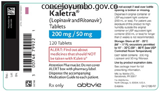
Kaletra dosages: 250 mg
Kaletra packs: 60 pills, 120 pills, 180 pills, 240 pills, 300 pills, 360 pills

Cheap 250 mg kaletra otc
The lung is smaller than normal treatment of hemorrhoids kaletra 250 mg on line, owing to the presence of fewer acini or a decrease of their measurement symptoms of dehydration 250 mg kaletra discount fast delivery. The lesion could also be accompanied by hypoplasia of the bronchi and pulmonary vessels if the insult happens early in gestation, as in congenital diaphragmatic hernia. The lesion often impacts one lobe of the lung and consists of multiple cyst-like areas, that are lined by bronchiolar epithelium and separated by loose fibrous tissue. Some sufferers with congenital cystic adenomatoid malformation produce other congenital anomalies. In the new child, a bronchogenic cyst could compress a serious airway and trigger respiratory misery. Secondary infection of the cyst in older patients may lead to hemorrhage and perforation. Airway Infections are Caused by Diverse Organisms the agents inflicting pulmonary infections are discussed in detail in Chapter 9. Many infectious brokers that involve the intrapulmonary airways tend to have an effect on the extra peripheral airways (bronchiolitis). All are more severe in malnourished children and populations not ordinarily exposed to these agents. Severe symptomatic diseases with these agents are more commonly encountered in infants and children, and restoration is the rule. Bronchioles could become obliterated or occluded by free fibrous tissue (obliterative bronchiolitis). It is three to four times as widespread in males as in females and is associated with other anomalies in two thirds of patients. In older children, it might come to medical attention due to recurrent bronchopulmonary infections. On gross examination, the sequestered pulmonary tissue shows the outcome of chronic recurrent pneumonia, with end-stage fibrosis and honeycomb cystic modifications. Microscopically, the cystic spaces are principally lined by cuboidal or columnar epithelium, and the lumen incorporates foamy macrophages and eosinophilic materials. Interstitial chronic irritation and hyperplasia of lymphoid follicles is commonly outstanding. The wall of this bronchiole shows an intense continual inflammatory infiltrate with native extension into the encircling peribronchial tissue. Severe overdistention may be discovered with out obvious bronchiolar obstruction, probably due to displacement of surfactant from the bronchiolar surface. Similar to adenovirus, it might end in bronchiolar obliteration and bronchiectasis. With the arrival of a pertussis vaccine, the illness has become uncommon in the United States, but the disease remains to be a problem in nonvaccinated populations. Recent enhance in the frequency of illness in the United States has been linked to failure to vaccinate and to a newly introduced vaccine, which has a decrease frequency of unwanted effects, however produces extra restricted immunity. Bronchocentric granulomatosis may also be a manifestation of rheumatoid arthritis, ankylosing spondylitis and granulomatosis with polyangiitis (formerly Wegener granulomatosis). Patients with bronchocentric granulomatosis of either the allergic or nonallergic kind generally reply well to corticosteroid remedy. Constrictive Bronchiolitis May Obliterate the Airway Constrictive (obliterative) bronchiolitis is an unusual disorder in which an preliminary inflammatory bronchiolitis is followed by bronchiolar scarring and fibrosis, resulting in constrictive narrowing and ultimately full obliteration of the airway lumen. Oxidants derive from the motion of sunlight on car exhaust fumes and are necessary in main city areas which have temperature inversions. The results of low concentrations of those brokers on normal persons are unsure, but in persons with persistent pulmonary illness, the scenario is completely different. Air pollution could exacerbate symptoms in asthmatic individuals and in those with established respiratory illness. In high concentrations corresponding to encountered in industrial accidents, irritant gases produce serious morphologic and functional results. Silo employees inhaling excessive concentrations of the fuel develop a type of lung injury generally known as silo-filler illness. Most sufferers recover however some develop progressive bronchiolitis obliterans and should die of respiratory failure. Secondary inflammation may result in in depth bronchiectasis, in part from bronchiolar obliteration and partially from direct damage to the bronchi. Bronchial Obstruction Leads to Atelectasis Bronchial obstruction in adults is most often the consequence of the endobronchial extension of major lung tumors, though mucinous plugs, aspirated gastric contents or overseas bodies could additionally be responsible, particularly in children. Areas distal to the obstruction are also prone to pneumonia, pulmonary abscess and bronchiectasis (see below). If the air provide is obstructed, the lack of gasoline from the alveoli to the blood causes collapse of the affected area. Atelectasis is an important postoperative complication of stomach surgical procedure, occurring due to (1) mucus obstruction of a bronchus and (2) diminished respiratory movement resulting from postoperative pain. Although atelectasis is often brought on by bronchial obstruction, it may additionally outcome from direct compression of the lung Such pressure may seriously compromise Bronchocentric Granulomatosis Usually Reflects Allergic Responses to Infection Bronchocentric granulomatosis refers to nonspecific granulomatous inflammation centered on bronchi or bronchioles. The principal inherited situations associated with generalized bronchiectasis are cystic fibrosis, the dyskinetic ciliary syndromes, hypogammaglobulinemias and deficiencies of particular immunoglobulin (Ig)G subclasses. Kartagener syndrome is among the immotile cilia (ciliary dyskinesia) syndromes and contains the triad of dextrocardia (with or without situs inversus), bronchiectasis and sinusitis. Other dyskinetic ciliary syndromes include radial spoke deficiency("Sturgess syndrome") and absence of the central doublet of cilia. In these ailments, cilia are deficient all through the body, with resultant female and male sterility. In the respiratory tract, ciliary defects lead to repeated upper and decrease respiratory tract infections within the lung and, thus, to bronchiectasis. Bronchi are dilated and have white or yellow thickened walls, and lumina regularly contain thick, mucopurulent secretions. In long-standing atelectasis, the collapsed lung turns into fibrotic and bronchi dilate, in part, due to an infection distal to the obstruction. Obstructive bronchiectasis is localized to a phase of the lung distal to a mechanical obstruction of a central bronchus by a big selection of lesions, together with tumors, inhaled foreign bodies, mucus plugs (in asthma) and compressive lymphadenopathy. Nonobstructive bronchiectasis is usually a complication of respiratory infections or defects in the defense mechanisms that defend the airways from an infection. Localized nonobstructive bronchiectasis was as soon as common, usually resulting from childhood bronchopulmonary infections. Although decreased in frequency by antibiotics and childhood immunizations, one half to two thirds of all instances still observe a bronchopulmonary an infection. Generalized bronchiectasis is, for the most part, secondary to inherited impairment in host protection mechanisms or acquired situations that permit introduction of infectious organisms into the airways. The resected higher lobe reveals widely dilated bronchi, with thickening of the bronchial partitions and collapse and fibrosis of the pulmonary parenchyma. With the consequent collapse of distal lung parenchyma, the damaged bronchi dilate.
Discount kaletra 250 mg fast delivery
Others are related to the actions of poisons symptoms webmd order kaletra 250 mg line, medication or physical agents (particularly ultraviolet light) treatment toenail fungus kaletra 250 mg generic otc. A wide selection of cataracts are inherited, and a few of them are related to other ocular or systemic abnormalities. Fortunately, in the United States, most cataractous lenses are surgically eliminated and changed by the implantation of prosthetic lenses. In other instances, spectacles or contact lenses may be offered to allow gentle to give attention to the retina. Inflammation of the uvea (uveitis) also encompasses inflammation of the iris (iritis), the ciliary physique (cyclitis) and the iris plus the ciliary physique (iridocyclitis). Inflammation of the iris and ciliary physique typically causes a red eye, photophobia, moderate ache, blurred vision, a pericorneal halo, ciliary flush and slight miosis. Synechiae (adhesions between the iris and either the lens or anterior chamber angle) are problems of iritis and may cause glaucoma. Perforating ocular damage and prolapse of uveal tissue often lead to this disorder in both eyes. Uveitis develops within the initially injured eye (exciting eye) and the uninjured eye (sympathizing eye) after a latent interval of four to eight weeks. The antigen responsible for sympathetic ophthalmitis is reported to reside in the photoreceptors of the retina. The degenerated lens materials exerts osmotic pressure, inflicting the damaged lens to swell by imbibing water. Such a swollen lens could hinder the pupil and trigger glaucoma (phacomorphic glaucoma). Unilateral blurred imaginative and prescient lasting a few minutes (amaurosis fugax) occurs with small retinal emboli. Hemorrhage in the nerve fiber layer spreads between axons and causes a flame-shaped look on funduscopy, whereas deep retinal hemorrhages are likely to be round. When positioned between the retinal pigment epithelium and Bruch membrane, blood appears as a dark mass and clinically may resemble a melanoma. After accidental or surgical perforation of the globe, choroidal hemorrhages may detach the choroid and displace the retina, vitreous physique and lens. Central Retinal Vein Occlusion Central retinal vein occlusion leads to flame-shaped hemorrhages within the nerve-fiber layer of the retina, especially across the optic disc. Edema of the optic disc and retina occurs because of impaired absorption of interstitial fluid. An intractable, closed-angle glaucoma, with extreme pain and repeated hemorrhages, generally ensues 2 to 3 months after central retinal vein occlusion (so-called 100-day glaucoma, thrombotic glaucoma or neovascular glaucoma). Certain issues of the center and main vessels, such because the carotid arteries, predispose to emboli that lodge in the retina and are evident on funduscopic examination at factors of vascular bifurcation. By funduscopy, abnormal retinal arterioles appear as parallel white strains at websites of vascular crossings (arterial sheathing). Initially, the narrowed lumen of the retinal vessels decreases the visibility of the blood column and makes it appear orange on ophthalmoscopic examination (copper wiring). However, as the blood column eventually becomes completely obscured, light reflected from the sclerotic vessels seems as threads of silver wire (silver wiring) Lumina of the thickened retinal arterioles turn into narrowed, increasingly tortuous and of irregular caliber. At sites the place the arterioles cross veins, the latter appear kinked (arteriovenous nicking). The kinked look of the vein reflects sclerosis within the venous partitions, as a outcome of the retinal arteries and veins share a typical adventitia at sites of arteriovenous crossings, somewhat than compression by a taut sclerotic artery. Small superficial or deep retinal hemorrhages usually accompany retinal arteriolosclerosis. Malignant hypertension is characterized by a necrotizing arteriolitis, with fibrinoid necrosis and thrombosis of the precapillary retinal arterioles. These spherical spots, that are seldom wider than the optic disc, include aggregates of swollen axons within the nerve-fiber layer of the retina. Central Retinal Artery Occlusion Like neurons in the the rest of the nervous system, those within the retina are extraordinarily vulnerable to hypoxia. Central retinal artery occlusion might comply with thrombosis of the retinal artery, as in atherosclerosis, large cell arteritis or embolization to that vessel. Intracellular edema, manifested by retinal pallor, is prominent, particularly within the macula, where ganglion cells are most numerous. The foveola, the middle of the macula, stands out in sharp distinction as a prominent cherry-red spot due to the underlying vascularized choroid. The generally associated arteriolosclerosis impacts the appearance of the retinal microvasculature. The relationship between retinal microvascular illness and blood glucose levels in sort 2 diabetes is much less clear, and other parameters Retinal ischemia can account for most options of diabetic retinopathy, including cotton-wool spots, capillary closure, microaneurysms and retinal neovascularization. Ischemia outcomes from narrowing or occlusion of retinal arterioles (as from arteriolosclerosis or platelet and lipid thrombi) or from atherosclerosis of the central retinal or ophthalmic arteries. On funduscopy, the primary discernible scientific abnormality is engorged retinal veins with localized sausage-shaped distentions, coils and loops. Neovascularization of the retina is a outstanding feature of diabetic retinopathy and of other situations attributable to retinal ischemia Tortuous new vessels first appear on the floor of the retina and optic nerve head and then develop into the vitreous cavity. The newly fashioned friable vessels bleed easily, and resultant vitreal hemorrhages obscure vision. Neovascularization is associated with proliferation and immigration of astrocytes, which grow around the new vessels to form delicate white veils (gliosis). The proliferating fibrovascular and glial tissue contracts, usually causing retinal detachment and blindness. Diabetic retinopathy, glaucoma and age-related maculopathy are the leading causes of irreversible blindness within the United States. Blindness in diabetic retinopathy outcomes when the macula is concerned, however it additionally follows vitreous hemorrhage, retinal detachment and glaucoma. Once blindness ensues, it heralds an ominous future for the affected person as a outcome of demise from ischemic heart illness or renal failure often follows. Laser phototherapy and strict glycemic control early in the course of proliferative retinopathy have proved effective in controlling these issues. Several yellowish "onerous" exudates (straight arrows), which are rich in lipids, are evident, together with a number of comparatively small retinal hemorrhages (curved arrows). Microaneurysms (arrows) and an exudate (arrowhead) are evident in a area of retinal nonperfusion. However, the sensory retina readily separates from the retinal pigment epithelium when fluid (liquid vitreous, hemorrhage or exudate) accumulates within the potential house between these buildings. Laser therapy has greatly improved the prognosis for patients with detached retina. This scenario causes the photoreceptors to degenerate, after which cyst-like extracellular areas appear within the retina.

250 mg kaletra buy mastercard
In such situations medications prolonged qt order 250 mg kaletra mastercard, each chief cell hyperplasia and multiple small adenomas are seen in the identical gland medicinenetcom symptoms purchase kaletra 250 mg otc. In half of patients, one gland is noticeably bigger than the others, which can make the excellence from adenoma troublesome. Microscopically, the conventional glandular adipose tissue is changed by hyperplastic chief cells organized in sheets or trabecular or follicular patterns. An necessary function that distinguishes hyperplasia from adenoma is the dearth of pleomorphism in the former. It is normally a functioning tumor, and most sufferers current with symptoms of hyperparathyroidism. The incidence of the illness varies from about 1 in 10,000 among whites to 1 in 500 in Alaskan Eskimos. A deficiency in this enzymatic exercise impairs cortisol biosynthesis, and amassed precursors are instead transformed to androgens. Female newborns uncovered to a big extra of adrenal androgens in utero exhibit pseudohermaphroditism and are born with fused labia, an enlarged clitoris and a urogenital sinus which might be mistaken for a penile urethra Microscopically, the cortex is widened between the medulla and the zona glomerulosa. In most cases, the zona glomerulosa is also hyperplastic, although to not the extent of the opposite zones, especially the zona fasciculata. Eventually, the excessive levels of adrenal androgens result in the premature closure of epiphyses and brief stature. As a result, hypoaldosteronism develops inside the first few weeks of life in two thirds of newborns, which is manifested as hyponatremia, hyperkalemia, dehydration, hypotension and increased renin secretion. Reconstructive surgical procedure may be essential for virilized girls with ambiguous genitalia. In idiopathic Addison disease, the biochemical defect of adrenoleukodystrophy is usually detected (see Chapter 28). The illness has a slight female predominance and is seen in older kids and adolescents. Premature ovarian failure, hypothyroidism, malabsorption syndromes, pernicious anemia, chronic hepatitis, alopecia totalis and vitiligo are also encountered. Autoimmune thyroiditis and occasionally Hashimoto thyroiditis and Graves disease occur in more than two thirds of cases. Autoimmune adrenalitis results in pale, irregular, shrunken glands, weighing 2 to 3 g or less. The medulla is intact but surrounded by fibrous tissue containing small islands of atrophic cortical cells. Depending on the stage of the illness, lymphoid infiltrates, predominantly T cells, of varying density are encountered. The gene for eleven -hydroxylase (on chromosome 8) catalyzes terminal hydroxylation in cortisol biosynthesis. It causes weakness, weight reduction, gastrointestinal symptoms, hypotension, electrolyte imbalance and hyperpigmentation. Adrenal disaster is nearly invariably fatal unless the affected person is promptly and aggressively handled with corticosteroids and supportive measures. Adrenal Hyperfunction Excess corticosteroid secretion happens in adrenal hyperplasia or neoplasia. Such hyperfunction may take one of two forms, specifically, hypercortisolism (Cushing syndrome) or hyperaldosteronism (Conn syndrome), issues reflecting the two main classes of adrenal steroid hormones. The constellation of scientific features is comprised of weight problems, hypertrichosis and amenorrhea, which reflects excessive glucocorticoid ranges. A section of the adrenal gland from a patient with Addison illness reveals chronic irritation and fibrosis in the cortex, an island of residual atrophic cortical cells and an intact medulla. A diffuse tan pigmentation often develops on the skin, and dark patches might appear on the mucous membranes. A number of gastrointestinal symptoms, including vomiting, diarrhea and belly ache, affects most patients and could be the presenting grievance. Patients with Addison illness usually exhibit marked personality adjustments and even natural brain syndromes. With glucocorticoid and mineralocorticoid replacement, patients reside normal lives. Symptoms are associated more to mineralocorticoid deficiency than to insufficient glucocorticoids. Adrenal crisis occurs in three settings: Abrupt withdrawal of corticosteroid therapy in patients with adrenal atrophy due to long-term administration of these steroids. Sudden, devastating worsening of continual adrenal insufficiency may be precipitated by the stress of infection or surgical procedure. Waterhouse-Friderichsen syndrome is acute, bilateral, hemorrhagic infarction of the adrenal cortex, most commonly secondary to meningococcal or Pseudomonas septicemia (see Chapter 7). The illness usually results from corticotroph microadenomas of the pituitary or, in a couple of sufferers, diffuse corticotroph hyperplasia. A typical adenoma is encapsulated, firm, yellow and barely lobulated, measuring about four cm in diameter. These tumors often weigh 10 to 50 g, though weights as much as a hundred g have been recorded. The cut floor is mottled yellow and brown and infrequently black, owing to the deposition of lipofuscin pigment. Microscopically, adenomas exhibit clear, lipid-laden (fasciculata type) cells arranged in sheets or nests, usually with interspersed clusters of compact, Adrenal Adenoma Adrenocortical adenomas can produce hormones, the most common being cortisol and aldosterone. The nontumorous cortexes of the concerned and contralateral gland are generally atrophic. Thus, hypertension and hirsutism, features generally seen in Cushing syndrome as a result of adrenal hyperplasia or neoplasia, are often absent on this iatrogenic disorder. Adrenal Cortical Carcinoma Adrenal cortical carcinoma is a uncommon and aggressive tumor that has an incidence of one case per million per 12 months. The tumor metastasizes to lung, liver and lymph nodes, and native recurrences are widespread. The minimize surface is variegated pink, brown or yellow, typically with necrosis, hemorrhage and cystic change. Local invasion is common, and remnants of normal adrenal are difficult to identify. Enlargement of the stomach and other areas of fat deposition stretches the thin skin and produces purplish striae, which symbolize venous channels which might be visible by way of the attenuated dermis. Back pain is common, and up to one fifth of patients with Cushing syndrome have radiologic evidence of vertebral compression fractures. The fusion gene is ectopically and constitutively activated in the zona fasciculata, and bilateral hyperplasia of this zone outcomes. Aldosterone hypersecretion enhances renal tubular sodium reabsorption, thereby rising physique sodium. Hypertension is triggered not only by retention of sodium and consequent volume growth but also by elevated peripheral vascular resistance. On microscopic examination, the dominant cells are clear and lipid-rich, resembling the zona fasciculata, and are organized in cords or alveoli. All types of Cushing syndrome are characterized by increased glucocorticoid levels.


Buy 250 mg kaletra overnight delivery
As the mass of micro organism adhering to the surface of tooth (dental plaque) ages symptoms 9 weeks pregnant kaletra 250 mg buy discount, it mineralizes to kind calculus (tartar) medicine for bronchitis order kaletra 250 mg on-line. Adult periodontitis is mostly associated with Bacteroides gingivalis, Bacteroides intermedius, Actinomyces species and Haemophilus species. Agranulocytosis causes necrotizing ulcers wherever in the oral and pharyngeal mucosa, but particularly within the gingiva. Infectious mononucleosis usually ends in gingivitis and stomatitis, with exudate and ulceration. Necrosis and ulceration of the gingiva predispose to severe superimposed infection, which can trigger lack of tooth and alveolar bone. This newly formed dentin is opposite the world of tooth destruction and was produced by the stimulated odontoblasts. Odontogenic Tumors: Ameloblastoma Ameloblastomas are tumors of odontogenic epithelia and are the most common clinically important odontogenic tumor. The abundant serous discharge then becomes mucopurulent, after which the surface epithelium is shed. Chronic rhinitis is characterised by nasal mucosal thickening as a outcome of persistent hyperemia, mucous gland hyperplasia and lymphocyte and plasma cell infiltration. Often known as hay fever, allergic rhinitis could also be acute and seasonal or persistent and perennial (see Chapter 4). A widespread histologic pattern is characterised by islands of odontogenic epithelium with a central stellate reticulum-like area, surrounded by basal cells with a "picket fence" appearance, due to subnuclear vacuoles. The etiology entails multiple elements, including allergy, infections, diabetes mellitus, cystic fibrosis and aspirin intolerance. Sinonasal inflammatory polyps are lined externally by respiratory epithelium and comprise mucous glands within a unfastened mucoid stroma, which is infiltrated by plasma cells, lymphocytes and lots of eosinophils. Microscopically, ameloblastoma resembles the enamel organ in its numerous phases of differentiation, and a single tumor might show numerous histologic patterns. Accordingly, tumor cells resemble ameloblasts on the periphery of epithelial nests or cords, the place columnar cells are oriented perpendicularly to the basement membrane. Some may spread and but stay histologically benign (metastasizing ameloblastoma). The causes range from the frequent cold to unusual infections, similar to diphtheria and anthrax. The virus replicates in epithelial cells, inflicting the degenerating cells to be shed. The mucosa is edematous and engorged and is infiltrated by neutrophils and mononuclear cells. Abundant mucus secretion and increased vascular permeability produce rhinorrhea (free discharge of a thin nasal mucus). Viral rhinitis could additionally be adopted within a number of days by or foreign physique that interferes with sinus drainage or aeration renders it liable to an infection. If the ostium of a sinus is blocked, secretions or exudate accumulate behind the obstruction. Maxillary sinusitis may also be attributable to odontogenic infections, by which case micro organism from the roots of the primary and second molars penetrate the skinny bony plate that separates them from the ground of the maxillary sinus. Chronic sinusitis is a sequel of acute irritation, either as a result of incomplete decision of an infection or because of recurrent acute issues. In contrast to acute sinusitis, the purulent exudate in continual sinusitis virtually all the time contains anaerobic micro organism. Rhinoscleroma is endemic in some Mediterranean nations and in components of Asia, Africa and Latin America. Microscopically, the granulation tissue is strikingly rich in plasma cells, lymphocytes and foamy macrophages. The characteristic massive macrophages, referred to as Mikulicz cells, include lots of phagocytosed bacilli. Serologic checks are useful in establishing the prognosis of rhinoscleroma as a outcome of particular antibodies are present in many sufferers. They arise from the sinonasal mucosa, the ectodermally derived lining of the sinonasal tract (Schneiderian membrane). Three morphologically distinct benign papillomas are acknowledged: inverted, oncocytic (cylindrical or columnar cell) and fungiform (exophytic, septal) papillomas. They are composed of a uniform cellular proliferation, which shows an inflammatory cell infiltrate and scattered microcysts. As the name implies, they present characteristic inversions of the surface epithelium into the underlying stroma. Unless surgical resection extends beyond the boundaries of grossly visible lesions, they frequently recur. Several industrial chemical substances including nickel and chromium have been reported to increase the danger of cancer of the nose and sinuses. Squamous tumors in nickel staff normally come up from the center turbinate, with latencies from 2 to 32 years. Cancers of the nasal cavity and sinuses grow relentlessly and invade adjoining buildings. Follicular tonsillitis is characterised by pinpoint exudates that can be extruded from the crypts. Pseudomembranous tonsillitis refers to a necrotic mucosa coated by a coat of exudate, for example, in diphtheria or in Vincent angina (see above). However, repeated infections could cause enlargement of tonsils and adenoids to a degree that obstructs air passages. In kids, repeated bouts of streptococcal tonsillitis may progress to rheumatic fever or glomerulonephritis, and patients may profit from tonsillectomy. Peritonsillar abscess (quinsy) is a set of purulent materials behind the posterior capsule of the tonsil, usually as a outcome of an infection with - and -hemolytic streptococci. Untreated, peritonsillar abscesses could lead to a quantity of life-threatening situations, similar to rupture into the airway, weakening of the carotid artery wall or penetration into the mediastinum, the bottom of the cranium or the cranial vault. Adenoids symbolize persistent inflammatory hyperplasia of the pharyngeal lymphoid tissue. Enlarged adenoids could cause partial or complete obstruction of the eustachian tube, leading to otitis media. The tumor infiltrate is characteristically polymorphic and surrounds small- to medium-sized blood vessels (angiocentric), infiltrates via vascular partitions (angioinvasive) and often occludes vessel lumina like a thrombus, inflicting necrosis in adjacent tissues (ischemictype). Ulcers are lined by a black crust, underneath which lesions progress to erode cartilage and bone, inflicting defects in the nasal septum, exhausting palate and nasopharynx. Death is because of secondary bacterial infection, aspiration pneumonia or hemorrhage from eroded large blood vessels.

Purchase kaletra 250 mg with amex
Poliomyelitis the term poliomyelitis describes any irritation of the gray matter of the spinal wire treatment modality definition 250 mg kaletra discount overnight delivery, but in widespread utilization symptoms questions 250 mg kaletra buy, it implies an an infection by poliovirus. Dogs, wolves, foxes and skunks are the principle reservoirs, but bats and domestic animals, including cattle, goats and swine, may also carry the illness. In the United States, the place canine are routinely vaccinated in opposition to rabies, the few human rabies infections (one to five per year) normally end result from publicity to rabid bats. However, in areas the place rabies is endemic, most human infections end result from dog bites, and rabies kills greater than 50,000 people annually. Latent intervals vary in proportion to the space of transport, from 10 days to even longer than 3 months. Eosinophilic cytoplasmic viral inclusion bodies within the hippocampus, brainstem and cerebellar Purkinje cells (Negri bodies) verify the prognosis. Infected cells could endure chromatolysis, a reversible course of involving neuronal swelling, cytoplasmic swelling and eccentric nuclear positioning. Initial inflammatory responses transiently embrace neutrophils, that are followed by lymphocytes that surround blood vessels within the spinal twine and brainstem. Sections of spinal twine in cases of healed poliomyelitis present a paucity of neurons, with secondary degeneration of corresponding ventral roots and peripheral nerves. The growth within the 1950s of effective vaccines against poliovirus has largely eliminated the disease in many of the world. Urgent rabies vaccination and hyperimmune globulin are administered for postexposure prophylaxis. The encephalitis is fulminant and the temporal lobes turn into swollen, hemorrhagic and necrotic. Intranuclear eosinophilic inclusions, usually surrounded by a halo (Cowdry A), occur in each neurons and glial cells. The infected neurons display intranuclear, eosinophilic viral inclusions (Cowdry A inclusions) that fill the nuclei (arrows). Togaviridae, Bunyaviridae and Flaviviridae account for most of the arboviruses that cause human encephalitis. Arbovirus infections are zoonoses of animals, and people are contaminated when bitten by virus-harboring arthropods. The varied encephalitides brought on by arboviruses are named principally for the location where they were first noted (Table 28-1). West Nile encephalitis has numerically eclipsed all different arbovirus encephalitides in the United States since its preliminary look in 1999. West Nile encephalitis has a propensity for the spinal wire and may produce a syndrome clinically indistinguishable from classical poliomyelitis. Mild cases of arbovirus encephalitis could entail solely a mild flulike syndrome and will not be diagnosed as encephalitis. In more extreme instances, onset is abrupt, often with excessive fever, headache, vomiting and meningeal signs, adopted by lethargy and coma. Death is extra likely at the extremes of age, and these who survive could additionally be left with cognitive impairment and seizures. Microscopically, the specimen exhibits pronounced perivascular lymphocytic irritation. The illness occurs mainly in childhood and is characterized by cognitive and behavioral decline over months to years, finally leading to dying. The course is protracted, and irritation occurs primarily in cerebral grey matter. Intranuclear inclusions are distinguished in neurons and oligodendroglia, as are marked gliosis in affected gray and white matter, patchy loss of myelin and ubiquitous perivascular lymphocytes and macrophages. They are characteristically spherical and a quantity of other millimeters in diameter with a central area largely devoid of myelin. Axons are retained, a couple of oligodendrocytes are seen and the lesion is infiltrated by macrophages. At the sting of the demyelinated space, there are oligodendrocytes with enlarged nuclei occupied by homogeneously dense, hyperchromatic, "ground-glass" intranuclear inclusions missing a halo. Astrocytes are additionally contaminated, however as an alternative of dying, they present extreme pleomorphism. If the host turns into immunocompromised, viremia ensues, with specific viral strains having a propensity for neurovirulence. Rather, these cells are injured indirectly by cytokines or neurotoxic viral proteins, which elicit oxidant-mediated cell harm. Spongiform degeneration of the gray matter is characterized by individual and clustered vacuoles, with no evidence of irritation. In addition, myelin pallor, reflecting diffuse demyelination, intense astrogliosis and loss of neurons, is frequent. It decimated the cattle business within the United Kingdom and has spread to other regions of the world and to different species including zoo animals, pets and people. Uniquely, conversion of the native protein to the pathogenic form is autocatalyzed by the pathogenic kind itself. Prion Diseases (Spongiform Encephalopathies) Are Transmissible Neurodegenerative Diseases Caused by Particles Containing Modified Proteins Prion diseases are characterised clinically by rapidly progressive ataxia and dementia and pathologically by the buildup of fibrillar or insoluble prion proteins, degeneration of neurons and vacuolization, termed spongiform encephalopathy. The spongiform encephalopathies are biologically exceptional as a result of the causative infectious entity is devoid of nucleic acids. Other traits include neuron degeneration, gliosis and accumulations of insoluble prions forming extracellular plaques These occur most in cortical grey matter but also involve deeper nuclei of the basal ganglia, thalamus, hypothalamus and cerebellum. Central myelin is made by oligodendrocytes, whereas peripheral myelin is synthesized by Schwann cells. This transition often happens about 2 to 3 mm after a cranial nerve or spinal root exits the brainstem or spinal wire. It becomes symptomatic at a mean age of 30 years, and women are troubled almost twice as often as males. Plaques, hardly ever more than 2 cm in diameter, accumulate in great numbers within the mind and spinal cord They are discrete, with easily rounded contours, and are often in white matter, although they could breach the gray matter. The evolving plaque is marked by (1) selective loss of myelin in a region of relative axonal preservation, (2) lymphocytes that cluster about small veins and arteries, (3) an influx of macrophages and (4) appreciable edema. Neuronal bodies within the boundaries of a plaque are remarkably spared, but the axons lose their myelin abruptly and may degenerate. As plaques age, they turn out to be more discrete, much less edematous, dense and ultimately gliotic. This sequence emphasizes the focal nature of the injury, its selectivity and its severity, as a end result of demyelination is complete within a plaque. This myelin-stained coronal complete mind part of the brain of a affected person with long-standing a number of sclerosis exhibits many areas of myelin loss-plaques (arrows)-with characteristic periventricular demyelination particularly distinguished on the superior angles of the lateral ventricles. Leukodystrophies Are Inherited Disturbances in Myelin Formation and Preservation these problems typically impact both central and peripheral myelin and often manifest in infancy or childhood, though milder adult phenotypes might happen. Disruption of central myelin leads to blindness, spasticity and lack of developmental milestones, whereas loss of peripheral myelin progresses to weakness and lack of reflexes. The disease typically begins with signs relating to lesions within the optic nerves, brainstem or spinal cord.
Cheap kaletra 250 mg amex
It is often encountered in males in the fifth decade and is commonly accompanied by a rash treatment centers order 250 mg kaletra overnight delivery. Endomyocardial illness is suspected to end result from myocardial injury produced by eosinophils medicine naproxen cheap kaletra 250 mg without a prescription, probably mediated by cardiotoxic granule components. Many circumstances of restrictive cardiomyopathy are categorized as idiopathic, with interstitial fibrosis as the one histologic abnormality. Amyloidosis the guts is affected in most types of generalized amyloidosis (see Chapter 23). Microscopically, amyloid accumulation is most outstanding in interstitial, perivascular and endocardial regions. Endocardial involvement is widespread in the atria, where nodular endocardial deposits typically impart a granular look and gritty texture to the endocardial floor. On minimize section of the ventricle, endocardial fibrosis spreads into the inside one third to one half of the wall. When the right ventricle is involved, the complete cavity may exhibit endocardial thickening, which may penetrate as far as the epicardium. Storage Diseases the assorted lysosomal storage illnesses are mentioned intimately in Chapter 6. The practical changes are those of a restrictive type of cardiomyopathy, and the standard reason for demise is cardiac failure. Cardiac illness outcomes from lysosomal accumulation of mucopolysaccharides (glycosaminoglycans) in numerous cells. In common, pseudohypertrophy of the ventricles develops, and contractility gradually diminishes. The coronary arteries might Endomyocardial Disease Endomyocardial illness includes two geographically separate disorders. The illness can additionally be sometimes seen in other tropical and subtropical areas of the world. The diploma of iron deposition within the coronary heart varies and solely roughly correlates with that in different organs. Cardiac involvement has features of each dilated and restrictive cardiomyopathy, with systolic and diastolic impairment. Congestive coronary heart failure happens in as many as one third of sufferers with hemochromatosis. The brown color seen on gross examination correlates with iron deposition in cardiac myocytes. However, the severity of myocardial dysfunction seems to be proportional to the quantity of iron deposited. Almost all rhabdomyomas are multiple and involve both ventricles and, in one third of circumstances, the atria as properly. In half of cases, the tumor projects into a cardiac chamber and obstructs the lumen or valve orifices. The tumor seems as a glistening, gelatinous, polypoid mass, often 5 to 6 cm in diameter, with a brief stalk. Microscopically, cardiac myxoma has a unfastened myxoid stroma containing ample proteoglycans. Polygonal stellate cells are found throughout the matrix, occurring singly or in small clusters. One third of patients with myxomas of the left atrium or left ventricle die from tumor embolization to the mind. Microscopically, tumor cells show small central nuclei and ample glycogenrich clear cytoplasm, during which fibrillar processes containing sarcomeres radiate to the margin of the cell ("spider cell"). Still, solely a minority of patients with these tumors will show cardiac metastases. Of all tumors, the one most probably to metastasize to the heart is malignant melanoma. Pericarditis associated with myocardial infarction and rheumatic fever is mentioned above. However, speedy accumulation of as little as 150 to 200 mL of pericardial fluid or blood may considerably enhance intrapericardial pressure and prohibit diastolic filling, especially of the right ventricle. Cardiac tamponade is the syndrome produced by the rapid accumulation of pericardial fluid, which restricts the filling of the center. The commonest kind is fibrinous pericarditis, in which the conventional clean glistening look of the pericardial surfaces turns into replaced by a dull, granular fibrin-rich exudate. The tough texture of the infected pericardial surfaces produces the attribute friction rub heard by auscultation. The effusion fluid in fibrinous pericarditis is normally rich in protein, and the pericardium contains primarily mononuclear inflammatory cells. Bacterial infection results in a purulent pericarditis, by which the pericardial exudate resembles pus and incorporates many neutrophils. Idiopathic or viral pericarditis is a self-limited dysfunction, though it might occasionally lead to constrictive pericarditis. Serous pericardial effusion is usually a complication of a rise in extracellular fluid volume, as occurs in congestive coronary heart failure or the nephrotic syndrome. Chylous effusion (fluid containing chylomicrons) results from a communication of the thoracic duct with the pericardial house secondary to lymphatic obstruction by tumor or infection. The commonest trigger is ventricular free wall rupture on the site of a myocardial infarct. Less frequent causes are penetrating cardiac trauma, rupture of a dissecting aneurysm of the aorta, infiltration of a vessel by tumor or a bleeding diathesis. The hemodynamic consequences vary from a minimally symptomatic condition to abrupt cardiovascular collapse and demise. As the pericardial stress increases, it reaches and then exceeds central venous stress, thereby limiting return of blood to the heart. Acute cardiac tamponade is almost invariably deadly unless the strain is relieved by eradicating pericardial fluid, by either needle pericardiocentesis or surgical procedures. In most instances, the cause for acute pericarditis is obscure and (as in myocarditis) is attributed to an undiagnosed viral an infection. The coronary heart of a affected person who died in uremia displays a shaggy, fibrinous exudate overlaying the visceral pericardium. The situation is rare at present and, in developed international locations, is predominantly idiopathic. Prior radiation therapy to the mediastinum and cardiac surgical procedure account for more than one third of circumstances. Constrictive pericarditis could observe tuberculous infection, which continues to be the most important cause in underdeveloped areas.
250 mg kaletra purchase with amex
Mycobacteria are is dependent upon age and immune competence treatment centers near me kaletra 250 mg discount free shipping, in addition to the whole burden of organisms medicine etodolac kaletra 250 mg purchase without prescription. Some sufferers have solely an indolent, asymptomatic an infection, whereas in others, tuberculosis is a damaging, disseminated illness. Active tuberculosis denotes the subset of tuberculous infections manifested by destructive and symptomatic disease. Contaminated feces are deposited on the pores and skin or clothes of a second host, penetrate an abrasion or are inhaled. Some of these cells carry organisms from the lung to regional (hilar and mediastinal) lymph nodes, from which they could disseminate by the bloodstream. Clones of sensitized T cells proliferate, produce interferon- and activate macrophages, thereby increasing their concentrations of lytic enzymes and augmenting their capability to kill mycobacteria. If an contaminated individual is immunologically competent, a vigorous granulomatous reaction is produced. Primary tuberculosis (in a person lacking earlier contact or immune responsiveness). Secondary (cavitary) tuberculosis outcomes from reactivation of dormant endogenous bacilli or reinfection with exogenous bacilli. Miliary tuberculosis is attributable to dissemination of tubercle bacilli to produce quite a few, minute, yellowwhite lesions (resembling millet seeds) in distant organs. Primary tuberculosis occurs on first publicity to the organism and can pursue both an indolent or aggressive course Secondary tuberculosis develops long after a main an infection, largely as a outcome of reactivation of a main an infection. Photomicrograph of a hilar lymph node exhibits a tuberculous granuloma with central caseation. B Secondary (Cavitary) Tuberculosis Results from Release of a Contained Infection the mycobacteria in secondary tuberculosis could also be either dormant organisms from old granulomas (which is normally the case) or newly acquired bacilli. A cross-section of lung shows a quantity of tuberculous cavities full of necrotic, caseous materials. In both lungs and lymph nodes, the Ghon complex heals, present process shrinkage, fibrous scarring and calcification-the last of which is seen radiographically. In immunologically immature topics (a young youngster or immunosuppressed patient), granulomas are poorly formed or not fashioned in any respect, and an infection progresses on the major site in the lung, within the regional lymph nodes or in multiple websites of dissemination. This process produces progressive major tuberculosis, during which the immune response fails to management the tubercle bacilli. The lungs, lymph nodes, kidneys, adrenals, bone marrow, spleen and liver are widespread websites of miliary lesions. There, the bacilli proliferate and elicit an inflammatory response, inflicting localized consolidation. These cavities include necrotic materials teeming with mycobacteria and are surrounded by a granulomatous response. Tubercle bacilli may spread throughout the physique via the lymphatics and bloodstream to cause miliary tuberculosis. Leprosy is transmitted from individual to particular person, often as a result of years of intimate contact. The mode of an infection is unclear however in all probability includes inoculation of bacilli into the respiratory tract or open wounds. Although leprosy is now uncommon in developed countries, half a million persons are reported to be infected worldwide, primarily in tropical areas, together with tropical Africa, Brazil and Southeastern Asia. Vigorous worldwide programs aimed at discovery and remedy have been profitable in lowering the incidence of latest cases. In the United States, about a hundred circumstances are recognized yearly, largely in immigrants from endemic areas. Humans probably purchase it from the setting by inhaling aerosols from contaminated water sources. Lesions differ from the small, insignificant and self-healing macules of tuberculoid leprosy to the diffuse, disfiguring and sometimes deadly lesions of lepromatous leprosy. Susceptible people span a broad immunologic spectrum, from anergy to hyperergy and may develop symptomatic infection. At one finish of the spectrum, anergic sufferers have little or no resistance and develop lepromatous leprosy, whereas hyperergic patients with excessive resistance contract tuberculoid leprosy. The bacilli replicate, fill the cells and unfold first to other macrophages after which all through the physique via the lymphatics and bloodstream. Progressive small bowel involvement produces malabsorption and diarrhea, typically with abdominal ache. Of these, most are "opportunists"-that is, they only infect individuals with impaired immune mechanisms. They range from 2 to a hundred m and are eukaryotes meaning that they possess nuclear membranes and cytoplasmic organelles, corresponding to mitochondria and endoplasmic reticulum. Tuberculoid leprosy is characterized by a single lesion or only a few lesions of the pores and skin, often on the face, extremities or trunk. Microscopically, lesions show well-formed, circumscribed, dermal, noncaseating granulomas with epithelioid macrophages, Langhans large cells and lymphocytes. Skin lesions kind well-demarcated, hypopigmented or erythematous, dry, hairless patches, with raised outer edges which might be characterised by decreased sensation. Lepromatous leprosy reveals multiple, tumor-like lesions of the pores and skin, eyes, testes, nerves, lymph nodes and spleen. Foamy macrophages comprise numerous organisms, which seem as aggregates of acid-fast material, referred to as "globi. Involvement of the higher airways leads to continual nasal discharge and voice change. In individuals with intact cell-mediated immunity, an infection is quickly contained with out producing symptoms. There is diffuse involvement, together with a leonine face, lack of eyebrows and eyelashes and nodular distortions, especially on the face, ears, forearms and hands-the exposed (cool) elements of the physique. A attribute "clear zone" of uninvolved dermis separates the dermis from tumor-like accumulations of macrophages, each containing quite a few lepra bacilli (Mycobacterium leprae). Skin from the raised "infiltrated" margin of the plaque accommodates discrete granulomas that stretch to the basal layer of the epidermis (without a clear zone). A silver stain reveals crescent-shaped organisms, which are collapsed and degenerated. Microscopically, the alveoli comprise a frothy eosinophilic material, composed of alveolar macrophages and cysts and trophozoites of P. The progressive filling of alveoli prevents adequate gasoline trade, and the patient slowly suffocates. Therapy is with trimethoprim-sulfamethoxazole, pentamidine, atovaquone or a number of different regimens. Many Candida species are endogenous human flora, well adapted to life on or within the human body.

Generic 250 mg kaletra otc
Secondary polycythemia can also occur underneath certain circumstances unrelated to generalized tissue hypoxia 7 medications that can cause incontinence cheap kaletra 250 mg amex. Initially medicine expiration dates kaletra 250 mg buy online, platelets adhere to the vascular endothelium and subsequently kind aggregates which might be stabilized by fibrin after the coagulation cascade is activated. Defects of the system for sustaining fluid blood passage via intact vessels fall into two categories: hemostatic problems and thrombotic problems. Failure of the hemostatic system to restore the integrity of an injured vessel causes bleeding. The clinical manifestations of hemorrhage related to problems of each part of the hemostatic system are inclined to be distinctive. Platelet abnormalities lead to both petechiae and purpura within the skin and mucous membranes. Deficiencies of coagulation elements are related to hemorrhage into muscle tissue, viscera and joint areas. A monoclonal antibody inhibiting the activation of complement part 5 (eculizumab) has prolonged the 5-year survival of sufferers to that of unaffected individuals. Hemostatic Disorders of Blood Vessels Reflect Dysfunction of Extravascular or Vascular Tissues Extravascular Dysfunction Resulting in Hemostatic Defects Dysfunction of the extravascular tissues is of restricted clinical significance. Senile purpura options superficial, sharply demarcated, persistent purpuric spots on the forearms and other sun-exposed areas. The decrease the platelet count, the higher the chance of traumatic and perioperative bleeding. Marrow infiltration with leukemic cells, metastatic cancer, bone marrow failure in patients with aplastic anemia, radiotherapy or chemotherapy produce pancytopenia, together with thrombocytopenia. Megaloblastic anemia and myelodysplasia could cause severe thrombocytopenia because of ineffective megakaryopoiesis. Increased platelet destruction may replicate immunemediated harm and removal of circulating platelets, as in idiopathic thrombocytopenic purpura and drug-induced thrombocytopenia. Alternatively, intravascular platelet aggregation might produce thrombocytopenia Vascular Dysfunction Resulting in Hemostatic Defects Vascular dysfunction and associated hemostatic defects replicate the following: Intrinsic genetic defects: Hereditary hemorrhagic telangiectasia (Rendu-Osler-Weber syndrome) is an autosomal dominant dysfunction of blood vessel partitions (venules and capillaries), which outcomes in tortuous, dilated vessels (telangiectasias). Patients with hereditary hemorrhagic telangiectasia have recurrent hemorrhages that occur spontaneously or following trivial trauma. Arteriovenous fistulas in the lung, mind and retina could also be troublesome and result in hemorrhage or clinically vital shunting of blood. IgA and complement complexes flow into in the blood and are often seen in vessel partitions. Deposition of Ig fragments in the vessel wall: Amyloidosis, cryoglobulinemia and paraproteinemias may all lead to injury to the vascular system (see Chapters sixteen and 23). Certain types of arteritis additionally injure the vessel wall and should cause hemorrhage (see Chapter 10). Table 20-4 Principal Causes of Thrombocytopenia Decreased Production Aplastic anemia Bone marrow infiltration (neoplastic, fibrosis) Bone marrow suppression by medication or radiation Platelet Disorders Impair Hemostasis Patients with platelet issues may have a historical past of simple bruising or life-threatening bleeding. Bleeding can happen in any broken vascular bed, however a pattern of mucocutaneous bleeding, together with gingival bleeding, epistaxis and menorrhagia, is common. More extreme manifestations are bleeding into the gastrointestinal tract, genitourinary tract and mind. Petechiae, which are characteristic of platelet problems, are nonblanching red lesions less than 2 mm in size. They usually occur in the decrease extremities, in dependent regions of the physique, on the buccal mucosal and taste bud and at strain points (waistband, wristwatch band). These people develop profound consumptive thrombocytopenia, platelet activation and consequently a hypercoagulable state. Platelet aggregation predisposes patients to arterial and venous thromboembolic occasions that might be lethal. In most sufferers, these autoantibodies are of the IgG class, although IgM antiplatelet antibodies often occur. Surface-bound complement causes platelets to be lysed within the blood or phagocytosed and destroyed by splenic and hepatic macrophages. Peripheral blood smears present quite a few massive platelets, which reflect accelerated launch of young platelets by a bone marrow actively engaged in platelet production. Accordingly, bone marrow examination reveals compensatory will increase in megakaryocytes. The pathology of those disorders outcomes from widespread platelet aggregation and the deposition of hyaline thrombi in the microcirculation. Fragmented erythrocytes (schistocytes) are always evident in peripheral blood smears, as are numerous reticulocytes. The drug often forms a fancy with a plateletrelated protein to make a neoepitope that elicits antibody manufacturing. By distinction, chemotherapeutic brokers, ethanol and thiazides cause thrombocytopenia by suppression of platelet production. Most patients current with neurologic symptoms, including seizures, focal weak point, aphasia and alterations in the state of consciousness. The manufacturing of a Shiga-like toxin damages the endothelium and initiates platelet activation, followed by platelet aggregation. Hereditary Disorders of Platelets Hereditary issues of platelets are uncommon and contain either quantitative or qualitative defects in membrane glycoprotein receptors or defects in platelet granules. Nonsteroidal analgesics, such as indomethacin or ibuprofen, impair platelet perform, but as their inhibition of cyclooxygenase is reversible, their effect on platelets is transient. Renal failure: End-stage kidney disease is usually accompanied by a qualitative platelet defect that ends in a chronic bleeding time and an inclination toward hemorrhage. Cardiopulmonary bypass: Platelet dysfunction due to platelet activation and fragmentation occurs within the extracorporeal circuit throughout bypass surgery. Coagulopathies Are Caused by Deficient or Abnormal Coagulation Factors Quantitative and qualitative problems of all of coagulation factors have been recognized. Hemophilia is an X-linked recessive dysfunction of blood clotting that leads to spontaneous bleeding, notably into joints, muscle tissue and inner organs. Bernard-Soulier syndrome manifests in infancy or childhood, with a bleeding sample attribute of abnormal platelet operate, namely ecchymoses, epistaxis and gingival bleeding. The lack of aggregation and clot retraction impairs hemostasis and causes bleeding despite a traditional platelet depend. The illness manifests shortly after birth, and older sufferers might undergo sudden hemorrhage after trauma or surgery. Alpha storage pool illness (gray platelet syndrome): the disease is a rare inherited malady characterized by the absence of morphologically recognizable alpha granules in platelets. Delta storage pool illness: the illness is heterogeneous and impacts the dense granules of platelets. The most frequent complication of hemophilia A is a deforming arthritis attributable to repeated bleeding into many joints. Although now uncommon, bleeding into the brain was previously the most common explanation for death. Many different mutations, from single-base substitutions to gross deletions, have been linked to hemophilia B. Severe liver disease could cause impaired secretion of these proteins as a manifestation of a basic defect in protein synthesis.


