
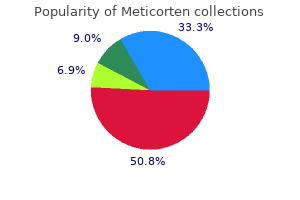
Meticorten
Meticorten dosages: 40 mg, 20 mg, 10 mg, 5 mg
Meticorten packs: 30 pills, 60 pills, 90 pills, 120 pills, 180 pills, 270 pills, 360 pills

Cheap 5 mg meticorten with amex
The paraventricular drug allergy treatment guidelines buy meticorten 20 mg with amex, dorsomedial allergy testing alcat purchase 10 mg meticorten visa, and arcuate nuclei of the hypothalamus additionally play a major position in regulating meals intake. For example, lesions of the paraventricular nuclei typically trigger excessive consuming, whereas lesions of the dorsomedial nuclei often depress consuming conduct. As mentioned later, the arcuate nuclei are the websites in the hypothalamus the place multiple hormones launched from the gastrointestinal tract and adipose tissue converge to regulate food intake, in addition to energy expenditure. Much chemical cross speak happens among the neurons on the hypothalamus, and collectively, these centers coordinate the processes that management consuming behavior and the perception of satiety. These hypothalamic nuclei also influence the secretion of a number of hormones that are necessary in regulating power steadiness and metabolism, including these from the thyroid and adrenal glands, as well as the pancreatic islet cells. The hypothalamus receives (1) neural indicators from the gastrointestinal tract that provide sensory details about stomach filling; (2) chemical alerts from vitamins within the blood (glucose, amino acids, and fatty acids) that signify satiety; (3) alerts from gastrointestinal hormones; (4) indicators from hormones launched by adipose tissue; and (5) alerts from the cerebral cortex (sight, smell, and taste) that influence feeding habits. The hypothalamic feeding and satiety centers have a high density of receptors for neurotransmitters and hormones that influence feeding conduct. A few of the many substances which have been shown to alter urge for food and feeding habits in experimental research are listed in Table 72-2 and are typically categorized as (1) orexigenic substances that stimulate feeding or (2) anorexigenic substances that inhibit feeding. Neurons and Neurotransmitters in the Hypothalamus That Stimulate or Inhibit Feeding. Ghrelin is released by the abdomen, especially during fasting, and stimulates urge for food. In truth, the neurons of the arcuate nuclei appear to be a web site of convergence of most of the nervous and peripheral signals that regulate power shops. Activation of those receptors reduces meals consumption whereas growing vitality expenditure. The hypothalamic melanocortin system plays a robust function in regulating energy stores of the body, and defective signaling of this pathway is related to extreme weight problems. Some research counsel that this activation could play a task in inflicting the anorexia related to extreme infections, most cancers tumors, or uremia. Another side of feeding is the she eat an amount of meals that approximates dietary needs. Several types of fast suggestions indicators are important for these functions, as described within the following sections. If the brain is sectioned beneath the hypothalamus but above the mesencephalon, the animal can still perform the basic mechanical options of the feeding course of. Therefore, the actual mechanics of feeding are controlled by facilities within the mind stem. The perform of the opposite centers in feeding, then, is to management the amount of food intake and to excite these facilities of feeding mechanics to activity. Neural facilities higher than the hypothalamus additionally play essential roles in the management of feeding, significantly in the control of urge for food. These facilities embrace the amygdala and the prefrontal cortex, which are intently coupled with the hypothalamus. It shall be recalled from the discussion of the sense of scent in Chapter 54 that portions of the amygdala are a major part of the olfactory nervous system. Destructive lesions within the amygdala have demonstrated that a few of its areas increase feeding, whereas others inhibit feeding. In addition, stimulation of some areas of the amygdala elicits the mechanical act of feeding. An important impact of destruction of the amygdala on either side of the brain is a "psychic blindness" in the selection of meals. In other phrases, the animal (and presumably the human being as well) loses or a minimum of partially loses the appetite management that determines the sort and high quality of meals it eats. Ghrelin is a hormone launched primarily by the oxyntic Short-Term Regulation of Food Intake When a person is pushed by hunger to eat voraciously and quickly, what turns off the desire to eat when he or she has eaten sufficient Blood ranges of ghrelin rise throughout fasting, peak simply before eating, and then fall quickly after a meal, suggesting a potential role in stimulating feeding. Also, administration of ghrelin increases meals intake in animal studies, additional supporting the possibility that it could be an orexigenic hormone. Chapter seventy two DietaryBalances;RegulationofFeeding;ObesityandStarvation;VitaminsandMinerals Oral Receptors Meter Food Intake. When an animal with an esophageal fistula is fed giant portions of food, even though this meals is straight away lost again to the exterior, the diploma of hunger is decreased after an affordable quantity of food has handed via the mouth. However, the inhibition attributable to this metering mechanism is considerably much less intense and of shorter duration-usually lasting for less than 20 to forty minutes-than is the inhibition caused by gastrointestinal filling. This phenomenon is caused by interaction throughout the hypothalamus between the temperature-regulating system (see Chapter 74) and the meals intake�regulating system. This is necessary as a end result of increased meals intake in a chilly animal (1) increases its metabolic price and (2) offers increased fats for insulation, both of which tend to correct the chilly state. Conversely, an animal that has been force-fed for a number of weeks eats little or no when allowed to eat according to its own desires. Thus, the feeding management mechanism of the physique is geared to the dietary status of the physique. Effect of Blood Concentrations of Glucose, Amino Acids, and Lipids on Hunger and Feeding. It has long been known that a lower in blood glucose concentration causes hunger, which has led to the so-called glucostatic concept of starvation and feeding regulation. Similar research have demonstrated the identical impact for blood amino acid focus and blood concentration of breakdown products of lipids such as the keto acids and some fatty acids, resulting in the aminostatic and lipostatic theories of regulation. That is, when the provision of any of the three major kinds of food decreases, the desire for feeding is elevated, finally returning the blood metabolite concentrations back towards regular. The following observations from neurophysiological research of perform in specific areas of the mind additionally support the glucostatic, aminostatic, and lipostatic theories: (1) A rise in blood glucose stage increases the rate of firing of glucoreceptor neurons in the satiety middle within the ventromedial and paraventricular nuclei of the hypothalamus, and (2) the identical increase in blood glucose level simultaneously decreases the firing of glucosensitive neurons within the starvation middle of the lateral hypothalamus. In addition, some amino acids and lipid substances have an effect on the charges of firing of those identical neurons or different carefully associated neurons. What regulates this energy reserve, and why is there a lot variability among people Studies in people and in experimental animals indicate that the hypothalamus senses energy storage through the actions of leptin, a peptide hormone released from adipocytes. When the quantity of adipose tissue increases (signaling excess vitality storage), the adipocytes produce increased quantities of leptin, which is released into the blood. Thus, leptin is a crucial means by which the adipose tissue signals the mind that enough power has been stored and that consumption of meals is no longer needed. In mice or people with mutations that render their fat cells unable to produce leptin or mutations that trigger defective leptin receptors within the hypothalamus, marked hyperphagia and morbid obesity happen. Even though our data on the different feedback elements in longterm feeding regulation is imprecise, we can make the following common assertion: When the energy stores of the body fall beneath regular, the feeding facilities of the hypothalamus and different areas of the brain turn out to be highly lively, and the particular person displays elevated starvation, as nicely as the conduct of searching for food. Conversely, when the vitality shops (mainly the fat stores) are already plentiful, the particular person usually loses the feeling of hunger and develops a state of satiety. The influence of obesity on the chance for numerous problems similar to cirrhosis, hypertension, coronary heart attack, stroke, and kidney illness seems to be extra intently related to elevated visceral (abdominal) adiposity than with increased subcutaneous fats storage, or storage of fats within the decrease components of the physique such as the hips. Therefore, many clinicians measure waist circumference as an indicator of abdominal obesity. In the United States a waist circumference larger than 102 centimeters in men and 88 centimeters in ladies or a waist/hip ratio of greater than zero.
Generic 5 mg meticorten with mastercard
The kidney is surrounded by a troublesome kinds of allergy shots meticorten 5 mg buy low price, fibrous capsule that protects its delicate inner constructions allergy symptoms for cats meticorten 40 mg discount overnight delivery. If the kidney is bisected from prime to bottom, the 2 major regions that may be visualized are the outer cortex and the inner medulla areas. The medulla is divided into 8 to 10 cone-shaped lots of tissue called renal pyramids. The base of every pyramid originates on the border between the cortex and medulla and terminates in the papilla, which projects into the area of the renal pelvis, a funnel-shaped continuation of the higher finish of the ureter. The outer border of the pelvis is divided into open-ended pouches called main calyces that stretch downward and divide into minor calyces, which collect urine from the tubules of each papilla. Chapter 19, the kidneys play a dominant function in long-term regulation of arterial strain by excreting variable quantities of sodium and water. The kidneys additionally contribute to short-term arterial pressure regulation by secreting hormones and vasoactive elements or substances. The kidneys contrib- ute to acid-base regulation, along with the lungs and physique fluid buffers, by excreting acids and by regulating the physique fluid buffer stores. The kidneys are the only technique of eliminating from the body certain forms of acids, such as sulfuric acid and phosphoric acid, generated by the metabolism of proteins. The kidneys secrete erythropoietin, which stimulates the production of red blood cells by hematopoietic stem cells in the bone marrow, as discussed in Chapter 33. The kidneys usually account for almost all the erythropoietin secreted into the circulation. The distal ends of the capillaries of every glomerulus coalesce to type the efferent arteriole, which outcomes in a second capillary community, the peritubular capillaries, that surrounds the renal tubules. The renal circulation is exclusive in having two capillary beds, the glomerular and peritubular capillaries, which are organized in sequence and separated by the efferent arterioles. These arterioles help regulate the hydrostatic strain in both units of capillaries. High hydrostatic stress in the glomerular capillaries (about 60 mm Hg) causes fast fluid filtration, whereas a a lot decrease hydrostatic stress within the peritubular capillaries (about thirteen mm Hg) permits fast fluid reabsorption. By adjusting the resistance of the afferent and efferent arterioles, the kidneys can regulate the hydrostatic strain in both the glomerular and the peritubular capillaries, thereby changing the rate of glomerular filtration, tubular reabsorption, or both in response to body homeostatic calls for. The peritubular capillaries empty into the vessels of the venous system, which run parallel to the arteriolar vessels. The blood vessels of the venous system progressively kind the interlobular vein, arcuate vein, interlobar vein, and renal vein, which leaves the kidney beside the renal artery and ureter. Therefore, with renal damage, illness, or regular aging, the number of nephrons steadily decreases. After age 40 years, the number of functioning nephrons often decreases about 10 % each 10 years; thus, at age eighty years, many people have 40 percent fewer functioning nephrons than they did at age 40 years. The glomerulus incorporates a community of branching and anastomosing glomerular capillaries that, in contrast with different capillaries, have high hydrostatic pressure (about 60 mm Hg). From the proximal tubule, fluid flows into the loop of Henle, which dips into the renal medulla. The partitions of the descending limb and the decrease end of the ascending limb are very skinny and subsequently are called the thin segment of the loop of Henle. The collecting ducts merge to kind progressively bigger ducts that finally empty into the renal pelvis by way of the ideas of the renal papillae. In each kidney, there are about 250 of the very giant collecting ducts, every of which collects urine from about 4000 nephrons. Distal tubule Cortical accumulating tubule Loop of Henle Regional Differences in Nephron Structure: Cortical and Juxtamedullary Nephrons. At the top of the thick ascending limb is a short phase that has in its wall a plaque of specialised epithelial cells, often identified as the macula densa. As discussed later, the macula densa plays an essential function in controlling nephron operate. Beyond the macula densa, fluid enters the distal tubule, which, like the proximal tubule, lies within the renal cortex. The distal tubule is adopted by the connecting tubule and the cortical collecting tubule, which result in the cortical collecting duct. The initial elements of eight to 10 cortical collecting ducts be a part of to form a single larger collecting duct that runs downward into the 326 ron has all of the elements described earlier, there are some variations, relying on how deep the nephron lies inside the kidney mass. About 20 to 30 % of the nephrons have glomeruli that lie deep within the renal cortex near the medulla and are referred to as juxtamedullary nephrons. These nephrons have long loops of Henle that dip deeply into the medulla, in some instances all the best way to the ideas of the renal papillae. The vascular structures supplying the juxtamedullary nephrons also differ from these supplying the cortical nephrons. For the cortical nephrons, the whole tubular system is surrounded by an in depth network of peritubular capillaries. For the juxtamedullary nephrons, lengthy efferent arterioles prolong from the glomeruli down into the outer medulla and then divide into specialized peritubular capillaries known as vasa recta that reach downward into the medulla, lying facet by side with the loops of Henle. Like the loops of Henle, the vasa recta return toward the cortex and empty into the cortical veins. This specialized community of capillaries in the medulla plays a vital role in the formation of a concentrated urine and is mentioned in Chapter 29. Schematic of relations between blood vessels and tubular structures and variations between cortical and juxtamedullary nephrons. This course of includes two major steps: First, the bladder fills progressively till the stress in its partitions rises above a threshold degree. This pressure elicits the second step, which is a nervous reflex referred to as the micturition reflex that empties the bladder or, if this fails, at least causes a acutely aware desire to urinate. Although the micturition reflex is an autonomic spinal wire reflex, it can additionally be inhibited or facilitated by centers within the cerebral cortex or mind stem. The lower part of the bladder neck is also known as the posterior urethra because of its relation to the urethra. Its muscle fibers lengthen in all instructions and, when contracted, can improve the strain in the bladder to 40 to 60 mm Hg. Smooth muscle cells of the detrusor muscle fuse with one another in order that low-resistance electrical pathways exist from one muscle cell to the other. Therefore, an action potential can spread throughout the detrusor muscle, from one muscle cell to the following, to cause contraction of the entire bladder at once. On the posterior wall of the bladder, mendacity immediately above the bladder neck, is a small triangular area called the trigone. At the lowermost apex of the trigone, the bladder neck opens into the posterior urethra and the two ureters enter the bladder on the uppermost angles of the trigone. The trigone can be recognized by the fact that its mucosa, the internal lining of the bladder, is smooth, in contrast to the remaining bladder mucosa, which is folded to type rugae.

Effective 10 mg meticorten
When the thumb is eliminated allergy shots dog discount 5 mg meticorten with visa, a pit is left in the pores and skin for a quantity of seconds till the fluid flows again from the encircling tissues allergy symptoms cough dry purchase 20 mg meticorten mastercard. This sort of edema is along with much bigger collagen fibrils in the interstitial areas, act as a "spacer" between the cells. The proteoglycan filaments also stop fluid from flowing too simply through the tissue spaces. When too much fluid accumulates in the interstitium, as occurs in edema, this further fluid creates large channels that permit the fluid to circulate readily by way of the intersti tium. Therefore, when severe edema occurs within the legs, the edema fluid often could be decreased by simply elevat ing the legs. Increased Lymph Flow as a Safety Factor Against Edema A major perform of the lymphatic system is to return to the circulation the fluid and proteins filtered from the capillaries into the interstitium. Without this steady return of the filtered proteins and fluid to the blood, the plasma quantity would be quickly depleted, and interstitial edema would occur. The lymphatics act as a safety issue in opposition to edema because lymph circulate can enhance 10 to 50fold when fluid begins to accumulate in the tissues. This elevated lymph move permits the lymphatics to carry away giant amounts of fluid and proteins in response to increased capillary filtration, stopping the interstitial strain from rising into the constructive stress range. The security issue brought on by increased lymph move has been calculated to be about 7 mm Hg. In most tissues the protein concen tration of the interstitium decreases as lymph flow is elevated, as a end result of bigger quantities of protein are carried away than may be filtered out of the capillaries; the reason for this phenomenon is that the capillaries are relatively impermeable to proteins compared with the lymph vessels. Therefore, the proteins are "washed out" of the interstitial fluid as lymph circulate will increase. Because the interstitial fluid colloid osmotic strain attributable to the proteins tends to draw fluid out of the capil laries, lowering the interstitial fluid proteins lowers the net filtration pressure across the capillaries and tends to forestall further accumulation of fluid. The protein must be eliminated through lymphatics or other channels and returned to the circulation. Each potential house is either instantly or indirectly connected with lymph vessels. In some cases, such as the pleural cavity and peritoneal cavity, large lymph vessels come up instantly from the cavity itself. The safety issue brought on by low tissue compliance in the adverse strain range is about three mm Hg. The safety issue brought on by washdown of proteins from the interstitial areas is about 7 mm Hg. This means that the capillary pressure in a peripheral tissue might theoretically rise by 17 mm Hg, or roughly double the normal worth, earlier than marked edema would happen. Virtually all these potential spaces have surfaces that almost contact each other, with solely a thin layer of fluid in between, and the surfaces slide over each other. To facili tate the sliding, a viscous proteinaceous fluid lubricates the surfaces. The floor membrane of a possible tissues adjoining to the potential area, edema fluid often collects within the potential area as properly; this fluid is recognized as effusion. Thus, lymph blockage or any of the a number of abnormalities that can cause excessive capillary filtration can cause effusion in the identical way that interstitial edema is caused. The stomach cavity is especially prone to collect effusion fluid, and on this instance, the effusion known as ascites. The other potential areas, such because the pleural cavity, pericardial cavity, and joint areas, can turn out to be significantly swollen when generalized edema is current. Also, damage or local an infection in any one of many cavities usually blocks the lymph drainage, causing isolated swelling in the cavity. The dynamics of fluid trade within the pleural cavity are discussed in detail in Chapter 39. These dynamics are mainly consultant of all the other potential spaces as properly. The normal fluid pressure in most or all of the potential spaces within the nonedematous state is negative in the identical means that this strain is adverse (subatmo spheric) in free subcutaneous tissue. For instance, the interstitial fluid hydrostatic pressure is generally about -7 to -8 mm Hg in the pleural cavity, -3 to -5 mm Hg in the joint areas, and -5 to -6 mm Hg in the pericardial cavity. Consequently, fluid within the capillaries adjoining to the potential house diffuses not solely into the interstitial fluid but additionally into the potential house. Planas-Paz L, Lammert E: Mechanical forces in lymphatic vascular developmentanddisease. For water and nearly all electrolytes within the physique, the balance between consumption (due to ingestion or metabolic production) and output (due to excretion or metabolic consumption) is maintained largely by the kidneys. This regulatory function of the kidneys maintains the steady inner setting essential for the cells to carry out their numerous activities. The kidneys perform their most essential functions by filtering the plasma and removing substances from the filtrate at variable rates, depending on the needs of the physique. Ultimately, the kidneys "clear" unwanted substances from the filtrate (and therefore from the blood) by excreting them in the urine whereas returning substances that are needed back to the blood. Although this chapter and the subsequent few chapters focus mainly on the management of renal excretion of water, electrolytes, and metabolic waste products, the kidneys serve many necessary homeostatic functions, together with the following: � Excretion of metabolic waste merchandise and overseas chemical compounds � Regulation of water and electrolyte balances � Regulation of body fluid osmolality and electrolyte concentrations � Regulation of arterial strain � Regulation of acid-base stability � Regulation of erythrocyte manufacturing � Secretion, metabolism, and excretion of hormones � Gluconeogenesis Excretion of Metabolic Waste Products, Foreign Chemicals, Drugs, and Hormone Metabolites. The kidneys additionally remove most toxins and other overseas substances that are both produced by the body or ingested, corresponding to pesticides, medication, and meals components. For kidneys are the primary means for eliminating waste products of metabolism which are not needed by the body. These products embody urea (from the metabolism upkeep of homeostasis, excretion of water and electrolytes should exactly match intake. If intake exceeds excretion, the quantity of that substance within the body will enhance. If consumption is lower than excretion, the quantity of that substance within the body will lower. Although short-term (or cyclic) imbalances of water and electrolytes could occur in various physiological and pathophysiological conditions related to altered consumption or renal excretion, the upkeep of life is decided by restoration of water and electrolyte steadiness. Within 2 to 3 days after elevating the sodium intake, renal excretion additionally increases to about 300 mEq/day in order that a stability between intake and output is quickly re-established. The capacity of the kidneys to alter sodium excretion in response to adjustments in sodium intake is big. Experimental research have proven that in many individuals, sodium intake could be increased to 1500 mEq/day (more than 10 occasions normal) or decreased to 10 mEq/day (less than one-tenth normal) with comparatively small adjustments in extracellular fluid volume or plasma sodium focus. The kidneys produce the lively form of vitamin D, 1,25-dihydroxyvitamin D3 (calcitriol), by hydroxylating this vitamin at the "number one" position. Calcitriol is essential for regular calcium deposition in bone and calcium reabsorption by the gastrointestinal tract. As discussed in Chapter eighty, calcitriol plays an necessary role in calcium and phosphate regulation. In the following few chapters, we focus on the precise mechanisms that let the kidneys to carry out these superb feats of homeostasis. As discussed in amino acids and other precursors during prolonged fasting, a course of referred to as gluconeogenesis.
10 mg meticorten buy with amex
Peritonitis brought on by unfold of infection from the uterus and fallopian tubes allergy medicine and erectile dysfunction 5 mg meticorten purchase fast delivery, generally ensuing from instrumental abortion performed under unsterile situations 2 allergy medicine expired meticorten 20 mg with mastercard. Peritonitis ensuing from rupture of the gastrointestinal system, generally caused by intestinal disease and sometimes by wounds three. Generalized bodily infection resulting from unfold of a pores and skin an infection such as streptococcal or staphylococcal an infection four. Generalized gangrenous an infection resulting particularly from gas gangrene bacilli, spreading first via peripheral tissues and eventually by means of the blood to the interior organs, especially the liver 5. Infection spreading into the blood from the kidney or urinary tract, usually attributable to colon bacilli Special Features of Septic Shock. Often marked vasodilation all through the body, particularly in the infected tissues 3. High cardiac output in maybe half of sufferers, caused by arteriolar dilation in the contaminated tissues and by excessive metabolic rate and vasodilation elsewhere in the body, ensuing from bacterial toxin stimulation of mobile metabolism and from excessive body temperature four. Sludging of the blood, caused by purple cell agglutination in response to degenerating tissues 5. Development of micro-blood clots in widespread areas of the body, a situation known as disseminated tors that may cause lack of vasomotor tone embrace the next: 1. Deep common anesthesia typically depresses the vasomotor center enough to trigger vasomotor paralysis, with ensuing neurogenic shock. Spinal anesthesia, particularly when this extends all the way up the spinal twine, blocks the sympathetic nervous outflow from the nervous system and is usually a potent cause of neurogenic shock. Also, even though mind ischemia for a couple of minutes nearly always causes excessive vasomotor stimulation and elevated blood strain, extended ischemia (lasting longer than 5 to 10 minutes) could cause the opposite effect-total inactivation of the vasomotor neurons in the mind stem, with consequent lower in arterial stress and growth of severe neurogenic shock. It outcomes primarily from an antigen-antibody reaction that rapidly occurs after an antigen to which the particular person is sensitive enters the circulation. One of the principal results is to cause the basophils within the blood and mast cells in the pericapillary tissues to release histamine or a histaminelike substance. The histamine causes (1) a rise in vascular capability because of venous dilation, thus causing a marked lower in venous return; (2) dilation of the arterioles, leading to tremendously decreased arterial stress; and (3) significantly elevated capillary permeability, with fast lack of fluid and protein into the tissue spaces. The web effect is a good discount in venous return and sometimes such severe shock that the particular person may die inside minutes. As the infection turns into more extreme, the circulatory system normally turns into concerned either due to direct extension of the infection or secondarily as a end result of toxins from the bacteria, with resultant loss of plasma into the contaminated tissues through deteriorating blood capillary partitions. There lastly comes a point at which deterioration of the circulation becomes progressive in the identical means that progression occurs in all other types of shock. Certain bacteria secrete dextran as a by-product of their development, and business dextran may be manufactured using a bacterial tradition process. By various the growth circumstances of the bacteria, the molecular weight of the dextran could be managed to the desired worth. Few poisonous reactions have been noticed when using purified dextran to provide colloid osmotic stress; due to this fact, solutions containing this substance have been used as an alternative to plasma in fluid replacement therapy. These drugs include norepinephrine, epinephrine, and a lot of long-acting medicine which have the identical effect as epinephrine and norepinephrine. In two types of shock, sympathomimetic medicine have proved to be especially helpful. The first of those is neurogenic shock, by which the sympathetic nervous system is severely depressed. Administering a sympathomimetic drug takes the place of the diminished sympathetic actions and can usually restore full circulatory operate. The second sort of shock by which sympathomimetic medicine are valuable is anaphylactic shock, by which excess histamine performs a prominent position. The sympathomimetic medicine have a vasoconstrictor effect that opposes the vasodilating impact of histamine. Therefore, epinephrine, norepinephrine, or different sympathomimetic medication are often lifesaving. The reason is that in this kind of shock, the sympathetic nervous system is nearly always maximally activated by the circulatory reflexes already; so much norepinephrine and epinephrine are already circulating within the blood that sympathomimetic drugs have basically no further beneficial impact. If an individual is in shock brought on by hemorrhage, the absolute best therapy is often transfusion of entire blood. If the shock is brought on by plasma loss, one of the best therapy is administration of plasma. When dehydration is the trigger, administration of an appropriate electrolyte answer can appropriate the shock. Plasma can often substitute adequately for whole blood as a outcome of it will increase the blood quantity and restores regular hemodynamics. In these situations, various plasma substitutes have been developed that perform almost exactly the same hemodynamic features as plasma. To stay within the circulation, the plasma substitute must include some substance that has a big enough molecular measurement to exert colloid osmotic strain. When the stress falls too low in most forms of shock, especially in hemorrhagic and neurogenic shock, putting the affected person with the top a minimal of 12 inches decrease than the toes helps in selling venous return, thereby also rising cardiac output. This head-down position is the primary essential step within the therapy of many kinds of shock. Because a serious deleterious effect of most kinds of shock is simply too little delivery of oxygen to the tissues, giving the affected person oxygen to breathe may be of profit in some cases. Glucocorticoids- adrenal cortex hormones that control glucose metabolism-are incessantly given to sufferers in extreme shock for a number of reasons: (1) experiments have proven empirically that glucocorticoids frequently improve the power of the heart in the late stages of shock; (2) glucocorticoids stabilize lysosomes in tissue cells and thereby forestall launch of lysosomal enzymes into the cytoplasm of the cells, thus preventing deterioration from this source; and (3) glucocorticoids might assist in the metabolism of glucose by the severely damaged cells. In this experiment, the brain was often capable of withstand up to 30 minutes of circulatory arrest with out everlasting brain harm. Also, administration of heparin or streptokinase (to forestall blood coagulation) earlier than cardiac arrest was shown to enhance the survivability of the mind as a lot as two to 4 instances longer than ordinary. Annane D, Sebille V, Charpentier C, et al: Effect of remedy with low doses of hydrocortisone and fludrocortisone on mortality in patientswithsepticshock. This condition can happen, for instance, on account of cardiac arrest or ventricular fibrillation. Ventricular fibrillation can usually be stopped by sturdy electroshock of the center, the fundamental ideas of that are described in Chapter 13. In general, greater than 5 to eight minutes of whole circulatory arrest may cause no much less than some degree of permanent brain damage in more than half of sufferers. Circulatory arrest for as lengthy as 10 to quarter-hour nearly all the time completely destroys significant amounts of mental power. For a few years, it was taught that this detrimental impact on the mind was caused by the acute cerebral hypoxia that happens throughout circulatory arrest. Some of the commonest and essential issues in clinical drugs arise because of abnor malities within the management systems that keep this relative constancy of the body fluids. In this chapter and in the following chapters on the kidneys, we discuss the general regulation of physique fluid quantity, constituents of the extracellular fluid, acidbase steadiness, and control of fluid trade between extracellular and intracellular compartments. Insensible water loss through the pores and skin happens indepen dently of sweating and is current even in people who are born with out sweat glands; the average water loss by dif fusion via the pores and skin is about 300 to four hundred ml/day.
Cheap 10 mg meticorten with amex
Hyponatremia is the commonest electrolyte disor der encountered in clinical apply and will occur in up to allergy forecast rochester ny generic meticorten 10 mg with mastercard 15% to 25% of hospitalized patients allergy questions meticorten 40 mg cheap free shipping. Primary lack of water from the extracellular fluid results in hypernatremia and dehydration. This situation can happen from an lack of ability to secrete antidiuretic hormone, which is required for the kidneys to conserve water. As a result of lack of antidiuretic hormone, the kidneys excrete giant amounts of dilute urine (a disorder referred to as "central" diabetes insipidus), inflicting dehydration and increased concentration of sodium chloride in the further mobile fluid. Hypernatremia can also occur when excessive sodium chloride is added to the extracellular fluid. This typically leads to hypernatremia-overhydration as a outcome of excess extracellular sodium chloride is often related to at least some extent of water retention by the kidneys as nicely. For example, excessive secretion of the sodiumretaining hormone aldosterone may cause a gentle degree of hypernatremia and overhydration. Thus, in analyzing abnormalities of plasma sodium focus and deciding on proper remedy, one ought to first determine whether or not the abnormality is brought on by a main loss or gain of sodium or a main loss or achieve of water. For instance, when blood move to a tissue is decreased, the delivery of oxygen and nutrients is lowered. If the blood flow turns into too low to preserve normal tissue metabolism, the cell membrane ionic pumps turn out to be depressed. When the pumps turn into depressed, sodium ions that normally leak into the interior of the cell can now not be pumped out of the cells and the excess intracellular sodium ions cause osmosis of water into the cells. Sometimes this course of can enhance intracellular volume of a tissue area-even of a whole ischemic leg, for example-to two to three times regular. Inflammation often increases cell membrane permea bility, allowing sodium and other ions to diffuse into the interior of the cell, with subsequent osmosis of water into the cells. One purpose for this phenomenon is that hypernatremia promotes intense thirst and stimu lates secretion of antidiuretic hormone, which each shield in opposition to a large enhance in plasma and extracel lular fluid sodium, as discussed in Chapter 29. However, severe hypernatremia can occur in patients with hypotha lamic lesions that impair their sense of thirst, in infants who might not have ready entry to water, in aged patients with altered psychological standing, or in persons with diabetes insipidus. Correction of hypernatremia may be achieved by administering hypoosmotic sodium chloride or dextrose options. These defense mechanisms are reverse to people who happen for hyponatremia and consist of mechanisms that increase the intracellular concentra tion of sodium and other solutes. There are two common causes of extracellular edema: (1) abnormal leakage of fluid from the plasma to the interstitial spaces throughout the capillaries, and (2) failure of the lymphatics to return fluid from the interstitium again into the blood, typically called lymphedema. The most common scientific cause of interstitial fluid accumulation is excessive capillary fluid filtration. In most cases, edema occurs primarily within the extracellular fluid compartment, but it could possibly contain intra cellular fluid as properly. The rise in protein focus raises the colloid osmotic stress of the interstitial fluid, which draws even more fluid out of the capillaries. Blockage of lymph move can be particularly extreme with infections of the lymph nodes, such as happens with infec tion by filaria nematodes (Wuchereria bancrofti), that are microscopic, threadlike worms. The grownup worms live within the human lymph system and are spread from person to particular person by mosquitoes. People with filarial infections can have severe lymphedema and elephantiasis and men can have swelling of the scrotum, known as hydrocele. Lymphatic filariasis impacts greater than one hundred twenty million individuals in 80 international locations throughout the tropics and subtropics of Asia, Africa, the Western Pacific, and components of the Caribbean and South America. For instance, massive numbers of lymph vessels are eliminated throughout radical mastectomy, impairing removing of fluid from the breast and arm areas and causing edema and swelling of the tissue spaces. A few lymph vessels ultimately regrow after this type of surgery, and thus the interstitial edema is normally temporary. Congenital absence or abnormality of lym phatic vessels Edema Caused by Heart Failure. The following is a partial listing of circumstances that may cause extracellular edema by these two types of abnormalities: I. Failure of venous pumps (a) Paralysis of muscles (b) Immobilization of components of the physique (c) Failure of venous valves C. In coronary heart failure, the heart fails to pump blood usually from the veins into the arteries, which raises venous strain and capillary strain, inflicting elevated capillary filtra tion. In addition, the arterial strain tends to fall, inflicting decreased excretion of salt and water by the kidneys, which causes nonetheless more edema. Thus, in individuals with untreated coronary heart failure, all these components acting together cause critical generalized extracellular edema. Consequently, all the pulmo nary vascular pressures, including pulmonary capillary pressure, rise far above regular, inflicting critical and life threatening pulmonary edema. When untreated, fluid accumulation in the lungs can rapidly progress, causing demise within a couple of hours. Most sodium chloride added to the blood remains in the extracellular compartment, and solely small quantities enter the cells. Therefore, in kidney illnesses that compromise urinary excretion of salt and water, massive amounts of sodium chloride and water are added to the extracellular fluid. Most of this salt and water leaks from 317 Unit V the Body Fluids and Kidneys the blood into the interstitial spaces, however some remains in the blood. The major results of this are (1) widespread will increase in interstitial fluid volume (extracellular edema) and (2) hypertension due to the rise in blood volume, as explained in Chapter 19. This leads to elevated capillary filtration throughout the body and extracellular edema. One of crucial causes of decreased plasma protein concentration is loss of proteins within the urine in sure kidney ailments, a situation referred to as nephrotic syndrome. Multiple forms of renal diseases can harm the membranes of the renal glomeruli, causing the membranes to turn out to be leaky to the plasma proteins and sometimes allowing massive portions of these proteins to move into the urine. When this loss exceeds the ability of the physique to synthesize proteins, a discount in plasma protein focus occurs. Serious generalized edema occurs when the plasma protein focus falls below 2. Cirrhosis of the liver is one other situation that causes a discount in plasma protein focus. Cirrhosis means growth of large quantities of fibrous tissue among the many liver parenchymal cells. Another way liver cirrhosis causes edema is that the liver fibrosis typically compresses the belly portal venous drainage vessels as they pass through the liver before emptying back into the general circulation. Blockage of this portal venous outflow raises capillary hydrostatic pressure all through the gastrointestinal area and additional will increase filtration of fluid out of the plasma into the intraabdominal areas. When this occurs, the combined results of decreased plasma protein concentra tion and excessive portal capillary pressures cause transuda tion of large quantities of fluid and protein into the stomach cavity, a situation referred to as ascites. Failure negative pressure vary, (2) the flexibility of lymph move to improve 10 to 50fold, and (3) "washdown" of interstitial fluid protein concentration, which reduces interstitial fluid colloid osmotic strain as capillary filtration increases. Safety Factor Caused by Low Compliance of the Interstitium within the Negative Pressure Range In Chapter 16, we famous that interstitial fluid hydrostatic strain in most loose subcutaneous tissues of the body is barely lower than atmospheric pressure, averaging about -3 mm Hg.
Proven 5 mg meticorten
That is allergy shots dosage schedule cheap 10 mg meticorten visa, part of the variation outcomes from readjust ments within the construction of the receptor allergy treatment by homeopathy trusted meticorten 5 mg, and half results from an electrical sort of lodging within the terminal nerve fibril. Slowly Adapting Receptors Detect Continuous Stimulus Strength-the "Tonic" Receptors. That is, when a continuous sensory stimulus is applied, the receptor responds at a excessive impulse fee at first after which at a progressively slower rate until lastly the speed of action potentials decreases to very few or usually to none at all. Note that the Pacinian corpuscle adapts very quickly, hair receptors adapt within a second or so, and a few joint capsule and muscle spindle receptors adapt slowly. For instance, the Pacinian corpuscles adapt to "extinction" within a number of hundredths of a second, and the receptors on the bases of the hairs adapt to extinction within a second or more. Therefore, they hold the mind con stantly apprised of the status of the body and its relation to its surroundings. For occasion, impulses from the muscle spindles and Golgi tendon apparatuses allow the nervous system to know the standing of muscle contraction and cargo on the muscle tendon at every prompt. Rapidly Adapting Receptors Detect Change in Stimulus Strength-the "Rate Receptors," "Movement Receptors," or "Phasic Receptors. Therefore, these receptors are referred to as price receptors, motion receptors, or phasic receptors. Thus, in the case of the Pacinian corpuscle, sudden stress utilized to the tissue excites this receptor for a few milliseconds, after which its excitation is over even though the pressure continues. For instance, the receptors of the semicircular canals in the vestibu lar equipment of the ear detect the rate at which the pinnacle begins to turn when one runs round a curve. Using this data, an individual can predict how a lot she or he will flip inside the next 2 seconds and may regulate the motion of the legs ahead of time to keep from losing steadiness. Likewise, receptors positioned in or near the joints help detect the rates of movement of the totally different elements of the body. For instance, when one is operating, information from the joint rate receptors allows the nervous system to predict where the feet might be during any exact fraction of the subsequent second. Nerve Fibers That Transmit Different Types of Signals and Their Physiological Classification Some signals have to be transmitted to or from the central nervous system extremely rapidly; otherwise, the informa tion could be ineffective. An example of this is the sensory alerts that apprise the mind of the momentary positions of the legs at every fraction of a second during working. In the overall classification, the fibers are divided into varieties A and C, and the kind A fibers are additional subdivided into, and fibers. Type A fibers are the everyday large and mediumsized myelinated fibers of spinal nerves. Type C fibers are the small unmyelinated nerve fibers that conduct impulses at low velocities. The C fibers constitute a couple of half of the sensory fibers in most peripheral nerves, as well as all the postganglionic autonomic fibers. Therefore, the following clas sification is incessantly utilized by sensory physiologists. Fibers from the annulospiral endings of muscle spindles (about 17 microns in diameter on aver age; these fibers are sort A fibers within the general classification). Fibers from the Golgi tendon organs (about 16 micrometers in diameter on common; these fibers also are kind A fibers). Fibers from most discrete cutaneous tactile receptors and from the flowerspray endings of the muscle spindles (about 8 micrometers in diameter on average; these fibers are and sort A fibers within the common classification). Unmyelinated fibers carrying pain, itch, temperature, and crude contact sensations (0. The completely different gradations of depth can be trans mitted either by utilizing growing numbers of parallel fibers or by sending more action potentials along a single fiber. These two mechanisms are known as, respectively, spatial summation and temporal summation. This figure shows a piece of pores and skin innervated by a lot of parallel ache fibers. Each of these fibers arborizes into hundreds of minute free nerve endings that function ache receptors. The whole cluster of fibers from one ache fiber regularly covers an area of skin as large as 5 centimeters in diameter. The number of endings is large within the center of the sector but diminishes towards the periphery. One can even see from the determine that the arborizing fibrils overlap these from other pain fibers. Therefore, a pinprick of the pores and skin usually stimulates endings from many different ache fibers simultaneously. To the left is the effect of a weak stimulus, with only a single nerve fiber in the midst of the bundle stimulated strongly (represented by the redcolored fiber), whereas a number of adjacent fibers are stimulated weakly (halfred fibers). The other two views of the nerve cross section present the impact of a moderate stimulus and a robust stimulus, with progressively more fibers being stimulated. A second means for transmit ting indicators of increasing power is by growing the frequency of nerve impulses in each fiber, which is called temporal summation. For instance, the whole cerebral cortex could presumably be considered to be a single giant neuronal pool. Other neuronal pools include the different basal ganglia and the specific nuclei in the thalamus, cerebellum, mesencephalon, pons, and medulla. Also, the entire dorsal grey matter of the spinal cord could be considered one long pool of neurons. Each neuronal pool has its own special organization that causes it to process signals in its own unique method, thus allowing the entire consortium of pools to obtain the multitude of functions of the nervous system. Yet, despite their differences in function, the swimming pools even have many similar rules of function, described in the following sections. The neuronal area stimulated by each incoming nerve fiber known as its stimulatory field. Note that giant numbers of the terminals from each enter fiber lie on the closest neuron in its "field," but progressively fewer terminals lie on the neurons farther away. Instead, large numbers of input terminals should discharge on the same neuron both concurrently or in speedy succession to trigger excitation. Note that enter fiber 1 has greater than sufficient terminals to trigger neuron a to discharge. Input fiber 1 also contributes terminals to neurons b and c, however not sufficient to cause excitation. Nevertheless, discharge of those terminals makes both these neurons extra prone to be excited by alerts arriving through different incoming nerve fibers. Therefore, the stimuli to these neurons are mentioned to be subthreshold, and the neurons are mentioned to be facilitated. Similarly, for enter fiber 2, the stimulus to neuron d is a suprathreshold stimulus, and the stimuli to neurons b and c are subthreshold, however facilitating, stimuli.
Diseases
- Laryngeal cleft
- Okamuto Satomura syndrome
- Uhl anomaly
- Xeroderma pigmentosum, variant type
- Ischiopatellar dysplasia
- Juvenile macular degeneration hypotrichosis
- Focal dermal hypoplasia
Generic meticorten 10 mg without a prescription
This effect is reverse to the effect in the convex lens allergy vaccine meticorten 5 mg buy amex, and it causes the peripheral light rays to diverge from the sunshine rays that move through the center of the lens allergy symptoms chest purchase 20 mg meticorten. Thus, the concave lens diverges light rays, but the convex lens converges gentle rays. Note that the cylindrical lens bends light rays from the two sides of the lens however not from the highest or the bottom-that is, bending happens in one aircraft but not the other. Conversely, gentle rays that pass through the spherical lens are refracted in any respect edges of the lens (in both planes) towards the central ray, and all of the rays come to a focal point. If the take a look at tube is positioned in a beam of daylight and a bit of paper is introduced progressively nearer to the opposite aspect of the tube, a sure distance will be discovered at which the sunshine rays come to a focal line. If such a lens is positioned in a beam of daylight and a bit of paper is brought progressively nearer to the lens, the sunshine rays will impinge on a standard focus at an acceptable distance. Concave cylindrical lenses diverge gentle rays in only one aircraft in the identical manner that convex cylindrical lenses converge mild rays in a single plane. Cylindrical Lens Bends Light Rays in Only One Plane- Comparison with Spherical Lenses. The vertical cylindrical lens converges the sunshine rays that cross through the two sides of the lens, and the horizontal lens converges the top and bottom rays. In other phrases, two cylindrical lenses crossed at proper angles to each other perform the identical operate as one spherical lens of the identical refractive energy. Focal Length of a Lens the distance beyond a convex lens at which parallel rays converge to a common focal point known as the focal size of the lens. In other phrases, when rays of sunshine which are already diverging enter a convex lens, the space of concentrate on the other facet of the lens is farther from the lens than is the focal size of the lens for parallel rays. In this diagram, the distance from the lens at which the sunshine rays come to focus is strictly the same as that from the lens in the first diagram, by which the lens is much less convex however the rays coming into it are parallel. This demonstrates that both parallel rays and diverging rays could be focused on the similar distance past a lens, provided the lens changes its convexity. Because gentle rays move by way of the middle of a convex lens with out being refracted in both direction, the sunshine rays from every point supply of sunshine are proven to come to a degree give attention to the other aspect of the lens immediately according to the point supply and the middle of the lens. Any object in entrance of the lens is, in actuality, a mosaic of point sources of light. Some of these points are very brilliant and some are very weak, and they range in color. Each point source of sunshine on the item involves a separate level focus on the alternative side of the lens in line with the lens center. However, this image is the incorrect means up with respect to the original object, and the two lateral sides of the image are reversed. Measurement of the Refractive Power of a Lens-"Diopter" the more a lens bends light rays, the greater is its "refractive energy. The refractive power in diopters of a convex lens is equal to 1 meter divided by its focal length. A lens able to converging parallel mild rays to a focal point only 10 centimeters (0. Thebottomlens has much more refractive power than either of the other two lenses. A, Two level sources of sunshine focused at two separate factors on reverse sides of the lens. Likewise, if the concave lens diverges gentle rays as much as a +10-diopter lens converges them, this lens is claimed to have a power of -10 diopters. Thus, inserting a 1-diopter concave lens instantly in front of a 1-diopter convex lens leads to a lens system with zero refractive energy. The strengths of cylindrical lenses are computed in the same method as the strengths of spherical lenses, except that the axis of the cylindrical lens must be stated along with its power. If a cylindrical lens focuses parallel mild rays to a line focus 1 meter past the lens, it has a strength of +1 diopter. Conversely, if a cylindrical lens of a concave kind diverges mild rays as much as a +1-diopter cylindrical lens converges them, it has a energy of -1 diopter. It has a lens system, a variable aperture system (the pupil), and a retina that corresponds to the movie. The lens system of the eye is composed of 4 refractive interfaces: (1) the interface between air and the anterior surface of the cornea, (2) the interface between the posterior surface of the cornea and the aqueous humor, (3) the interface between the aqueous humor and the anterior floor of the lens of the eye, and (4) the interface between the posterior floor of the lens and the vitreous humor. Consideration of All Refractive Surfaces of the Eye as a Single Lens-The "Reduced" Eye. If all of the refrac- tive surfaces of the attention are algebraically added collectively 638 Chapter 50 TheEye:I. OpticsofVision after which thought-about to be one single lens, the optics of the traditional eye may be simplified and represented schematically as a "reduced eye. In the decreased eye, a single refractive floor is taken into account to exist, with its central point 17 millimeters in front of the retina and a complete refractive power of 59 diopters when the lens is accommodated for distant imaginative and prescient. About two thirds of the fifty nine diopters of refractive energy of the eye is supplied by the anterior floor of the cornea (not by the attention lens). The whole refractive energy of the inner lens of the attention, because it normally lies within the eye surrounded by fluid on all sides, is just 20 diopters, about one third the whole refractive energy of the attention. However, the importance of the internal lens is that, in response to nervous indicators from the brain, its curvature may be elevated markedly to provide "lodging," which is discussed later in the chapter. However, the mind perceives objects within the upright place regardless of the upside-down orientation on the retina as a result of the brain is trained to consider an inverted image as normal. To make this lodging, the form of the lens is changed from that of a reasonably convex lens to that of a really convex lens. In a young person, the lens consists of a robust elastic capsule filled with viscous, proteinaceous, but transparent fluid. When the lens is in a relaxed state with no tension on its capsule, it assumes an almost spherical form, owing mainly to the elastic retraction of the lens capsule. These ligaments are continually tensed by their attachments at the anterior border of the choroid and retina. The tension on the ligaments causes the lens to stay comparatively flat under regular conditions of the eye. Also positioned on the lateral attachments of the lens ligaments to the eyeball is the ciliary muscle, which itself has two separate units of easy muscle fibers-meridional fibers and round fibers. The meridional fibers extend from the peripheral ends of the suspensory ligaments to the corneoscleral junction. The round fibers are organized circularly all the way around the ligament attachments so that once they contract, a sphincter-like action happens, lowering the diameter of the circle of ligament attachments; this action also allows the ligaments to pull less on the lens capsule. Thus, contraction of both set of smooth muscle fibers in the ciliary muscle relaxes the ligaments to the lens capsule, and the lens assumes a extra spherical shape, like that of a balloon, because of the natural elasticity of the lens capsule. The ciliary muscle is managed nearly completely by parasympathetic nerve indicators transmitted to the eye through the third cranial nerve from the third nerve nucleus within the mind stem, as defined in Chapter 52.
Purchase meticorten 20 mg line
Most odorants cause depolarization of the olfactory cell membrane allergy medicine red eyes meticorten 40 mg trusted, lowering the adverse potential within the cell from the conventional degree of -55 millivolts to -30 millivolts or less-that is zopiclone allergy symptoms 20 mg meticorten purchase with amex, changing the voltage within the optimistic direction. Along with this, the number of motion potentials increases to 20 to 30 per second, which is a high fee for the minute olfactory nerve fibers. Over a wide range, the speed of olfactory nerve impulses adjustments roughly in proportion to the logarithm of the stimulus energy, which demonstrates that the olfactory receptors obey rules of transduction similar to those of different sensory receptors. The olfactory receptors adapt about 50 percent within the first second or so after stimulation. Yet everyone knows from our own expertise that scent sensations adapt almost to extinction inside a minute or so after entering a strongly odorous ambiance. The following neuronal mechanism for the adaptation is postulated: Large numbers of centrifugal nerve fibers move from the olfactory regions of the brain backward alongside the olfactory tract and terminate on special inhibitory cells within the olfactory bulb, the granule cells. It has been postulated that after the onset of an olfactory stimulus, the central nervous system rapidly develops sturdy suggestions inhibition to suppress relay of the odor alerts via the olfactory bulb. For instance, the substance methylmercaptan could be smelled when only one 25 trillionth of a gram is current in each milliliter of air. Because of this very low threshold, this substance is mixed with natural gas to give the gas an odor that may be detected when even small amounts of fuel leak from a pipeline. Although the brink concentrations of gear that evoke odor are extremely slight, for a lot of (if not most) odorants, 690 Chapter fifty four TheChemicalSenses-TasteandSmell concentrations solely 10 to 50 times above the threshold evoke most depth of scent. This range of intensity discrimination is in contrast to most other sensory methods of the body, during which the ranges of depth discrimination are tremendous-for instance, 500,000 to 1 in the case of the eyes and 1 trillion to 1 in the case of the ears. This distinction may be defined by the fact that scent is anxious more with detecting the presence or absence of odors somewhat than with quantitative detection of their intensities. In truth, part of the brain that originally subserved olfaction later developed into the basal brain structures that management emotions and other elements of human conduct; we name this technique the limbic system, as mentioned in Chapter 59. The olfactory bulb, exhibiting quick axons from the olfactory cells terminating in multiple globular buildings within the olfactory bulb referred to as glomeruli. Each bulb has a number of thousand such glomeruli, every of which is the terminus for about 25,000 axons from olfactory cells. Each glomerulus also is the terminus for dendrites from about 25 massive mitral cells and about 60 smaller tufted cells, the cell our bodies of which lie within the olfactory bulb superior to the glomeruli. These dendrites receive synapses from the olfactory cell neurons, and the mitral and tufted cells ship axons through the olfactory tract to transmit olfactory signals to higher levels in the central nervous system. It is possible that particular glomeruli are the true clue to the analysis of various odor alerts transmitted into the central nervous system. The medial olfactory space represents a really primitive olfactory system, whereas the lateral olfactory area is the input to (1) a much less old olfactory system and (2) a newer system. The medial olfactory area consists of a gaggle olfactory nerve fibers main backward from the bulb are referred to as cranial nerve I, or the olfactory tract. In actuality, each the tract and the bulb are an anterior outgrowth of brain tissue from the bottom of the mind; the bulbous enlargement at its end, the olfactory bulb, lies over the cribriform plate, separating the brain cavity from the higher reaches of the nasal cavity. The cribriform plate has multiple small perforations by way of which an equal variety of small nerves cross upward from the olfactory membrane within the nasal cavity to enter the olfactory bulb within the cranial cavity. This is the mind area most concerned with fundamental conduct (described in Chapter 59). The significance of this medial olfactory space is greatest understood by considering what happens in animals when the lateral olfactory areas on either side of the mind are removed and solely the medial system remains. The removal of those areas hardly affects the extra basic responses to olfaction, such as licking the lips, salivation, and other feeding responses caused by the odor of food or by basic emotional drives associated with scent. Conversely, removing of the lateral areas abolishes the more sophisticated olfactory conditioned reflexes. An necessary characteristic of the lateral olfactory space is that many signal pathways from this space also feed directly into an older a half of the cerebral cortex referred to as the paleocortex in the anteromedial portion of the temporal lobe. This space is the one space of the complete cerebral cortex where sensory indicators pass directly to the cortex without passing first through the thalamus. A newer olfactory pathway that passes through the thalamus, passing to the dorsomedial thalamic nucleus after which to the lateroposterior quadrant of the orbitofrontale cortex, has been found. On the idea of research in monkeys, this newer system in all probability helps in the aware evaluation of odor. Lodovichi C, Belluscio L: Odorant receptors in the formation of the olfactory bulb circuitry. Mandairon N, Linster C: Odor perception and olfactory bulb plasticity in grownup mammals. Matsumoto I, Ohmoto M, Abe K: Functional diversification of taste cells in vertebrates. Nei M, Niimura Y, Nozawa M: the evolution of animal chemosensory receptor gene repertoires: roles of chance and necessity. These nerve fibers terminate on numerous small granule cells positioned among the many mitral and tufted cells in the olfactory bulb. Centrifugal Control of Activity within the Olfactory Bulb by the Central Nervous System. Instead, the circuits for these actions are within the wire, and the brain simply sends command indicators to the spinal wire to set into motion the walking course of. All this is done via "analytical" and "command" signals generated within the mind. However, the various neuronal circuits of the spinal twine that are the objects of the instructions are also required. These circuits provide all but a small fraction of the direct management of the muscles. Each segment of the spinal cord (at the level of each spinal nerve) has a number of million neurons in its gray matter. Aside from the sensory relay neurons mentioned in Chapters forty eight and 49, the opposite neurons are of two types: (1) anterior motor neurons and (2) interneurons. They give rise to the nerve fibers that leave the twine by means of the anterior roots and immediately innervate the skeletal muscle fibers. Sensory signals enter the wire almost completely through the sensory roots, also know because the posterior or dorsal roots. Motor and Integrative Neurophysiology Dorsal root ganglion Posterior horn the middle of the muscle spindle, which helps control primary muscle "tone," as mentioned later on this chapter. They are small and highly excitable, typically exhibiting spontaneous exercise and able to firing as rapidly as 1500 occasions per second. The interconnections among the interneurons and anterior motor neurons are responsible for a lot of the integrative capabilities of the spinal twine which are discussed within the the rest of this chapter. Essentially all of the several sorts of neuronal circuits described in Chapter forty seven are discovered in the interneuron pool of cells of the spinal twine, including diverging, converging, repetitive-discharge, and different forms of circuits. In this chapter, we look at many purposes of those completely different circuits within the efficiency of particular reflex actions by the spinal wire. Only a couple of incoming sensory indicators from the spinal nerves or indicators from the mind terminate instantly on the anterior motor neurons. Stimulation of a single alpha nerve fiber excites anywhere from three to several hundred skeletal muscle fibers, that are collectively called the motor unit. Transmission of nerve impulses into skeletal muscular tissues and their stimulation of the muscle motor units are mentioned in Chapters 6 and seven.
Meticorten 10 mg cheap free shipping
Any pathology of the intestines that obstructs motion of intestinal contents allergy symptoms dry mouth discount meticorten 5 mg on-line, such as tumors allergy treatment over the counter meticorten 10 mg order visa, adhesions that constrict the intestines, or ulcers, may cause constipation. Infants are seldom constipated, but a half of their training within the early years of life requires that they be taught to control defecation; this control is effected by inhib iting the natural defecation reflexes. For this purpose, if a person estab lishes common bowel habits early in life, defecating when the gastrocolic and duodenocolic reflexes trigger mass transfer ments within the large intestine, the event of constipa tion in later life is much less doubtless. Ulcerative colitis is a disease during which in depth areas of the partitions of the massive gut turn into inflamed and ulcerated. The motility of the ulcerated colon is often so great that mass actions occur much of the day rather than for the similar old 10 to half-hour. Some clini cians imagine that it results from an allergic or immune damaging effect, nevertheless it additionally may end result from a persistent bacterial infection not but understood. Once the situation has progressed far, the ulcers seldom will heal until an ileostomy is performed to permit the small intestinal contents to drain to the exte rior rather than to move by way of the colon. Even then the ulcers generally fail to heal, and the only resolution may be surgical removing of the whole colon. Paralysis of Defecation in Persons With Spinal Cord Injuries As mentioned in Chapter 64, defecation is generally initiated by accumulating feces in the rectum, which causes a spinal cord�mediated defecation reflex passing from the rectum to the conus medullaris of the spinal wire and then back to the descending colon, sigmoid, rectum, and anus. When the spinal cord is injured somewhere between the conus medullaris and the mind, the voluntary portion of the defecation act is blocked whereas the essential twine reflex for defecation remains to be intact. Nevertheless, loss of the vol untary aid to defecation-that is, loss of the increased stomach strain and relaxation of the voluntary anal sphincter-often makes defecation a difficult process within the individual with this type of upper cord damage. However, because the wire defecation reflex can nonetheless occur, a small enema to excite motion of this wire reflex, normally given in the morning shortly after a meal, can typically cause enough defecation. General Disorders of the Gastrointestinal Tract Vomiting Vomiting is the means by which the upper gastrointestinal tract rids itself of its contents when almost any part of the higher tract becomes excessively irritated, overdistended, and even overexcitable. Excessive distention or irritation of the duodenum provides an especially robust stimulus for vomiting. The sensory signals that provoke vomiting originate mainly from the pharynx, esophagus, stomach, and upper portions of the small intestines. In the early phases of extreme gastrointestinal irritation or overdisten tion, antiperistalsis begins to occur, usually many minutes earlier than vomiting appears. Antiperistalsis may begin as far down within the intestinal tract because the ileum, and the antiperistaltic wave travels backward up the intes tine at a price of 2 to 3 cm/sec; this course of can really push a large share of the decrease small intestine contents all the finest way again to the duodenum and stomach inside 3 to 5 minutes. Then, as these upper portions of the gastroin testinal tract, particularly the duodenum, become overly dis tended, this distention turns into the exciting issue that initiates the actual vomiting act. From right here, a particular vomiting act involving the belly muscle tissue takes over and expels the vomitus to the exterior, as defined under. Once the vomiting heart has been suf ficiently stimulated and the vomiting act has been insti tuted, the first results are (1) a deep breath, (2) raising of the hyoid bone and larynx to pull the higher esophageal sphincter open, (3) closing of the glottis to prevent vomitus flow into the lungs, and (4) lifting of the soft palate to close the posterior nares. Next comes a robust downward con traction of the diaphragm together with simultaneous con traction of all the abdominal wall muscle tissue, which squeezes the stomach between the diaphragm and the stomach muscles, constructing the intragastric strain to a high degree. Finally, the decrease esophageal sphincter relaxes fully, allowing expulsion of the gastric contents upward through the esophagus. Thus, the vomiting act outcomes from a squeezing action of the muscle tissue of the abdomen related to simultane ous contraction of the stomach wall and opening of the esophageal sphincters in order that the gastric contents could be expelled. Chemoreceptor "Trigger Zone" in the Brain Medulla for Initiation of Vomiting by Drugs or by Motion Sickness. Aside from the vomiting initiated by irritative Obstruction at pylorus causes acid vomitus Obstruction beneath duodenum causes neutral or primary vomitus Causes 1. Some frequent causes of obstruction are (1) most cancers, (2) fibrotic constriction ensuing from ulceration or from peritoneal adhesions, (3) spasm of a segment of the intestine, and (4) paralysis of a segment of the intestine. The irregular consequences of obstruction rely upon the purpose in the gastrointestinal tract that turns into obstructed. If the obstruction occurs at the pylorus, which frequently results from fibrotic constriction after peptic ulcer ation, persistent vomiting of stomach contents occurs. This vomiting depresses bodily nutrition; it also causes extreme lack of hydrogen ions from the abdomen and may end up in numerous levels of whole-body metabolic alkalosis. If the obstruction is beyond the stomach, antiperistaltic reflux from the small intestine causes intestinal juices to flow backward into the stomach, and these juices are vomited together with the abdomen secretions. He or she turns into severely dehydrated, but the loss of acid from the stomach and base from the small gut may be approximately equal, so little change in acidbase stability occurs. If the obstruction is close to the distal end of the massive intestine, feces can accumulate within the colon for a week or more. After the large intestine has become utterly stuffed and it finally turns into unimaginable for extra chyme to move from the small gut into the big gut, severe vomiting then occurs. Prolonged obstruction of the large gut can finally trigger rupture of the intestine or dehydration and circulatory shock ensuing from the extreme vomiting. Gases in the Gastrointestinal Tract (Flatus) Gases, known as flatus, can enter the gastrointestinal tract from three sources: (1) swallowed air, (2) gases shaped in the gut because of bacterial action, or (3) gases that diffuse from the blood into the gastrointestinal tract. This mechanism is especially true for a small space known as the chemoreceptor set off zone for vomiting, located within the area postrema on the lateral partitions of the fourth ventricle. Electrical stimulation of this space can initiate vomiting, but extra importantly, administration of sure medicine, includ ing apomorphine, morphine, and a few digitalis deriva tives, can immediately stimulate this chemoreceptor trigger zone and provoke vomiting. The mechanism for this phenom enon is the following: the motion stimulates receptors in the vestibular labyrinth of the inside ear, and from here impulses are transmitted mainly by the use of the brain stem vestibular nuclei into the cerebellum, then to the chemoreceptor trigger zone, and eventually to the vomiting center to trigger vomiting. Nausea is the aware recognition of unconscious exci tation in an space of the medulla closely associated with or part of the vomiting center. It could be brought on by (1) irritative impulses coming from the gastrointestinal tract, (2) impulses that originate in the lower brain related to movement sickness, or (3) impulses from the cerebral cortex to initiate vomiting. Vomiting occasionally occurs with out the prodromal sensation of nausea, which indicates that only sure portions of the vomiting heart are related to the sensation of nausea. Most gases in the abdomen are mixtures of nitrogen and oxygen derived from swallowed air. Only small quantities of gas usually happen in the small intestine, and far of this fuel is air that passes from the abdomen into the intes tinal tract. In the large gut, bacterial motion generates most of the gases, including particularly carbon dioxide, methane, and hydrogen. When methane and hydrogen become suit ably blended with oxygen, an precise explosive mixture is typically fashioned. Use of the electric cautery throughout sig moidoscopy has been recognized to cause a light explosion. Certain foods are recognized to trigger larger expulsion of flatus by way of the anus than others-beans, cabbage, onion, cauliflower, corn, and sure irritant meals corresponding to vinegar. Some of these foods function an acceptable medium for gasforming bacteria, particularly unabsorbed ferment ready types of carbohydrates. For instance, beans contain an indigestible carbohydrate that passes into the colon and becomes a superior meals for colonic bacteria. But in other situations, excess expulsion of gas results from irrita tion of the large gut, which promotes speedy peristal tic expulsion of gases by way of the anus before they can be absorbed. The quantity of gases entering or forming within the large intestine each day averages 7 to 10 liters, whereas the average amount expelled through the anus is usually only about zero.
Purchase meticorten 40 mg amex
Notespecificallyareas1 allergy shots years discount meticorten 5 mg without prescription, 2 allergy symptoms eye twitch 10 mg meticorten order visa,and3,which constituteprimary somatosensory area I,andareas5and7A,which constitutethesomatosensory association area. Representation of the completely different areas of the body in somatosensoryareaIofthecortex. It is known that indicators enter this area from the mind stem, transmitted upward from either side of the physique. In addition, many alerts come secondarily from somatosensory area I, in addition to from other sensory areas of the mind, even from the visual and auditory areas. Thus, a lot of what we learn about somatic sensation seems to be explained by the features of somatosensory area I. Spatial Orientation of Signals from Different Parts of the Body in Somatosensory Area I. Note, however, that each lateral aspect of the cortex receives sensory information virtually solely from the other aspect of the physique. Some areas of the physique are represented by giant areas in the somatic cortex-the lips the best of all, followed by the face and thumb-whereas the trunk and decrease part of the physique are represented by relatively small areas. For instance, a nice number of specialised nerve endings are discovered in the lips and thumb, whereas just a few are current in the pores and skin of the body trunk. Note additionally that the pinnacle is represented in probably the most lateral portion of somatosensory space I, and the lower a part of the physique is represented medially. As would be expected, the neurons in each layer carry out capabilities totally different from those in other layers. This input primarily controls the overall stage of excitability of the respective regions stimulated. Those in layer V are typically larger and project to more distant areas, similar to to the basal ganglia, brain stem, and spinal wire, the place they management signal transmission. Functions of Somatosensory Area I Widespread bilateral excision of somatosensory space I causes loss of the next kinds of sensory judgment: 1. The particular person is unable to localize discretely the dif ferent sensations in the different components of the body. However, he or she can localize these sensations crudely, such as to a specific hand, to a major level of the body trunk, or to one of the legs. The particular person is unable to choose texture of supplies as a outcome of this sort of judgment depends on extremely crucial sensations brought on by motion of the fingers over the surface to be judged. Note that in the list nothing has been mentioned about loss of ache and temperature sense. In the precise absence of solely somatosensory area I, appreciation of these sensory modalities remains to be preserved each in high quality and depth. However, the sensations are poorly localized, indicating that ache and temperature localization depend greatly on the topographical map of the body in somato sensory area I to localize the supply. Each of those columns serves a single specific sensory modality; some columns reply to stretch receptors around joints, some to stimulation of tactile hairs, others to discrete localized strain points on the pores and skin, and so forth. At other levels of the columns, inter actions occur that provoke evaluation of the meanings of the sensory alerts. Many of the indicators from these sensory columns then spread anteriorly, directly to the motor cortex positioned immediately forward of the central fissure. These indicators play a major function in controlling the effluent motor indicators that activate sequences of muscle contraction. As one moves posteriorly in somatosensory area I, more and more of the vertical columns reply to slowly adapting cutaneous receptors, and then nonetheless farther pos teriorly, higher numbers of the columns are sensitive to deep strain. In probably the most posterior portion of somatosensory area I, about 6 p.c of the vertical columns reply solely when a stimulus moves across the pores and skin in a specific course. Thus, this is a still greater order of interpretation of sensory alerts; the process turns into even more complicated as the indicators spread farther backward from somatosensory area I into the parietal cortex, an space known as the somatosensory association area, as we talk about subsequently. Electrical stimulation in a somatosensory affiliation area can often cause an awake particular person to experi ence a fancy physique sensation, typically even the "feeling" of an object such as a knife or a ball. Therefore, it appears clear that the somatosensory association area combines information arriving from multiple factors within the major somatosensory area to decipher its which means. This incidence also suits with the anatomical prepare ment of the neuronal tracts that enter the somatosen sory affiliation space as a end result of it receives signals from (1) somatosensory area I, (2) the ventrobasal nuclei of the thalamus, (3) other areas of the thalamus, (4) the visible cortex, and (5) the auditory cortex. General Principles and Sensory Physiology complicated varieties felt on the opposite side of the body. In addition, she or he loses a lot of the sense of form of his or her own physique or body parts on the alternative aspect. Therefore, the individual additionally often forgets to use the other facet for motor features as nicely. Likewise, when feeling objects, the person tends to recognize just one facet of the thing and forgets that the opposite aspect even exists. The upper curves of the determine present that the cortical neurons that discharge to the greatest extent are those in a central a part of the cortical "area" for each respective receptor. In this check, two needles are pressed flippantly towards the skin at the similar time, and the particular person determines whether or not one level or two points of stimulus is/are felt. On the information of the fingers, a person can normally distinguish two separate points even when the needles are as shut collectively as 1 to 2 millimeters. The cause for this difference is the different numbers of spe cialized tactile receptors within the two areas. This figure reveals two adjacent factors on the skin that are strongly stimulated, as nicely as the areas of the somato sensory cortex (greatly enlarged) which are excited by alerts from the two stimulated factors. The blue curve shows the spatial sample of cortical excitation when each skin points are stimulated concurrently. These two peaks, separated by a valley, allow the sensory cortex to detect the presence of two stimulatory factors, somewhat than a single level. The functionality of the sensorium to distinguish this presence of two points of stimulation is strongly influenced by one other mechanism, lateral inhibition, as explained in the next section. GeneralOrganization,theTactileandPositionSenses Effect of Lateral Inhibition (Also Called Surround Inhibition) to Increase the Degree of Contrast within the Perceived Spatial Pattern. As identified in Chapter 47, nearly every sensory pathway, when excited, provides rise concurrently to lateral inhibitory indicators; these inhibitory alerts unfold to the edges of the excitatory sign and inhibit adjacent neurons. The importance of lateral inhibition is that it blocks lateral spread of the excitatory signals and, therefore, will increase the diploma of distinction in the sensory sample perceived in the cerebral cortex. In the case of the dorsal column system, lateral inhibi tory alerts occur at every synaptic level-for instance, in (1) the dorsal column nuclei of the medulla, (2) the ven trobasal nuclei of the thalamus, and (3) the cortex itself. At every of those ranges, the lateral inhibition helps to block lateral unfold of the excitatory sign. As a end result, the peaks of excitation stand out, and far of the encompass ing diffuse stimulation is blocked. The dorsal column system is also of particu lar importance in apprising the sensorium of rapidly altering peripheral circumstances. Based on recorded motion potentials, this technique can recognize changing stimuli that occur in as little as 1/400 of a second.