
Panmycin
Panmycin dosages: 500 mg, 250 mg
Panmycin packs: 20 pills, 30 pills, 60 pills, 90 pills, 120 pills, 180 pills, 270 pills, 360 pills

Panmycin 250 mg purchase on-line
This variant is normally discovered on the trunk of older males and is known as a Pinkus tumor or fibroepithelioma of Pinkus virus with diarrhea panmycin 500 mg cheap line. At instances antibiotic nitro panmycin 500 mg generic amex, the dimensions may project above the skin surface, producing a cutaneous horn. Like warts, they often occur on palms and toes however can even appear on the anogenital epithelium and oral mucosa. Verrucous carcinomas are sluggish growing and rarely metastasize, but they are often extraordinarily locally aggressive. A, Numerous basal carcinomas various from small to massive on the face and trunk of this patient with a quantity of hundred basal cell carcinomas. This aggressive tumor was already metastatic to the cervical lymph node on the time of presentation. Keratoacanthomas are epidermal tumors that just about invariably appear on sun-exposed pores and skin. In the previous, these lesions have been thought of to be a benign neoplasm, as their pure development is to spontaneously regress without remedy. Currently, the usual of care is to deal with keratoacanthomas as welldifferentiated squamous cell carcinomas, and many pathologists will describe their distinctive histology as "squamous cell carcinoma, keratoacanthoma type. The site of origin of keratoacanthomas is unsure, however experimental and epidemiologic studies implicate follicular epithelium. Karaa A: Keratoacanthoma: a tumor seeking a classification, Int J Dermatol forty six:671�678, 2007. B, Squamous cell carcinoma of the penis (erythroplasia of Queyrat) presenting as an erythematous, minimally indurated plaque. Actinic keratoses (solar keratoses) are sun-induced precancerous lesions of the pores and skin. They are quite common in sufferers with mild pores and skin color and vital sun publicity. Microscopically, actinic keratoses are characterized by a proliferation of cytologically atypical keratinocytes that bud off or substitute the bottom of the dermis. The rate of progression of untreated actinic keratoses to invasive squamous cell carcinomas varies widely in several studies from zero. Fuchs A, Marmur E: the kinetics of pores and skin most cancers: development of actinic keratosis to squamous cell carcinoma, Dermatol Surg 33:1099�1101, 2007. The overlying scale could also be intensive to the point that markedly exophytic cutaneous horns are produced. Actinic cheilitis is a term used for actinic keratoses that present on the sun-exposed vermilion lip. Many patients who complain of persistent dry decrease lips actually have in depth actinic cheilitis. Patients with multiple actinic keratoses can benefit from field remedy of an entire anatomic area with harmful modalities (dermabrasion, chemical peels, laser therapy), topical therapies (imiquimod, topical 5-fluorouracil, diclofenac, ingenol mebutate, and retinoids), or photodynamic remedy. Less common websites of primary melanoma happen within the mind, oral mucosa, nasopharyngeal area, and anal space. In terms of incidence, melanoma is the most quickly rising form of most cancers in the United States. For non-Hispanic white folks within the United States, the yearly incidence is 19 instances per one hundred,000 men and 14 cases per one hundred,000 women. More than 76,000 circumstances of invasive melanoma and roughly fifty five,000 new circumstances of melanoma in situ were diagnosed within the United States in 2012. Malignant melanoma is the fifth and sixth most common type of most cancers in women and men, respectively. The lifetime danger of growing melanoma has increased from 1 in 1500 for somebody born within the early 1900s to 1 in a hundred for individuals born in 1990 to an estimated 1 in forty for persons born in the 12 months 2007. In this regard, earlier than the age of forty, the incidence of melanoma is higher in young ladies; subsequently the incidence of melanoma increases quickly in males, but the price of enhance slows in women. The mortality charges for melanoma have stabilized or might need decreased in some populations. From 1975 to 1977, 1987 to 1989, and 2003 to 2009 the 5-year survival price for melanoma has been 82%, 88%, and 93%, respectively. The discrepancy between the charges of improve of incidence and mortality in melanoma has been attributed to improved early detection and the analysis of thinner lesions. However, there was a concurrent improve in thicker, extra superior melanoma lesions, suggesting a real increase in biologically aggressive disease. This issue is likely not going to be resolved till there are higher prognostic markers for malignant melanoma. Risk elements for melanoma could also be divided into genetic components, environmental elements, and the interplay between the 2. Variants differ of their associations with melanoma and nonmelanoma skin most cancers, but both red hair and non�red hair variants confer elevated danger. Ephelides (freckles), which are a reflection of sun publicity, are related to a two- to threefold increased threat of melanoma. In the previous, the event of melanoma has been modeled as a stepwise course of from a cutaneous melanocyte through nevus through atypical nevus levels to melanoma in situ and eventually invasive melanoma. However, around 40% to 75% of melanomas develop in normal skin de novo, and, of those melanomas that develop in affiliation with a preexisting nevus, greater than 50% of the nevi are atypical. It is assumed that the discrepancy between melanomas arising in preexisting nevi and de novo melanomas can finest be defined by the cancer stem cell principle. In this regard, the chance of melanoma related to nevi may be as a outcome of the potential for secondary mutations within nevi, in addition to due to the inherent properties of the stem cell inhabitants in people with quite a few moles. They are thought of the supply of the first tumor mass and are thought to be answerable for drug resistance and most cancers recurrence. La Porta C: Cancer stem cells: classes from melanoma, Stem Cell Rev 5(1):61�65, 2009. Melanoma has traditionally been divided into major subsets primarily based on the clinical and histologic appearance and diversified associations with pores and skin colour, solar publicity, and anatomic website. However, recent molecular investigations have demonstrated biologically distinct subsets of the disease that change in their relationship to anatomic web site, solar publicity, nevus phenotype, and mutational evaluation. Melanoma, probably the most aggressive malignant neoplasm arising within the skin, has one of the quickly rising incidence charges of all human malignancies. Cancer stem cells are uncommon cells inside tumors having the ability to self-renew and drive tumorigenesis. In distinction, those melanomas that develop on chronically sun-exposed skin in older sufferers reveal a higher share of N-Ras mutations (20% to 40%). Along the identical traces, melanomas that develop on acral and mucosal websites reveal a greater share of c-Kit mutations (<40%) compared to non�sun-exposed (0%) and sun-exposed (30%) melanomas. Further, acral and mucosal melanomas reveal a decrease share of B-Raf mutations (<20% and <10%, respectively).
Diseases
- Lutz Richner Landolt syndrome
- Pseudovaginal perineoscrotal hypospadias
- Acyl-CoA dehydrogenase, medium chain, deficiency of
- Sudden infant death syndrome
- Craniosynostosis, sagittal, with Dandy-Walker malformation and hydrocephalus
- Dwarfism deafness retinitis pigmentosa
Purchase panmycin 250 mg on line
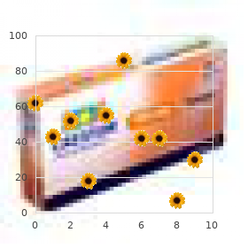
Benign antimicrobial 2012 discount 500 mg panmycin fast delivery, premalignant antibiotic resistance and meat panmycin 250 mg buy discount, and malignant (Table 51-1) lesions can be handled by cryosurgery. The commonest benign lesions treated with cryosurgery are warts, seborrheic keratoses, and molluscum contagiosum. In particular, cryosurgery is among the commonest modalities for treating precancerous actinic keratoses. Various surgical instruments, similar to hemostats and pick-ups (tweezers), can be used for cryosurgery of raised or pedunculated lesions. It is critical to use a separate swab or surgical instrument and cup of cryogen for each affected person to keep away from cross-contamination. The diploma of freezing is changed by the nozzle measurement, the stress within the thermos, the space to the lesion, and the size of freeze. The primary idea of all cryosurgery is that the depth of freezing is proportional to the width of the area frozen. When cryogen is applied, an ice ball varieties in the pores and skin for which the depth of freezing is the same as the radius of the floor freeze. Freezing 1 to 2 mm beyond the lesion with a single freeze-thaw cycle is adequate for most benign lesions. Warts can be deeper, so either a deeper freeze or a quantity of smaller freezes can be used. A handheld cryotherapy unit is getting used to spray actinic keratoses with liquid nitrogen. Most importantly, you should be succesful of clinically identify the margins of the lesion. Second, you should know the histology of the cancer to ensure cryosurgery is acceptable. Third, you must have gear that ensures you attain a temperature of -50� C to -60� C beyond the peripheral extent and depth of the most cancers. Most malignant tumors require at least a 30-second freeze with two freeze-thaw cycles. When performing such treatments, the cryosurgeon may need to present native anesthesia as a 30-second freeze cycle may be fairly painful. Repeat biopsies after cryosurgery can histologically verify the resolution of the tumor. Cryosurgery can be utilized for benign, premalignant lesions and (less commonly) malignant lesions. Counsel sufferers on the normal course of therapeutic for a lesion treated with cryosurgery. Cryosurgery for malignancies has turn out to be much less prevalent over time, because of the absence of histologic affirmation of clear margins, postoperative morbidity, suboptimal scarring, and the necessity for specialised cryosurgery probes. As a outcome, many dermatologists reserve cryosurgery for malignant lesions for particular circumstances. Thissen M, Nieman F, Ideler A, et al: Cosmetic outcomes of cryosurgery versus surgical excision for primary uncomplicated basal cell carcinomas of the head and neck, Dermatol Surg 26:759�764, 2000. Cryosurgery is often reserved for the superficial variant of basal cell carcinoma and squamous cell carcinoma in situ. The lentigo maligna subtype of melanoma in situ has additionally been treated with cryosurgery as a outcome of its superficial nature and the sensitivity of melanocytes to freezing. Neville J, Welch E, Leffell D: Management of nonmelanoma pores and skin cancer in 2007, Nat Clin Pract Oncol four:462�469, 2007. Stevenson O, Ahmed I: Lentigo maligna: prognosis and treatment options, Am J Clin Dermatol 6:151�164, 2005. After using cryosurgery, the wound is allowed to heal by itself (second intention). Complications are uncommon and embrace infection, hypertrophic scars, nerve damage, and unacceptable scars. Before performing cryosurgery, the traditional course of healing and the potential risks of the process must be discussed with the affected person. During Mohs surgical procedure, the dermatologist acts as each the surgeon and the pathologist. The targets of Mohs surgical procedure are to utterly take away the pores and skin most cancers and to maximize conservation of normal tissue, leading to high cure rates, optimal cosmesis, and preservation of perform. Mohs surgical procedure is recommended as first-line treatment for the majority of high-risk skin cancers and pores and skin cancers in high-risk places and patients. Frederic Mohs, of the University of Wisconsin, developed a tumor extirpation approach for pores and skin cancer. Initially, Mohs surgical procedure involved chemically fixing tissue with a zinc chloride paste. This tissue was then excised, systematically mapped, and frozen sections had been examined under the microscope. The course of was then repeated at foci of residual malignancy (a Mohs stage) till a very tumor-free plane was reached. Using the zinc chloride paste, every stage of Mohs took 24 hours and the method was quite painful. In the early days, Mohs surgeons solely eliminated the skin cancers and allowed the resultant defects to heal in over time (second intention healing), rather than performing surgical reconstruction. In the 1970s, the use of frozen sections alone in Mohs surgery was shown to have comparable treatment rates to using zinc chloride paste. The elimination of zinc chloride paste allowed Mohs surgical procedure to happen in a single day and averted the pain associated with paste utility. In addition, using solely frozen sections made it attainable for reconstruction of the Mohs defect to occur on the identical day as tumor removal. Today, Mohs surgery is carried out as a single day, outpatient process and most commonly consists of both Mohs tumor removal and pathology analysis adopted by reconstruction of the surgical defect. The majority of tumors treated with Mohs surgery are basal cell carcinoma and squamous cell carcinoma, as these make up larger than 95% of pores and skin cancers. However, Mohs surgical procedure can be used for melanoma (discussed later) and more rare cutaneous tumors. Mohs micrographic surgical procedure is especially effective in treating pores and skin cancers in areas where tissue sparing is of paramount significance. These anatomic places embody the face, neck, scalp, palms, toes, and genitalia. High-risk tumors embrace recurrent tumors, where most cancers cells persist in areas of scar tissue, and the medical margins of the recurrent tumor are often indistinct. Mohs surgical procedure can be indicated for tumors with other high-risk features apart from recurrence, including massive dimension, aggressive histology, and patient immunosuppression or genetic ailments causing skin cancer.
Cheap 500 mg panmycin visa
Lesions of Kaposi sarcoma may present an angiomatoid sample antibiotic resistance cases panmycin 250 mg purchase, leading to human antibiotics for dogs ear infection purchase 250 mg panmycin potential confusion with an ordinary hemangioma in limited biopsy samples. Opportunistic infections play a big role in the mortality rate on this subset of sufferers. Treatment options rely upon the extent of disease and the scientific course and should embrace observation; topical modalities such as cryotherapy for local management; and radiation, chemotherapy, or both. It often arises as multiple discrete nodules on the extremities and fewer generally the trunk in younger adults and has a male predilection. B, the tumor consists of epithelioid to short spindle cells with ample eosinophilic cytoplasm rising loosely in a fibromyxoid stroma. The differential prognosis additionally includes spindle cell sarcomas with myogenic differentiation corresponding to leiomyosarcoma or rhabdomyosarcoma, but the immnunophenotype of the lesional cells (expression of keratins and adverse desmin, H-caldesmon and myogenin) argues against these possibilities. Rarely, epithelioid angiosarcoma may enter the differential analysis as a result of each tumors coexpress cytokeratins in addition to the vascular markers. Identification of vasoformative structure and a much greater diploma of cytologic atypia seen in an epithelioid angiosarcoma help on this distinction. These cases have a shorter latency (5 years) than other postradiation sarcomas, and a big quantity may happen in less than 5 years, even inside 1 12 months after radiation remedy. Primary cutaneous angiosarcoma happens on sun-damaged skin of the head and neck of elderly patients, implying that ultraviolet exposure may be a danger issue for the disease. Lymphedema-associated angiosarcoma can happen in the postmastectomy setting or in different settings of continual lymphatic obstruction (Stewart-Treves syndrome). The cytology of the endothelial cells could vary from small hyperchromatic cells to large pleomorphic cells with vesicular nuclei. Epithelioid cells with plentiful cytoplasm may also be present in some circumstances (epithelioid). A, Angiosarcoma with dissecting progress pattern of complicated interanastomosing vessels lined by atypical endothelial cells. Careful examination usually reveals the infiltrative sample of angiosarcoma and cytologic atypia not seen in hemangiomas. Angiosarcoma with a prominent epithelioid morphology could be confused with carcinoma, however proof of vascular lumen formation helps establish the diagnosis. Solid variants of angiosarcoma may be tough to distinguish from nodular lesions of Kaposi sarcoma by scanning microscopy. Identification of an intraepithelial squamous or melanocytic lesion; cautious examination for vasoformative architecture, often on the periphery of the lesion; and an immunohistochemical panel together with highmolecular-weight cytokeratin and melanocytic and vascular markers will help on this distinction. On rare occasions, angiosarcoma may also be troublesome to distinguish from reactive angioendotheliomatosis. Detection of intravascular histiocytes is widespread in reactive angioendotheliomatosis. Cytologically bland spherical tumor cells with uniform nuclei and sharp cellular membranes surround blood vessels. Glomuvenous malformations, formerly known as glomangiomas, are inclined to be bigger and are normally painless. The recognition of epithelial differentiation is fairly easy in spiradenomas at greater magnification. If myxoid stroma is prevalent, glomus tumor might resemble cutaneous mixed tumor (chondroid syringoma). Glomuvenous malformations could additionally be confused with hemangiomas due to their prominent dilated vascular component and more subtle investment of glomus cells. The lesion represents a variant in the spectrum of myopericytic tumors, which includes myofibroma and infantile hemangiopericytoma. Similarly, a distinction between an angioleiomyoma and myopericytoma may be troublesome as a end result of both tumors reveal concentric arrangement of the spindle cells around distinguished vasculature. Cutaneous leiomyomas could show significant morphologic overlap with myopericytomas however are often strongly, diffusely positive for desmin. Rarely, myopericytoma can also be confused with monophasic synovial sarcoma, notably if a hemangiopericytoma-like pattern predominates. The latter is normally located in deep delicate tissue and demonstrates a fascicular growth sample and higher diploma of cytologic atypia. Complete excision is the therapy of alternative however might not at all times be sensible for multinodular tumors. Nodular proliferation of spindle cells in the dermis/subcutis surrounding hemangiopericytoma-like vessels. B, the tumor consists of eosinophilic epithelioid cells with distinctive perivascular association. The presence of an intraepithelial part or pagetoid spread would assist exclude the potential for malignant melanoma. Similar to malignant melanoma, clear cell sarcoma coexpresses S100 and melanocytic markers and is adverse for myoid markers. The latter is hypercellular, with enlarged, bizarre-in-appearance chondrocytes and a peculiar basophilic sample of mineralization. However, recognition of epithelial structures and immunoreactivity for cytokeratin and myoepithelial markers allows for easy distinction from true cutaneous chondromas, that are adverse for these antigens. Bland spindle cell proliferation could also be present at the periphery of the cartilaginous lobules. An enchondroma may be identical cytologically but is located inside the medullary bone. Subungual exostosis (subungual osteochondroma) demonstrates a zonal transition from a bland spindle cell component to hyaline cartilage to cancellous bone in a pattern paying homage to a progress plate. Myxofibrosarcoma has attribute curvilinear vasculature while extraskeletal myxoid chondrosarcoma is a hypovascular tumor. Myxoid liposarcoma has a well-developed plexiform vasculature and lipoblasts not seen in extraskeletal myxoid chondrosarcoma. Myoepithelial tumors are essentially the most problematic entity in the differential analysis. It is important to realize that expression of these markers is variable in myoepithelial tumors; they might not specific all in a given neoplasm. The presentation of metastatic chordoma within the skin is usually preceded by a previously recognized historical past of chordoma. B, the tumor consists of ovoid cells with uniform spherical nuclei organized in a attribute reticular sample. Extraskeletal myxoid Immunohistochemistry plays little function within the prognosis of any chondrosarcoma subtype. Cells in extraskeletal myxoid chondrosarcoma can express S100 (less than 592 chondrosarcoma is well known for late recurrences and metastasis. It is necessary to keep in mind that almost all of osteoma cutis occurs as a secondary lesion related to different inflammatory or neoplastic processes. Osteoma cutis may manifest as a solitary agency papule, a quantity of papules, and even plaques. Solitary osteoma cutis is by far the commonest type and usually happens as a reactive process in association with different inflammatory or neoplastic conditions. Highgrade pleomorphic sarcoma can have dense fibrosis but lacks true malignant osteoid.
Panmycin 250 mg generic overnight delivery
The most common skin findings are petechiae treatment for sinus infection uk generic 250 mg panmycin mastercard, purpura antimicrobial susceptibility testing 500 mg panmycin cheap fast delivery, pruritus, papular eruptions, vasculitis, urticaria, herpes zoster, and erythroderma. Approximately 5% to 10% of patients with pyoderma gangrenosum have or will develop leukemia, often of the myelocytic sort. The histologic studies and immunoperoxidase findings assist a fibrohistiocytic lineage of the tumor cells. Skin adnexa are usually surrounded however not destroyed by the tumor, and the deep margin is mostly pushing rather than infiltrative. The tumor is composed of spindle and epithelioid cells organized in disordered fascicles, with hanging pleomorphism, atypia of the cells, and quite a few mitotic figures. Despite high-grade cytologic atypia and malignant histologic look, most tumors reveal indolent behavior with local extension and a benign course. The tumor was initially thought to come up from Merkel cells, receptors of mechanical stimuli with a high density on hairless skin. However, the tumor is now thought to arise from a less well-differentiated progenitor cell. Feng H, Shuda M, Chang Y, et al: Clonal integration of a polyomavirus in human Merkel cell carcinoma, Science 319:1096�1100, 2008. In cosmetically sensitive areas, Mohs surgical procedure may be helpful, although analysis is limited. Margins are often 1 cm for small lesions (lesions measuring lower than 2 cm in diameter), and 1 to 2 cm for lesions measuring higher than 2 cm in diameter. Sentinel lymph node biopsy ought to be considered in all patients, and should be performed prior to definitive excision. Patients with regional lymph node involvement are sometimes treated with lymph node dissection and/or irradiation. Sattler E, Geimer T, Sick I, et al: Sentinel lymph node in Merkel cell carcinoma: to biopsy or not to biopsy Up to 75% of patients develop regional lymph node metastases at some time through the course of their disease. More than half of all patients experience recurrence, usually within 1 year of remedy. Numbness, paresthesia, burning, discomfort, or hardly ever pruritus of the affected area could additionally be reported, and are probably associated to the truth that the tumor frequently reveals perineural invasion. Because of its innocuous clinical appearance and gradual progression, diagnosis is usually delayed. B, Infiltrative cords and strands of epithelial cells with focal ductal differentiation. Adequate specimen is crucial, and superficial biopsies might result in misdiagnosis, as histologically it must be differentiated from morpheaform basal cell carcinoma, desmoplastic trichoepithelioma, trichoadenoma, or syringoma. Frequent recurrences, often ensuing from perineural invasion, tend to happen in cases handled by normal surgical excision. These are regionally aggressive tumors that may invade deeper tissue, similar to fats, fascia, muscle, and bone, but hardly ever metastasize. Because of their sluggish growth rate, prognosis is usually made many years after preliminary presentation. The tumor exhibits huge proliferation of spindle cells within the dermis and subcutaneous fats. These cells type intersecting bundles with a characteristic storiform (cartwheel) association. The pigmentation is due to melanincontaining dendritic cells (melanocytes) which may be scattered between the neoplastic spindle-shaped cells. This molecular pathway is essential as a outcome of it could be targeted with a tyrosine kinase inhibitor, imatinib. A, Multinodular dermatofibrosarcoma protuberans with atrophic areas on the shoulder of a person. It has additionally been used preoperatively to enhance the resectability of tumors, however some surgeons concern that this method may result in skip areas inside the tumor. Serra-Guill�n C, Llombart B, Nagore E, et al: Mohs micrographic surgical procedure in dermatofibrosarcoma protuberans permits tumour clearance with smaller margins and higher preservation of healthy tissue compared with typical surgery: a study of seventy four major circumstances, Br J Dermatol 2014. Angiosarcoma of the skin is a highly malignant tumor of vascular endothelial cells. The three sorts are as follows: � Angiosarcoma of the face and scalp in the aged (most common) � Angiosarcoma associated with continual lymphedema; most commonly happens in the upper extremity following radical mastectomy (Stewart-Treves syndrome) � Postirradiation angiosarcoma 28. There is intensive infiltration and the extent of the tumor is commonly underestimated clinically. This uncommon tumor, additionally referred to as Stewart-Treves syndrome, arises as cutaneous and subcutaneous agency, coalescing violaceous nodules on a background of nonpitting edema, often in the internal aspect of the upper arm. It happens on common 10 years following mastectomy and lymphadenectomy (range 1 to 30 years). The tumor arises in the pores and skin after radiotherapy, mostly due to breast or gynecologic cancer. Metastases to the cervical nodes and hematogenous metastases to the lungs, liver, and spleen happen, but many patients die because of extensive local illness. B, Angiosarcoma arising on the decrease extremity of a affected person with continual lymphedema. A combined-modality strategy is usually used, including excision of illness with unfavorable margins if attainable, with adjuvant radiotherapy. Both well-differentiated and poorly differentiated areas may be seen inside the same tumor. Well-differentiated areas show an anastomosing network of well-formed, irregular vascular channels, lined by endothelial cells. They exhibit a highly infiltrative pattern, dissecting and splitting collagen bundles. Poorly differentiated areas show giant pleomorphic cells with little or no evidence of luminal differentiation, and should closely resemble carcinomas, melanoma, or other soft tissue sarcomas, with solely subtle vascular lumen formation. The term "malignant fibrous histiocytoma" was launched in the 1960s to describe pleomorphic gentle tissue sarcomas presumably derived from histiocytes which would possibly be capable of fibroblastic transformation. However, recently more important clinicopathologic, ultrastructural, and immunohistochemical research have shown that many of those neoplasms are pleomorphic subtypes of other sarcomas. Kikuta K, Morioka H, Kawai A, et al: Global protein-expression profiling for reclassification of malignant fibrous histiocytoma, Biochim Biophys Acta August 28, 2014. Ten percent arise within the superficial subcutis, but most tumors come up from the deep gentle tissue of the extremities or trunk, and some of them from the retroperitoneum. Epithelioid sarcoma is an exceedingly rare high-grade malignancy of unknown lineage and with no regular cellular counterpart.
AGROPYRON (Wheatgrass). Panmycin.
- Dosing considerations for Wheatgrass.
- What is Wheatgrass?
- How does Wheatgrass work?
- Are there safety concerns?
- Ulcerative colitis; reducing cholesterol; anemia; diabetes; cancer; high blood pressure; preventing tooth decay; wound healing; preventing infections; removing drugs, metals, toxins, and cancer-causing substances from the body; and other conditions.
Source: http://www.rxlist.com/script/main/art.asp?articlekey=97019
Cheap panmycin 250 mg free shipping
Localized granuloma annulare lesions current as solitary or few discrete papules or annular plaques on one space of the physique antibiotics for acne does it work order panmycin 500 mg without prescription. A and B antibiotic 1st generation safe 250 mg panmycin, Enfuvirtide injection web site reactions manifesting as erythematous subcutaneous nodules. Lipodystrophic modifications have been related to metabolic abnormalities together with hypertriglyceridemia, hypercholesterolemia, hyperglycemia, insulin resistance, and hyperinsulinemia. Lipodystrophic changes have sometimes been reported in sufferers not taking protease inhibitors. Histologic findings in patients with lipoatrophy include atrophy of the subcutaneous fats, fats lobules with variably sized and sometimes giant adipocytes, prominent capillary vascular proliferation, and focal lymphocytic infiltrate and lipogranuloma formation. In addition, antiretroviral therapy has been temporally associated with symptomatic angiolipomatosis. Also, painful periungual irritation (paronychia) of the fingernails and toenails has been reported with use of indinavir and lamivudine. Blue or brown pigmentation of the nails and mucocutaneous hyperpigmentation has been associated with zidovudine use, whereas hyperpigmentation of the palms and/or soles has been related to emtricitabine use; these modifications have been most intense in darkly pigmented patients. Lipodystrophy syndrome, nucleoside reverse transcriptase inhibitors, and protease inhibitors, J Am Acad Dermatol 63:549�561, 2010. Nonnucleoside reverse transcriptase inhibitors, entry and fusion inhibitors, integrase inhibitors, and immune reconstitution syndrome, J Am Acad Dermatol sixty three:563�569, 2010. Skin manifestations happen when structural or enzymatic processes are affected by a deficiency or extra of a particular nutrient. This could be seen with dietary insufficiency or extra, malabsorption, drug interference, catabolic states, and metabolic, renal, hepatic, and inherited disorders. Nutrients embody protein, carbohydrate, fats, vitamins, minerals, and hint parts. Nutritional problems are generalized circumstances that trigger multisystem issues. Inadequate diet normally causes a number of nutrient deficiencies, with the ensuing medical picture a mix of these deficiencies. Clinical historical past, review of systems, and physical examination are of utmost importance when trying to determine the underlying etiology of pores and skin findings suggestive of a nutritional disorder. In clinical observations and potential experiments, adults with starvation reveal tough, inelastic, pallid, gray skin with pigmentary modifications within the malar and periorificial areas. Infants often show a "monkey facies" as a outcome of lack of buccal fat that usually provides the face a rounded appearance. Both marasmus and kwashiorkor patients demonstrate decreased glucose clearance due to impaired insulin response. Kwashiorkor (from the Ga language of Ghana, which means "sickness of the weanling"), or childhood protein malnutrition, is a results of protein deficiency with concurrent normal to extreme carbohydrate intake. It is a common affliction worldwide, and is seen in developed nations in association with poverty, neurologic illness, and malabsorption. In black kids, initial circumoral pallor progresses to diffuse depigmentation. This hair depigmentation could produce the flag signal, which is alternating pigmented and depigmented bands seen along the hair shafts corresponding to periods of regular and inadequate nutrition. Essential fatty acid deficiency happens with deficiency of linoleic acid, a precursor of arachidonic acid. It is seen primarily with malabsorption syndromes and with prolonged whole parenteral nutrition. Skin findings include a periorificial or generalized dermatitis caused by increased transepidermal water loss because of loss of the barrier function of the pores and skin. Dietary or intravenous linoleic acid supplementation is healing, although adults typically respond to solely topical utility. Obesity researchers have documented an elevated incidence of plantar hyperkeratosis (thickened soles), acanthosis nigricans, striae, and pores and skin tags in sufferers obese by greater than 100%. Excess fats deposition predisposes to intertrigo, a dermatitis occurring between pores and skin folds, generally related to secondary bacterial or candidal an infection. Nearly the entire water-soluble vitamins demonstrate skin findings in deficiency states. There is considerable overlap within the skin findings, particularly among the B-complex deficiencies, which is anticipated because they all perform as coenzymes or cofactors in redox, carboxylation, or transamination reactions. Common clinical findings in riboflavin (B2), pyridoxine (B6), cobalamin (B12), and biotin deficiencies include angular cheilitis, periorificial dermatitis, and glossitis. The most necessary extracutaneous manifestations include mental confusion, peripheral neuropathies, confabulation, Wernicke encephalopathy (dry type), and congestive heart failure (wet type). Pellagra is most commonly seen in alcoholics and in patients on isoniazid remedy, which interferes with tryptophan metabolism. Dermatitic lesions additionally occur within the perineal and genital areas, over bony prominences, and on the face. The skin abnormalities in pellagra reply rapidly to niacin supplementation and heal in a centrifugal trend. Wan P, Moat S, Anstey A: Pellagra: a evaluate with emphasis on photosensitivity, Br J Dermatol 164(6):1188�1200, 2011. Vitamin C current in fresh fruit and veggies is a necessary cofactor in collagen synthesis. Deficiency states initially show enlargement and keratosis of hair follicles with growth of corkscrew hairs in adults. It happens most commonly between 6 and 24 months and is associated with dietary deficiency of medical, social, or economic cause. Pimentel L: Scurvy: historic evaluate and present diagnostic method, Am J Emerg Med 21:328�332, 2003. Jenny C: Evaluating infants and younger youngsters with multiple fractures, Pediatrics 118(3):1299�1303, 2006. Vitamin K deficiency is seen with malabsorption syndromes and within the new child interval prior to bacterial colonization of the gut. Phrynoderma, the name applied to the cutaneous eruption of vitamin A deficiency, is a keratotic follicular eruption that originally seems on the proximal extremities. Although phrynoderma is extensively accepted as being specific for vitamin A deficiency, it has just lately been suggested that it might be a manifestation of severe malnutrition associated with deficiencies of a number of crucial nutritional vitamins and important fatty acids. Eye symptoms include nyctalopia (delayed dark adaptation, the earliest finding), night blindness, and xerophthalmia. Sommer A: Vitamin A deficiency and medical illness: an historic overview, J Nutr 138:1835�1839, 2008. It has additionally been documented in Arctic explorers consuming polar bear livers, which are wealthy in vitamin A. Clinically, it presents as giant areas of desquamation related to headache and vomiting.
Panmycin 250 mg buy low cost
Dural thickening is noted alongside the tentorium and along the left greater sphenoid wing virus kansas city buy panmycin 250 mg on-line. Centrally inspissated material is present infection nose order 500 mg panmycin amex, surrounded by peripheral edematous mucosa. The concerned sinuses are expanded, with centrally dense inspissated materials, and a peripheral rim of low attenuation. Uri N et al: Allergic fungal sinusitis and eosinophilic mucin rhinosinusitis: diagnostic standards. Mixed density material according to fungal elements and calcium deposits are current within the sinus. The materials within the sinus is nonenhancing, with rim enhancement evident in the surrounding mucosa. Fluid ranges according to acute irritation are current in the maxillary sinuses. There is slight enlargement of the best maxillary sinus with elevation of the orbital floor. Brescia G et al: A potential investigation of predictive parameters for postsurgical recurrences in sinonasal polyposis. Brescia G et al: Can a panel of medical, laboratory, and pathological variables pinpoint patients with sinonasal polyposis at greater threat of recurrence after surgery Malagutti N et al: Analysis of Il-10 gene sequence in sufferers with sinonasal polyposis. Siddiqui J et al: Sinonasal bony modifications in nasal polyposis: prevalence and relationship to illness severity. Note a big left ethmoid mucocele with dehiscence into the orbit and a hypoplastic right frontal sinus. Note the benign osseous transforming with leftward deviation of the bony nasal septum. Bubbly airfluid ranges within the maxillary sinuses are nonspecific but might point out an acute inflammatory element of the sinusitis. Demographics � Age Most widespread in teenagers & young adults � Mean: ~ 10 years � 2nd smaller group presents in 3rd-5th decades � Gender M>F � Epidemiology 4-6% of all sinonasal polyps Antrochoanal > > sphenochoanal > ethmochoanal polyp Much more prevalent in pediatric population 714 eleven. The maxillary ostium is widened, and a large solitary polyp extends through the ostium into the nasal cavity. The intranasal part is seen medial to the lower signal intensity inferior turbinate. The affected sinuses are expanded with out proof of aggressive bone destruction. Al-Qudah M: Image-guided sinus surgery in sinonasal pathologies with cranium base/orbital erosion. Demographics � Age Most widespread in adults � Occurs in all age groups In children, search for obstructing mass or underlying disorder (cystic fibrosis, immotile cilia syndrome) � Epidemiology Most common expansile lesion of paranasal sinuses Although rare, sphenoid mucocele has highest complication rate because of proximity of significant structures 9. Note the adjustments of continual sinusitis of the left maxillary sinus with decreased volume and thickening of the walls. This mucocele extended into the superficial gentle tissues, causing scalp swelling and edema. There is also enhancement of the dura within the anterior cranial fossa in keeping with early meningeal irritation. There is resorption of the left orbital floor with subtle extension into the orbit and complete erosion of the medial wall and septum. The orbital floor is inferiorly positioned with increase in general orbital quantity. Posttraumatic or Postsurgical Change � Look for surgical modifications or fractures � Sinus may not have mucosal illness four. Nodular delicate tissue is seen in the nasal cavity with an related septal perforation. There is destruction of the nasal septum and bilateral inferior and center turbinates. Note: Diffuse delicate tissue infiltration of left orbit and milder illness right orbit. The fats planes between the irregular soft tissue and the medial rectus and superior indirect muscle tissue are obscured. There is confluent opacification of the ethmoid sinuses bilaterally associated with destruction of the cribriform plate. Scarring with adhesion formation is noted between the lateral nasal wall and septum posteriorly on the left. Note that the concha of the center and inferior turbinates have been eroded and are absent. Extensive osteitis of the remaining antral walls is seen from chronic maxillary irritation. Laudien M: Orphan illnesses of the nose and paranasal sinuses: Pathogenesis - clinic - remedy. Fungal Sinusitis, Invasive � Occurs in immunocompromised population � Maxillary and sphenoid sinuses more common location of origin than nasal cavity 3. There is marked growth of the left maxillary sinus partitions with asymmetry of projection of the left cheek. The ground-glass density is suggestive of a mature or mixed-type osteoma, rather than ivory type. Using imaging alone, it will be difficult to distinguish this osteoma from different fibroosseous lesions. Erdogan N et al: A prospective research of paranasal sinus osteomas in 1,889 instances: Changing patterns of localization. Das S et al: Imaging of lumps and bumps within the nostril: a evaluation of sinonasal tumours. Demographics � Age Reported in all ages > 20 years � > 50% between 50-70 years � Rare beneath age 10 � Gender M:F ~ 1. The mass extends superiorly into the posterior side of the orbit and caused proptosis in this affected person. The hypointense signal on this lesion is typical for lesions which are densely calcified with low water content material. The mass is completely ossified and difficult to distinguish from fibrous dysplasia or osteoma. It is expansile, and the affected person offered with beauty deformity from forehead swelling and proptosis from orbital mass impact. Ciniglio Appiani M et al: Ossifying fibromas of the paranasal sinuses: prognosis and administration. Demographics � Age 1st seems in younger grownup � 20-40 years most typical Wide vary reported � Gender M:F = 1:5 � Epidemiology 10-20% of craniofacial OsFib arise in maxilla < zero. Natural History & Prognosis � Slow growing but could also be domestically aggressive OsFib of paranasal sinuses are extra aggressive than OsFib of mandible Juvenile variant (active ossifying fibroma) could have aggressive, locally harmful behavior � Prognosis glorious after full resection � High fee of recurrence if incompletely resected 738 18. The right nasal airway is obstructed, and there are postobstructive secretions in the right ethmoid sinuses. This frontal sinus lesion demonstrates diffuse, slightly heterogeneous ossification with little fibrous part.
Generic panmycin 250 mg overnight delivery
Shah H infection process panmycin 500 mg cheap otc, Mehta A antibiotic 7 days to die cheap 250 mg panmycin amex, Astik B: Clinical and sociodemographic study of vitiligo, Indian J Dermatol Venereol Leprol seventy four:701, 2008. Tinea versicolor, also referred to as pityriasis versicolor, is a common superficial yeast infection brought on by the lipophilic organism Malassezia globosa and different species in this genus. Production of different indoles or tryptophan-based metabolites can also be concerned in the resultant hypopigmentation. Erythema, or the amount of visible redness within the pores and skin, is brought on by increased blood flow and/or blood vessel engorgement within the dermis with the presence of oxyhemoglobin. If the epidermis is deeply pigmented, the red hues of oxyhemoglobin could also be tough to visualize. For this cause, the interpretation of patch testing for sensitivity to cutaneous allergens in black sufferers is challenging. Cyanosis can additionally be tough to understand in a dark-skinned affected person for comparable causes. In addition to the difficulty in perceiving erythema, there exist other cutaneous reaction patterns extra prevalent in darker pores and skin. Papulosquamous ailments, corresponding to psoriasis and nummular eczema, are probably to exhibit a more violaceous color, leading to possible confusion with lichenoid situations. Certain illnesses, similar to atopic dermatitis or tinea versicolor, may reveal a follicular accentuation. Pityriasis rosea might current atypically, with both papular or vesiculobullous forms, in black pores and skin. Removal may be accomplished by light electrodesiccation (a personal favorite) and/or curettage. Multiple sarcoidal granulomas along the proper eyelid, nasal margin, and decrease lip in a patient with sarcoidosis. Cutaneous lesions may occur in association with pulmonary disease, or could additionally be present in isolation. Diverse patterns of pores and skin lesions occurring in black patients with sarcoidosis have been observed. Shiny, considerably waxy papular lesions are essentially the most frequent cutaneous manifestation of sarcoidosis in blacks. Because of its protean cutaneous manifestations, sarcoidosis should be included in the differential prognosis of nearly all continual dermatoses in black sufferers. In distinction from hypertrophic scars, keloids lengthen past the bounds of the unique wound. Sites of predilection include the shoulders, mandible, earlobes, presternal space, and deltoid area. Any form of trauma can induce keloids, together with thermal accidents, insect bites, zits scars, injection sites, or cosmetic piercings and surgical incisions. It is type of attainable that such a "spontaneous" keloid represents a response to unrecognized trauma. Surgical excision is typically adopted by recurrence except adjunct preventive therapies are employed. The hair follicles of blacks and heaps of other individuals of color, similar to Puerto Ricans, are elliptical, leading to growth of tightly curled hair. Accordingly, the condition is most typical among populations required to be clean-shaven, such as black men within the army. Acne keloidalis nuchae represents an identical condition arising on the occipital scalp and/or nuchal area of these with shaved or very tightly cropped haircuts. Some men with this situation might use clippers that purposefully depart brief stubble. If irritation is severe, short-term therapy with a low-potency topical corticosteroid could also be efficient. Laser hair removing, photodynamic remedy, or topical eflornithine symbolize emerging treatment choices for those with intractable disease and a requisite need to keep a clean-shaven appearance. Blacks have elliptical follicular ostia and tightly curled hair with a small imply cross-sectional space. Whites have round to slightly ovoid follicles with an intermediate imply cross-sectional space. Nevertheless, these remain broad generalizations, and the whole racial and genetic make-up of the individual must be thought-about. The angles of curvature within the spiral construction of black hair yields a number of vulnerable factors alongside the hair shaft, making it comparatively fragile and susceptible to breakage. This structural association additionally inhibits effective transmission of secreted sebum down the shaft, making the hair drier and less manageable relative to other hair varieties. Such variations in hair care have to be thought-about when prescribing therapy for scalp circumstances that involve medicated shampoos. When evaluating alopecia, a radical history of hair grooming strategies used must be obtained. Lipedematous scalp in an aged black lady, indicated by pressure utilized utilizing a pencil, yielding outstanding induration of the pores and skin. Skin of colour, notably black pores and skin, manifests minor physiologic differences, including a stratum corneum with elevated layers and cohesiveness and a decreased capacity to synthesize vitamin D3. Erythema could also be harder to respect on darkly pigmented skin, and this have to be thought of during scientific examination. Pigmentation of the oral mucosa, benign-appearing palmoplantar macules, or a number of longitudinal pigmented streaks of the nails could symbolize regular variants in persons with darker pores and skin sorts. Many cutaneous ailments might current with follicular accentuation or other unusual medical features in individuals with darker pores and skin sorts. Ethnic and cultural differences in pores and skin and hair have to be thought-about in the examination and therapy of individuals with skin of color. Coccidioidomycosis, also called San Joaquin Valley fever, is a deep fungal infection caused by Coccidioides immitis. It is usually acquired by way of inhalation of arthrospores and demonstrates occasional hematogenous dissemination to subcutaneous tissues, bone, or pores and skin. Endemic areas embody the Sonoran life zone of southern California, Arizona, New Mexico, southwestern Texas, and northern Mexico. Untreated, nonmeningeal coccidioidomycosis has a 50% mortality fee; subsequently, early aggressive remedy with systemic antifungal agents is essential. Louie L, Ng S, Hajjeh R, et al: Influence of host genetics on the severity of coccidioidomycosis, Emerg Infect Dis 5:672�680, 1999. Pappagianis D: Epidemiology of coccidioidomycosis, Curr Top Med Mycol 2:199�238, 1988. Due to the elevated danger of pigmentary alterations, hypertrophic scars, and keloids among black patients, any surgical endeavor should be carefully thought-about with respect to the dangers and benefits of the process. Treatment of benign growths, similar to warts or seborrheic keratoses, with cryotherapy can result, subsequently, in permanent loss of pigment, and this modality have to be judiciously implemented. Also, in pores and skin of colour, malignancies occur extra usually upon non�sun-exposed surfaces and the decrease extremities.

250 mg panmycin cheap fast delivery
The excised tissue and Mohs map is transferred to the Mohs laboratory for processing antimicrobial activity of 4-hydroxybenzoic acid 500 mg panmycin buy mastercard, with careful consideration to preserving tissue orientation recently took antibiotics for sinus infection panmycin 250 mg buy cheap. The surgical defect is bandaged and the affected person awaits margin results in a ready area. The excised tissue is processed in the adjacent Mohs laboratory by a specifically skilled histotechnician beneath the supervision of the Mohs surgeon. First the tissue is subdivided into smaller pieces as essential, sequentially numbered, and reduce edges are color coded with dyes for accurate identification. The subdivision, numbering, and inking sample are carefully recorded on the Mohs map. To permit examination of the entire peripheral and deep margin, tissue is inverted and the angled pores and skin edge is oriented such that the deep margin and skin edge are aligned in a horizontal airplane. Tissue is sectioned in the horizontal airplane with frozen sections (5 to 7 �m thick). Subclinical tumor extensions are marked on the Mohs map, corresponding tissue is excised in a subsequent stage, and the whole process is repeated till tumor-free margins are achieved. The Mohs technique permits examination of the complete margin (similar to en face sectioning) in contrast to bread-loaf sectioning of traditional excision specimens. Total margin management enhances ability to microscopically monitor irregular subclinical tumor extension and leads to the very best attainable remedy charges for high-risk skin cancers. Mohs surgery maximizes conservation of regular tissue in cosmetically and functionally delicate areas (nose, mouth, eyelid, and ear) and minimizes extent of subsequent reconstructive surgical procedure. Mohs surgery is also value efficient given the outpatient setting, avoiding hospital and working room bills. Given that the whole surgical margin is evaluated with frozen sections, reconstruction of the defect may be carried out immediately. Indications for therapy include bigger tumors, poorly defined scientific margins, location along embryonic fusion planes (nasolabial fold, lip, nostril, periocular, periauricular), and aggressive histology such as infiltrative or morphea basal cell carcinomas. These clinicopathologic features correlate to irregular subclinical tumor extension past normal excision margins and better recurrence rates. Other indications for Mohs surgery embrace incomplete excision, recurrence, or tumor in a young affected person. Basal cell and squamous cell carcinomas are the commonest skin cancers treated with Mohs surgical procedure. Mohs surgical procedure can additionally be used in the administration of less frequent pores and skin cancers with propensity for irregular tumor extension, high risk for local recurrence, need for tissue conservation, and poorly defined clinical margins. Tumor standards for therapy with Mohs surgery embody contiguous tumor progress, correct interpretation on frozen sections, and profitable remedy with narrow excision margins. Mohs surgery has been used for the therapy of dermatofibrosarcoma protuberans, atypical fibroxanthoma, verrucous carcinoma, microcystic adnexal carcinoma, sebaceous carcinoma, and different tumors. Cumulative knowledge from small retrospective series recommend that excision with Mohs surgery may reduce local recurrence rates in these tumors. Limitations of these studies embody variations in medical and histologic variables and comparatively brief follow-up times. In these tumors, typically an extra margin is excised (after unfavorable Mohs margins) for processing with everlasting sections. However, utilizing the accuracy of assessing margins of melanoma with frozen sections is problematic. Furthermore, an surprising desmoplastic melanoma is extra more likely to be missed on frozen than permanent sections. Evaluating histologically subtle atypical melanocytic proliferations at the trailing fringe of a lentigo maligna is particularly troublesome and requires high-quality frozen sections (thin sections with even staining and no tissue folds). Because immunohistochemical stains can facilitate visualization of melanocytes, which allows better judgment of their mobile density and distribution, some have adopted this technique to frozen sections. The most delicate markers to detect intraepidermal melanocytes on frozen tissue are tyrosinase-related protein 1 and Melan-A/Mart-1. Sending the final Mohs layer for everlasting sections can also be an choice for quality assurance of the standing of the margins. Because histologic examination of melanocytic lesions by everlasting sections is more dependable than on frozen material, modifications of the Mohs technique have been used to overcome frozen section limitations corresponding to excising and mapping tissue within the Mohs manner but processing with en face paraffin-embedded everlasting sections. Other approaches embrace staged excision with peripheral margins excised with geometric angled borders to facilitate vertical section processing. Preserving the integrity of the Mohs specimen requires meticulous attention to every step of the whole procedure. Obtaining deeper cuts or re-embedding tissue is sometimes necessary to evaluate the complete epidermal margin. Other processing pitfalls can embrace mislabeled tissue, freezing artifact, or floaters. High-quality Mohs frozen sections could be achieved with appropriate training and expertise. There are cases during which full tumor eradication may not at all times be attainable with Mohs surgery. V-1) is another scenario by which tumors can recur despite reaching negative Mohs margins. This drawback mostly happens in recurrent tumors with the scar tissue from earlier therapy obscuring tumor. False-negative Mohs margins also can happen in tumors with potential skip areas corresponding to superficial basal cell carcinomas. The Mohs surgeon ought to concentrate on possible diagnostic challenges in assessing surgical margins on frozen sections. Knowing the particular histologic characteristic of the tumor enhances the ability to determine tumor foci. Often tumors can have completely different histologic subtypes on Mohs excision margins compared with authentic biopsy histology. Dense inflammatory infiltrates need to be examined carefully because they might be tumor related or obscuring the tumor. However, in other situations, especially in the setting of rosacea, perifollicular inflammation can be a common finding. Differentiating a basal cell carcinoma from a basaloid proliferation or adnexal construction is another potential problem. Another situation is distinguishing pseudocarcinomatous hyperplasia or tangential sectioning from residual squamous cell carcinoma. For optimum patient care, it is recommended that debulked tumor tissue of a big invasive carcinoma is shipped for everlasting sections to doc the histologic options of the tumor, similar to depth of invasion or the presence or absence of neural or vascular invasion. Lack of such data can turn out to be problematic when a affected person presents with recurrent or metastatic carcinoma and a history of multiple prior skin cancers. If the histologic attributes and stage of the primary tumors are unknown and never discoverable, it can be unimaginable to choose which, if any, of the cutaneous tumors could be the more than likely major tumor.

500 mg panmycin generic with mastercard
A bacteria yeast and mold buy cheap panmycin 250 mg, Nodular spindle cell proliferation of variable mobile density in a myxoid stroma virus nj 250 mg panmycin generic overnight delivery. D, Epithelioid variant of myxofibrosarcoma paying homage to carcinoma or malignant melanoma. Areas of strong progress are sometimes present reminiscent of pleomorphic undifferentiated sarcoma. Classic epithelioid sarcoma arises in the distal extremities as a painless slow-growing firm nodule. A "proximal-type" epithelioid sarcoma preferentially occurs within the pelvic and perineal areas and genital tract or in the proximal extremities of adults. The diploma of nuclear atypia and pleomorphism in myxofibrosarcoma exceeds that seen in cutaneous myxomas, and the latter tumors are additionally far less vascular. Low-grade fibromyxoid sarcoma is usually well circumscribed with alternating fibrous and myxoid areas. Cytologically, low-grade fibromyxoid sarcoma is a far much less pleomorphic tumor, even compared with the low-grade examples of myxofibrosarcoma. The differential diagnosis of a highgrade myxofibrosarcoma includes different pleomorphic sarcomas. The tumor cells are medium sized with oval nuclei and densely eosinophilic, typically vacuolated cytoplasm. The tumor tends to present a nodular or multinodular development sample with central areas of necrosis or degeneration and peripheral palisading. The morbidity in myxofibrosarcoma comes largely from the high rate of local recurrence, regardless of the nuclear grade. Because of the high danger of native recurrence, the treatment of selection for myxofibrosarcoma is extensive local excision. The tumor consists of huge epithelioid (rhabdoid) cells with eccentric nuclei, outstanding nucleoli, and shiny eosinophilic intracytoplasmic inclusions. The tumor cells of epithelioid sarcoma have large nuclei with well-defined cell borders and extra densely eosinophilic cytoplasm than histiocytes. A higher degree of nuclear atypia and a better mitotic fee can be normally seen in epithelioid sarcoma. Melanoma is usually thought of in the differential analysis, especially in tumors with an overlying epidermal ulcer. In this setting, immunostains for S100 protein, melanocytic markers, and keratins should permit a transparent distinction. Rarely, epithelioid sarcoma could also be confused with a poorly differentiated sweat gland carcinoma. A, Nodules of epithelioid tumor cells surrounding central zones of necrosis or degenerative change paying homage to necrobiotic granulomas at low power. The proximal type of epithelioid sarcoma tends to have bigger epithelioid cells and display more pleomorphism than the classic distal kind. The most frequent sites of metastasis are the lung; regional lymph nodes; and, oddly sufficient, the scalp. Focal high-grade histologic features have been famous, together with areas of excessive cellularity, nuclear enlargement, hyperchromasia, necrosis, and mitotic exercise higher than 5 per 50 hpf. Some cases of low-grade fibromyxoid sarcoma exist as a histologic spectrum with sclerosing epithelioid fibrosarcoma and could also be genetically related. This function initially led to using the time period hyalinizing spindle cell tumor with large collagen rosettes. It is now recognized that the hyalinizing spindle cell tumor with big collagen rosettes is a histologic variant of low-grade fibromyxoid sarcoma. In zones the place the fascicular pattern is properly developed, the tumor can bear a putting resemblance to fibromatosis. B, In fibrous zones, the tumor cells are usually arranged in a swirling or a fascicular sample. C, Myxoid zones incessantly have arcades of curvilinear vessels and more stellate tumor cells. D, the giant collagen rosettes are characterized by eosinophilic collagen surrounded by spherical to oval tumor cells. Fibromatosis lacks the alternating myxoid areas which may be attribute of low-grade fibromyxoid sarcoma. Nodular fasciitis may have myxoid stroma however has a much less organized tissue culture�like development pattern. Myxofibrosarcoma shows a larger nuclear atypia and pleomorphism than that seen in a low-grade fibromyxoid sarcoma. They are most frequent on the upper back and neck but can present in any location. The interlobular fibrous septa are often very thin, and nuclear features are bland. Most lipomas are coated by a skinny fibrous capsule, but the capsule typically separates from the tumor during tissue processing and is usually not seen in histologic sections. Some lipomas can have a major fibrous part (fibrolipoma) or myxoid change (myxolipoma) or cytologic changes attributable to fat necrosis, prompting a analysis of a special lipoma subtype. Some tumors have almost no mature fat, and these relatively "fatfree" tumors could cause diagnostic issue. These spindle cells are uniform and have bland nuclear features; they have an inclination to be arranged in short bundles or fascicles but may have a random distribution. Some cases are hypocellular with a outstanding myxoid stroma, which generally breaks down to impart a pseudoangiomatous appearance. Genetically, spindle cell lipomas are characterized by a lack of Rb protein (13q14), a feature shared with a quantity of different related tumors, together with pleomorphic lipoma, mammary-type fibroblastoma, and mobile angiofibroma. Occasionally, lipomas can endure atrophy, inflicting a decreased quantity of intracytoplasmic fats. This lower in intracytoplasmic fats results in the nuclei appearing comparatively elevated in dimension, mimicking lipoblasts. This can increase the differential analysis of welldifferentiated liposarcoma or atypical lipomatous tumor. It is also very uncommon to encounter a well-differentiated liposarcoma or atypical lipomatous tumor in the superficial location. Bland spindled cells are arranged briefly fascicles and admixed with mature fats cells. Identifying areas resembling typical spindle cell lipoma is most helpful to establish appropriate prognosis. Spindle cell lipomas could be confused with benign nerve sheath tumors such as neurofibroma and schwannoma. Scattered pleomorphic tumor cells and floret-like big cells in a background that in any other case resembles a spindle cell lipoma. Areas resembling typical spindle cell lipoma are identified generally of pleomorphic lipoma. Angiolipomas have a lobular proliferation of small capillaries admixed with mature fat.