
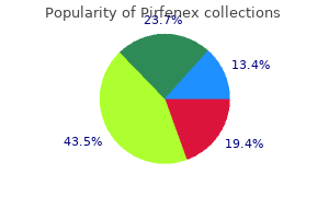
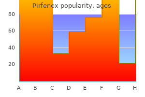
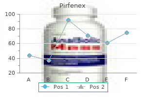
Pirfenex
Pirfenex dosages: 200 mg
Pirfenex packs: 90 pills, 180 pills, 270 pills, 360 pills

Pirfenex 200 mg cheap visa
In the past treatment yeast infection nipples breastfeeding pirfenex 200mg cheap without a prescription, polymyxins have been used systemically 911 treatment for hair pirfenex 200 mg buy visa, however due to poor distribution in tissues, neurotoxicity and nephrotoxicity, their common use has been outmoded by much less poisonous brokers. Long-term remedy is due to this fact a challenge for drug supply, and orally administrable medicine are consequently highly fascinating. There is renewed curiosity in polymyxins as a last-effort choice for treating multiresistant Gram-negative infections Polymyxins are lively against most Gram-negative organisms except Proteus spp. Concerns regarding the shortage of effective antibiotics for treating multidrug resistant Gram-negative micro organism (especially Pseudomonas and Acinetobacter spp. Resistance is as a end result of of chromosomally mediated alterations in membrane construction or antibiotic uptake. If the strain is susceptible to each isoniazid and rifampicin, ethambutol is then discontinued. The structure and mechanism of action of rifampicin have been described earlier this chapter. Treatment of leprosy the event of resistance during dapsone monotherapy for leprosy has led to its use in combination with rifampicin Infection caused by M. However, monotherapy has resulted in the emergence of resistance, and a mixture of dapsone, rifampicin and clofazimine, a phenazine compound (mechanism of action not properly understood), is now commonly used as multidrug therapy. Its bactericidal activity outcomes from inhibition of mycolic acid synthesis, which additionally accounts for its specificity. It is well absorbed after oral administration, and a single every day dose is normally prescribed besides in harder instances corresponding to meningitis or miliary tuberculosis. The main toxic effects in humans are neurological issues (which may be prevented by the concurrent administration of pyridoxine) and hepatitis. Susceptibility tests performed in the laboratory look at the interaction between antibiotics and bacteria in an isolated and rather artificial trend. Patient components such as age, underlying disease, website and type of infection, renal and liver impairment, and drug pharmacodynamics must be taken into account in the antibiotic management of an an infection. It acts by inhibiting the polymerization of arabinoglycan, a important constituent of the mycobacteria cell wall. An necessary toxic aspect effect is optic neuritis, and visible acuity ought to be monitored during therapy. Susceptibility tests Laboratory tests for antibiotic susceptibility fall into two main categories: � diskdiffusiontests � dilutiontests. Diffusion tests involve seeding the organism on an agar plate and making use of filter paper disks containing antibiotics the isolate to be tested is seeded over the complete surface of an agar plate, and filter paper disks containing the antibiotics are utilized. The quantity of antibiotic in the disk is related to , among other issues, the achievable serum concentration and subsequently differs for various antibiotics. In addition, antibiotics differ of their capacity to diffuse in agar, so the size of the inhibition zone (and not merely its presence) is an indicator of susceptibility of the isolate. Pyrazinamide Pyrazinamide is an artificial analogue of nicotinamide which seems to target mycolic acid synthesis. After oral administration, the drug is quickly absorbed from the gastrointestinal tract and properly distributed in body tissues and fluids. As with ethambutol, resistance throughout monotherapy requires that the drug be used in mixture with different first-line agents. Mycobacterial resistance Drug resistance and immunocompromised sufferers complicate tuberculosis therapy Despite the use of antibiotics together, the incidence of resistance among mycobacteria is a persistent and growing downside. Serial dilutions of the check antibiotic are ready in broth or agar medium and inoculated with a suspension of the test organism. These tests can be performed in a microtitre plate format and kind the basis of some automated susceptibility take a look at systems. An various method is the E-test, in which a filter paper strip impregnated with a gradient of antibiotic is laid on an agar plate seeded with the take a look at isolate. A number of the automated susceptibility check systems use a measure of bacterial viability. After in a single day incubation the organism grows and the antibiotics diffuse to produce a zone of inhibition that signifies the degree of susceptibility: disk susceptibility check indicating sulphonamide resistance. Disks containing sulphonamide and trimethoprim have been placed to demonstrate the synergistic exercise of those two agents against E. Synergy can be recognized by the truth that the zones of inhibition turn out to be steady between the 2 disks. When the disks are positioned far aside, nalidixic acid inhibits the check organism, but when placed close collectively this inhibition is antagonized by the presence of nitrofurantoin, as demonstrated by the foreshortening of the zone of inhibition. In these experiments a tradition of 2 � 106 colony forming U / mL was handled with antibiotics A and B alone and together. Compared with the untreated management, both A and B inhibit the growth of the bacterial tradition, however B is more active than A. The mixture also prevents the re-growth seen after 6�24 h when the antibiotics are used singly. Combining antibacterial brokers can lead to synergism or antagonism Hospital sufferers incessantly receive a couple of antibacterial agent, and these brokers could work together with one another (and also with other drugs such as diuretics). Both disk diffusion and dilution exams allow the action of combinations of antibiotics to be studied. Another example is the mix of penicillin (or ceftriaxone) with gentamicin in the remedy of endocarditis brought on by a penicillin-susceptible pressure, as this mix has been shown to be clearly superior to the effect of the beta-lactam alone (Box 34. The concentrations of such antibiotics should be monitored each to forestall toxicity and to ensure that therapeutic concentrations are achieved. Other much less poisonous agents must be monitored in some circumstances in some patients (Box 34. Antibiotic assays could also be performed by a variety of strategies similar to high-performance liquid chromatography and direct assays for organic exercise (bioassay). However, probably the most 472 Section Five � Diagnosis and management ease of taking these therapies has improved, by lowering pill burden by mixture therapy and by being in a position to provide single-tablet oral therapy. It can also be being investigated as a way of decreasing transmission when given as prophylaxis. As of 2017, there were 14 single-tablet two�four drug combined pills and 25 antiretroviral agents constituting six completely different lessons of drug (summarized in Table 34. The downside in growing new antivirals has been mostly as a result of the issue of interfering with viral activity in the cell with out adversely affecting the host. Reports have highlighted the importance of creating an early diagnosis in short-incubation-period viral infections, corresponding to influenza, in order for antiviral treatment to achieve success. Bearing in thoughts that antivirals can be used to deal with acute and chronic viral infections, and in the latter case could also be given for a number of years or for life, concerns embody the size of the remedy course, single versus combination remedy, drug pharmacokinetics and interactions, antagonistic results and antiviral resistance. Antiretroviral resistance is seen across all the main lessons of brokers � nucleoside reverse transcriptase inhibitors, non-nucleoside reverse transcriptase inhibitors, and protease inhibitors � with increasing frequency in resource-rich countries. One problem with antiviral resistance is that the replication health of the drug-resistant variants is commonly less than the wild-type pressure.
Pirfenex 200 mg cheap on line
Apparently healthy kids could continue to present depressed pulmonary perform or wheeze even 1�2 years after apparent recovery medications ordered po are discount 200 mg pirfenex amex. The purpose for recurrence medications made easy pirfenex 200 mg with amex, which can also be a characteristic of parainfluenza virus infection, is unknown. The bronchioles of a younger baby have such a fine bore that if their lining cells are swollen by inflammation the passage of air to and from the alveoli may be severely restricted. Infection ends in necrosis of the epithelial cells lining the bronchioles and results in peribronchial infiltration, which may unfold into the lung fields to give an interstitial pneumonia (see below). In most children, treatment is supportive, involving rehydration, bronchodilators and, if needing admission to hospital, oxygen. The antiviral agent ribavirin, given as an aerosol or orally, has been used successfully in a number of clinical settings, including kids with severe an infection and immunosuppressed individuals at threat of severe illness. Its surface spikes bear G protein (not haemagglutinin or neuraminidase) for attachment to the cell, and fusion (F) protein. The latter initiates viral entry by fusing the viral envelope to the cell membrane, and likewise fuses host cells to type syncytia. The sickness could be significantly extreme in babies, with peak mortality at three months of age, the virus invading the decrease respiratory tract by direct surface unfold to trigger bronchiolitis or pneumonia. At least 26 deaths had been reported secondary to pulmonary oedema, hypotension and cardiogenic shock. Ribavirin treatment has not been proven to be effective despite success treating patients with haemorrhagic fever with renal syndrome. The polymorph exudate fashioned in response to an infection clots within the alveoli and renders them strong. Infection could spread to adjoining alveoli till constrained by anatomical limitations between segments or lobes of the lung. The outcomes frequent to all these conditions are respiratory misery ensuing from the interference with air trade in the lungs, and systemic effects because of an infection in any part of the physique. A wide range of microorganisms could cause pneumonia Age is an important determinant (Table 20. Neonates born to moms with genital Chlamydia trachomatis infection could develop a chlamydial interstitial pneumonitis resulting from colonization of the respiratory tract during delivery. Children and younger adults with cystic fibrosis are very susceptible to lower respiratory tract an infection, caused characteristically by Staph. Pneumonia acquired in hospital tends to be brought on by a special spectrum of organisms, particularly Gram-negative micro organism. It is brought on by a wide range of microorganisms and the challenge lies not within the scientific prognosis of pneumonia, besides maybe in youngsters, in whom it might be more difficult to diagnose, but in the laboratory identification of the microbial trigger. Microorganisms reach the lungs by inhalation, aspiration or by way of the blood Microorganisms gain access to the decrease respiratory tract by inhalation of aerosolized material or by aspiration of the normal flora of the higher respiratory tract. The size of inhaled particles is essential in determining how far they travel down the respiratory tract; only these less than about 5 mm in diameter attain the alveoli. Less frequently, the lungs become seeded with organisms as a end result of unfold via the blood from other contaminated sites. Healthy individuals are vulnerable to an infection by a range of pathogens possessing adhesins, which permit the pathogens to connect specifically to the respiratory epithelium. Of 2488 adults, a pathogen was detected in 38%, a number of viruses in 23%, bacteria in 11%, bacterial and viral co-infections in 3% and a fungal or mycobacterial (should have been included in bacterial! The aged are more likely to be contaminated and have a tendency to have a more extreme sickness than younger adults. It is necessary that the specimen submitted for examination is truly sputum and not simply saliva. A physiotherapist could be of great assistance to sick sufferers who could also be unable to cough unaided. Moraxella catarrhalis is recognized more and more as a reason for pneumonia, significantly in patients with carcinoma of the lung or different underlying lung illness. The ordinary laboratory procedures on sputum specimens from patients with pneumonia are Gram stain and tradition Examination of the Gram-stained sputum can provide a presumptive prognosis within minutes if the movie reveals a bunch response in the type of plentiful polymorphs and the putative pathogen. The presence of organisms within the absence of polymorphs is suggestive of contamination of the specimen somewhat than an infection, however it is important to remember that immunocompromised sufferers may not be in a position to mount a polymorph leukocyte response. Also, do not forget that the causative brokers of atypical pneumonia, excluding L. Patients with pneumonia often current feeling unwell and with a fever Signs and symptoms of a chest an infection embrace: � chest pain, which may be pleuritic in nature (pain on inspiration) � acough,whichmayproducesputum � shortnessofbreath(dyspnoea). Patients with pneumonia often have shadows indicating consolidation (see above for descriptions of lobar, broncho- and interstitial pneumonia). However, cautious interpretation is required to differentiate between infection and non-infective processes similar to tumours. Pneumonia is the most typical explanation for death from infection within the elderly It can be an essential cause of dying in the younger and previously wholesome. For instance, the majority of sufferers with pneumococcal pneumonia have optimistic blood cultures, and pneumococcal meningitis could comply with pneumonia in the aged. A B Sputum samples are finest collected within the morning and earlier than breakfast Microscopic examination and tradition of expectorated sputum remain the mainstays of respiratory bacteriology, regardless of doubts about the worth of these procedures. Collection of sputum is non-invasive, but extra invasive methods, such as transtracheal aspiration, bronchoscopy and bronchoalveolar lavage and open lung biopsy, could yield more helpful outcomes. Special media or conditions are required for the causative brokers of atypical pneumonia, including L. Rapid non-cultural strategies have been applied efficiently to the analysis of pneumococcal pneumonia. Detection of pneumococcal antigen by agglutination of antibody-coated latex particles can be used with both sputum and urine specimens, as antigen is excreted within the urine. Pneumonia is handled with appropriate antimicrobial remedy Once the purpose for the pneumonia has been recognized, appropriate antimicrobial therapy may be given, though there are completely different pointers all over the world and the incidence of penicillin and different antibiotics resistance in pneumococci has increased in some countries (Table 20. For these reasons, the prognosis is usually confirmed by serological exams somewhat than by tradition. In some infections, IgM, antigen or genome detection is being used to make the analysis at an early stage. The classic techniques involve detection of a single high titre of specific antibodies, or ideally demonstration of a rising titre between the acute and convalescent part of the disease, however the prognosis is often made retrospectively. It is always better to reveal a rising titre between acute- and convalescent-phase sera than to depend on a single pattern. However, molecular diagnostic exams on respiratory samples have been developed which are more delicate and fast. Many of the resistant strains are still vulnerable to cephalosporins, and in countries with a high incidence of resistance these brokers might substitute amoxicillin, no less than till the outcomes of antibiotic susceptibility are identified.

Cheap pirfenex 200mg without prescription
Which of the next affected ligaments helps to stabilize the joints that are primarily responsible for transmitting the weight of the body to the hip bones They are liable for stabilizing this joint and transferring the load of the physique laterally through the pelvis medications 377 generic pirfenex 200 mg online. These ligaments are susceptible to medications you cant drink alcohol pirfenex 200 mg buy generic on-line the effects of the hormone relaxin, which is released throughout pregnancy. In later stages of being pregnant, pelvic ligaments loosen up, and joints can turn out to be malaligned, inflicting ache and discomfort in the pelvis. It was found that his prosthesis was improperly fitted and placing constant stress on his perineum on the ischial ramus. The genital structure that may most probably be broken is the: Correct answer = D. The crus of the penis attaches along the inferior border of the ischial ram us and extends medially and anteriorly to join the contralateral crus to kind a half of the physique of the penis. In this case, the prosthesis was putting fixed stress on the crus and limiting sexual operate. Her previous medical historical past contains four pure childbirths of full-term infants, all weighing at least 8 lbs. Which of the following buildings separates the pelvic cavity from the perineum and is most probably broken on this scenario The abscess is covered in parietal peritoneum and situated simply posterior to the posterior uterine body. Retropubic space Vesicouterine pouch Rectouterine pouch Rectovesical pouch Correct answer= D. Parietal peritoneum drapes over surfaces of the pelvic viscera, which created areas or pouches. Which of the following structures must be recognized during this procedure as a result of its close proximity to the uterine artery Recall that pelvic splanchnic nerves arise from S2-S4 and are parasympathetic preganglionic fibers. Parasympathetic innervation throughout sexual perform is primarily confined to the arousal/excitement and plateau phases. Therefore, engorgement of the corpora cavernosa of the penis could be under parasympathetic control. Which of the next constructions would nonetheless obtain enough blood flow on this state of affairs The ureter travels inferior to the uterine artery at the base of the broad ligament to reach the posterior bladder. This relationship must be recognized previous to ligation of the uterine artery and removal of the uterus, as it creates a high-risk scenario for ureter injury. The proper ovary would still be receiving adequate blood circulate from the proper ovarian artery, which is a direct department of the belly aorta proximal to the positioning of compression. These functions depend upon decrease limb energy instead of the increased vary of motion within the higher limb. Proximally, the pelvic girdle-composed of proper and left hip bones and the sacrum-connects the lower limbs and trunk and transfers physique weight distally. The lower limb may be divided into the following regions, from proximal to distal: gluteal, thigh, leg, ankle, and foot. The acetabulum, which serves as the socket portion of the hip joint, is shaped where all three bones intersect laterally. Right and left hip bones are joined posteriorly by the sacrum through the sacroiliac joint and anteriorly at the pubis symphysis-forming the ring-like pelvic girdle. Proximally, the femur is characterised by a round femoral head (ball portion of the hip joint) and inferolaterally angled neck. The neck connects the top to the shaft of the femur adjoining to the higher and lesser trochanters-two bony prominences that function muscle and ligament attachment sites. Lower Limb posteriorly by the intertrochanteric crest and anteriorly by the intertrochanteric line. The femoral shaft is primarily easy, apart from the raised ridge of bone posteriorly, the linea aspera. Distally, the femur has rounded medial and lateral condyles that articulate with the proximal tibia to type the knee joint. Medial and lateral epicondyles prolong superiorly from the condyles and serve as collateral ligament attachment websites. An adductor tubercle is located just superior to the medial epicondyle of the femur and serves as a tendon attachment site. Patella the patella (knee cap) is a triangular-shaped sesamoid bone that covers the anterior intercondylar surface of the femur and contributes to the kinetics of the knee joint. Between the tibial condyles are the intercondylar eminence and tubercles, which function ligament attachment websites. Anteriorly, the tibial tuberosity is a palpable mass onto which the patellar ligament attaches. The shaft of the tibia is marked by a lateral interosseous border and sharp anterior ridge that extends distally, where the bone tapers to articulate with the talus and distal fibula. The medial malleolus is the distal most part of the tibia and frames the ankle joint medially. Fibula the fibula is a skinny, non-weight-bearing bone within the leg that articulates laterally with the tibia. Distally, the fibula expands into the lateral malleolus, which frames the ankle joint laterally. Of the seven tarsal bones, only the talus articulates with the tibia and fibula to kind the ankle joint. The talus is characterized by a dome-shaped trochlea, which is framed by the medial and lateral malleoli. The calcaneus (heel bone) articulates with the physique of the talus inferiorly and with the cuboid anteriorly. Owing to the proximity of the circumflex femoral arteries and the neck, care have to be taken to determine any sort of intracapsular bleed. Vascular injury in this region could result in avascular necrosis of the femoral head. Tibial fractures are the most common kind of open fracture because of the superficial nature of the anterior tibial border. Fibular fractures are more typically associated with extreme inversion ankle sprains, during which the drive is so nice that the lateral ankle ligaments might trigger an avulsion of the lateral malleolus or, if torn utterly, the talus can translate laterally into the lateral malleolus, causing a fracture. The calcaneus has a shelf-like projection laterally, called the sustentaculum tali, which supports the talus, and a posteroinferior calcaneal tuberosity, which bears body weight within the hind foot. Medially, the boat-shaped navicular bone lies between the talus and three cuneiforms, whereas the cuboid lies laterally to full the distal tarsal row.
Buy pirfenex 200 mg fast delivery
Cysterna chyli: Efferents from preaortic nodes (celiac treatment goals for ptsd purchase pirfenex 200 mg with mastercard, superior mesenteric medications just for anxiety 200 mg pirfenex discount otc, inferior mesenteric) join to form right and left intestinal lymph trunks. These trunks coalesce at the abdominal confluence, the positioning of a small lymphatic sac known as the cysterna chyli. Although generally much less sac-like and extra plexiform in appearance, the cysterna chyli sits simply inferior to the diaphragm at the aortic hiatus (T12 level). Here, it transitions into the thoracic duct, which continues superiorly in the thoracic cavity. These autonomic buildings collectively represent the aortic plexus, which is described as prevertebral (preaortic), as opposed to the paravertebral sympathetic trunks that run on either facet of the vertebral our bodies. In basic, each fiber sorts travel into the stomach cavity as preganglionic fibers before synapsing on either prevertebral ganglia (sympathetic) or on ganglia within the walls of viscera (parasympathetic). Aortic plexus the aortic plexus spans between vertebral levels T12 to L3, primarily on the anterior surface of the abdominal aorta. Sympathetic ganglia and blended (sympathetic and parasympathetic) autonomic fibers that make up subsidiary plexuses are named by the arteries they surround. Named blended plexuses include celiac, renal, intermesenteric, inferior mesenteric, and superior hypogastric. Named sympathetic ganglia related to these plexuses embody celiac, superior mesenteric, aorticorenal, and inferior mesenteric. Postganglionic parasympathetic neurons are primarily positioned * Efferent vessels from lumbar and intestinal nodes converge to type lumbar and intestinal lymphatic trunks, respectively, earlier than draining into the cistern chyli. Autonomic fibers from these plexuses travel on vasculature to attain belly viscera. Although the sample of preganglionic sympathetic fiber distribution to prevertebral ganglia is nicely established, some fibers will bypass adjacent ganglia and travel through other plexuses to attain more lateral or inferior ganglia. General Parasympathetic Rules: Long preganglionic fibers could journey through prevertebral ganglia before synapsing in or close to the wall of the viscera. General Sympathetic Rules: Long preganglionic fibers synapse in prevertebral ganglia. Sympathetic Thoracic splanchnic nerves: Parasympathetic Vagal trunks: Anterior Posterior An interesting exception to the rule that preganglionic sympathetic fibers will synapse in prevertebral ganglia is the case of suprarenal medulla innervation. Preganglionic sympathetic fibers from the greater splanchnic nerve (T5-T9) bypass the celiac ganglion and travel to synapse immediately on chromaffin cells positioned within the suprarenal medulla. Typically, these preganglionic sympathetic fibers travel to prevertebral (preaortic) plexuses and synapse in associated ganglia. Thoracic splanchnic nerves: these nerves have cell our bodies that originate within the lateral horn (intermediolateral cell column) of the thoracic spinal twine (T5-Td. Greater splanchnic nerve: this nerve originates at T5-T9 and travels to synapse on postganglionic cell bodies within the celiac ganglion. These fibers distribute totally on branches of the celiac trunk to reach viscera. In common, these postganglionic fibers distribute to foregut constructions in the stomach. Lesser splanchnic nerve: this nerve originates at T10-T11 and travels to synapse on postganglionic cell bodies within the celiac and superior mesenteric ganglia. These fibers might distribute through celiac, superior mesenteric, and renal plexuses to reach viscera. In basic, these postganglionic fibers distribute to midgut structures and the kidneys. Lumbar splanchnic nerves (L1-L213) Anterior view Pelvic splanchnic nerve (S2-S4) Arteries 1. Least splanchnic nerve: this nerve originates at T12 and travels to synapse on postganglionic cell our bodies within the aorticorenal ganglia. Lumbar splanchnic nerves: these nerves have cell our bodies that originate within the lateral horn (intermediolateral cell column) of the lumbar spinal twine (L1-L213). Upper lumbar splanchnic nerves: these originate at L1-L2 and travel to synapse primarily in the inferior mesenteric ganglion. Postganglionic fibers travel by way of the intermesenteric plexus and distribute along branches of the inferior mesenteric artery to reach viscera. Lower lumbar splanchnic nerves: these originate at L1-L2 however exit the sympathetic trunk at L3-L4 levels. They feed into the superior hypogastric plexus, which provides a thruway for sympathetic fibers to reach pelvic viscera. Typically, these preganglionic parasympathetic fibers travel by way of prevertebral (preaortic) plexuses and synapse in the partitions of abdominal viscera. Vagus nerve: At the distal end of the esophagus, vagal fibers from the esophageal plexus converge to form left and right vagus nerves. With the rotation of the stomach, these nerves assume a extra anterior and posterior position, respectively. These preganglionic fibers will bypass prevertebral ganglia and travel within the periarterial plexuses to reach the partitions of the viscera. Vagal innervation covers foregut and midgut structures before terminating on the distal third of the transverse colon. Pelvic splanchnic nerves: Originating in the grey matter of spinal wire levels S2, S3, and S4, these preganglionic parasympathetic fibers exit the spinal nerve as these ranges be a part of the inferior hypogastric plexus, journey through the hypogastric nerves to the superior hypogastric plexus, and distribute along periarterial plexuses associated with hindgut structures (inferior mesenteric artery distribution). For example, visceral pain from the stomach will travel again to T6-T9 spinal cord ranges, which characterize a portion of the higher splanchnic nerve. Due to the overlap with somatic afferents at these ranges, visceral ache could be referred to somatic representations of concerned levels-T6-T9 dermatomes. Pain from viscera situated inferior to the midpoint of the sigmoid colon, as well as unconscious reflex sensations all through the belly viscera, journey back along the trail of the pelvic splanchnic nerves (origin: S2-S 4). In general, sympathetic visceromotor function consists of decreased peristalsis, decreased gland secretion, vasoconstriction, and closure of sphincters. Parasympathetic visceromotor function consists of elevated peristalsis, enhance gland secretion, vasodilation, and opening of sphincters. General digestive system formation During week 4, the primitive gut tube varieties as a result of the incorporation of the dorsal a part of the yolk sac into the embryo with craniocaudal folding 132 4. The primitive intestine tube extends from the oropharyngeal membrane cranially to the cloacal membrane caudally. The primitive intestine tube consists of endoderm lining the lumen and surrounding visceral mesoderm. Foregut derivatives the derivatives of the foregut embrace the esophagus, abdomen, liver, gallbladder/extrahepatic bile ducts, pancreas, and higher part of the duodenum. The foregut is split into the esophagus dorsally and the trachea ventrally by indentations of the visceral mesoderm referred to as the tracheoesophageal folds. When the tracheoesophageal folds fuse in the midline to form the tracheoesophageal septum, the foregut is divided into the trachea ventrally and the esophagus dorsally. The esophagus is initially quick but lengthens with descent of the heart and lungs.
Buy pirfenex 200mg visa
The species involved differ of their geographical distribution medicine over the counter buy pirfenex 200mg amex, in their predilection for various physique sites and in the degree of host response elicited in Superficial and cutaneous mycoses these are some of the most typical infections in people medicine 3d printing pirfenex 200 mg cheap with amex. Geophilic species such as Microsporum gypseum are uncommon causes of human disease, but are seen in people who have acceptable publicity, similar to gardeners and agricultural workers. They can survive in the setting for weeks or months earlier than infecting a new host. In anthropophilic and zoophilic species, these are shed from the primary host in pores and skin scales and hair. Dermatophytes invade skin, hair and nails the dermatophytes are keratin-loving organisms and invade the keratinized buildings of the physique. Dermatophytes have septate hyphae and kind arthrospores which adhere to keratinocytes, germinate and invade. In adapting to life within the pores and skin, they produce proteases to break down keratin, Lys M proteins to evade recognition by the host and kinases and pseudokinases to modulate host cell metabolism. The typical lesion is an annular or serpentine scaling patch with a raised margin. The degree of related irritation varies with the infecting species, normally being larger with zoophilic than with anthropophilic species. Similarly, dermatophyte species differ of their ability to elicit an immune response; some, such as Trichophyton rubrum, cause chronic or relapsing circumstances, whereas different species induce long-term resistance to re-infection. In some sufferers, circulating fungal antigens give rise to immunologically mediated hypersensitivity phenomena within the skin. Very rarely, dermatophytes invade the subcutaneous tissues by way of the lymphatics, inflicting granulomas, lymphoedema and draining sinuses. Most dermatophyte species fluoresce underneath ultraviolet light this function can be utilized as a diagnostic aid, notably for tinea capitis, in the clinic. Dermatophytes infecting hair present a characteristic distribution, which may be useful for identification: � Some,suchasmostMicrosporum species, form arthrospores on the skin of the hair shaft (ectothrix infections). Dermatophyte infections are treated topically if possible A range of brokers is out there for topical remedy (see Ch. Some dermatophytes fluoresce under ultraviolet gentle, and this may be an help to analysis. In order to forestall relapse, remedy must be continued for 1 to 2 weeks after resolution of clinical indicators. Systemic remedy with oral antifungal medicine is required for scalp an infection and is simpler than topical agents for nail infections. Candida and the skin Candida requires moisture for development the relative dryness of most areas of skin limits the growth of fungi similar to Candida that require moisture. Candida also colonizes the oral and vaginal mucosa and overgrowth could end in disease in these websites (thrush, see Ch. Clinical presentation depends on host immune standing, measurement and depth of inoculum, and pathogenicity and thermal tolerance of the infecting strain. Diagnosis is made by culture of drained or aspirated materials onto Sabouraud agar. Azole medicine are extremely effective and, itraconazole has changed treatment with oral potassium iodide. It is extra widespread in compromised sufferers similar to these with underlying carcinoma or sarcoidosis, however many instances occur in individuals in whom no underlying illness is acknowledged. Treatment with amphotericin B is indicated for induction remedy, adopted by itraconazole. Other species causing subcutaneous infections embrace Cladosporium and Phialophora (chromoblastomycosis). Here, infection has occurred between two apposing skin surfaces, which provide a suitably moist environment for this yeast to multiply. It most commonly entails the foot (hence Madura foot) however it can also have an result on the hand or different parts of the physique. As a results of international migration patterns, clinicians in areas considered non-endemic for mycetoma at the moment are seeing imported circumstances for the first time. Subcutaneous mycoses Subcutaneous fungal infections may be caused by numerous different species Lesions normally develop at sites of trauma (a thorn, a bite) the place the fungus turns into implanted. With the exception of sporotrichosis, subcutaneous fungal infections are uncommon, however comparable diseases could be caused by sure micro organism corresponding to Actinomyces and atypical mycobacteria, and therefore you will need to establish the aetiology so as to choose optimum remedy. The fungi concerned are troublesome to eradicate with antifungal brokers, and surgical intervention, in the type of excision and even amputation, is usually required. Pathological responses to parasites associated with the pores and skin vary from delicate to disablingly extreme. Leishmaniasis could additionally be cutaneous or mucosal (formerly termed mucocutaneous) Two main disease complexes attributable to the protozoan Leishmania affect the pores and skin and both are transmitted by the bite of sandfly vectors: � the cutaneous leishmaniases, which happen in each the Old World (Asia, Africa, Southern Europe) and New World (Central and South America), include conditions starting from localized self-healing ulcers to non-curing, disseminated lesions just like leprosy in appearance. Infection is acquired by the respiratory route, and the first web site of an infection is the lung. However, in continual blastomycosis the skin is the most common extrapulmonary website of infection. Biopsy for histopathology, bacterial and fungal culture is required to set up the diagnosis and determine the infecting organism. Eumycetoma requires surgical procedure in addition to extended antifungal remedy, usually with itraconazole and where attainable, broad local excision ought to be carried out. Repeat operation may be essential to deal with recurrent disease and amputation may be required in superior circumstances. Schistosome infection can cause a dermatitis Transmission of schistosomal infection to humans is achieved by way of lively pores and skin penetration by larvae (cercariae) launched into fresh water by the snail intermediate host (see Ch. It may be produced by the cercariae of bird schistosomes and is comparatively frequent the place natural water used for recreation is populated with aquatic birds. Cutaneous larval migrans is characterised by itchy inflammatory hookworm larvae trails Human hookworm (the nematodes Ancylostoma and Necator) invade the physique via the pores and skin, the infective larvae burrowing into the dermis, then migrating by way of the blood to finally reach the intestine. Invasion could cause dermatitis (known as ground itch) and this becomes more extreme upon repeated infection. Humans, nevertheless, may also be invaded by the larvae of the cat and dog species of Ancylostoma. Infection is acquired when exposed skin comes into contact with soil that has been contaminated by animals carrying the adult worms in their intestines. Eggs in the faeces hatch to produce the infective larvae, which stay viable for extended intervals. Systemic fungal infections with skin manifestations embody blastomycosis, coccidioidomycosis and cryptococcosis Skin lesions happen in 40�80% of circumstances of blastomycosis, a illness endemic in Central and North America and Africa and brought on by the dimorphic fungus Blastomyces dermatitidis. Infection is acquired by aspiration of the fungal spores and spreads from the first site within the lung. Other systemic fungal infections that will have skin manifestations are these caused by Coccidioides immitis and Cryptococcus neoformans.
Pirfenex 200mg mastercard
Prevention depends upon good aseptic apply in hospitals adhd medications 6 year old generic pirfenex 200mg without a prescription, avoidance of pointless or prolonged broad-spectrum antibiotic treatment and prophylaxis treatment quadriceps tendonitis purchase pirfenex 200 mg on-line. Curved Gram-Negative Rods There are several genera of curved Gram-negative rods containing species that occur in people as pathogens. Characteristics Laboratory identification Curved Gram-negative rods, extremely motile by the use of single polar flagellum. Grow in alkaline circumstances (can be selected from different gut flora in alkaline peptone water). Biochemical exams and use of specific antisera required for full identification. Chromosomally encoded subunit toxin produced after cells bind to intestinal epithelium enters cells and binds to ganglioside receptors activating adenyl cyclase and inflicting fluid loss, leading to huge watery diarrhoea. Prevention of cholera depends upon provision of a clear (chlorinated) water supply and enough sewage disposal. Diseases Transmission Pathogenesis Treatment and prevention Genus Campylobacter Curved Gram-negative rods as soon as categorised as vibrios, campylobacters are primarily pathogens of animals, but several species also trigger infections in humans. Campylobacter jejuni Characteristics Laboratory identification Slender, curved (seagull-shaped) Gram-negative rods. Full identification by biochemical tests and attribute antibiotic susceptibility pattern. First-line agents for remedy for invasive illness include fluoroquinolones or azithromycin. Helicobacter pylori Characteristics Laboratory identification Associated with gastritis and duodenal ulcers; originally named C. Organism in endoscopic biopsy specimens; positive urease take a look at from endoscopic biopsy specimens or labelled urea breath-test also very useful. Protease impacts gastric mucosa; urease produces ammonia and buffers abdomen acid. Diseases Transmission Pathogenesis Treatment and prevention Gram-Negative Non-Spore-Forming Anaerobes Historically, all brief Gram-negative anaerobic rods or coccobacilli have been classified within the genus Bacteroides and longer rods with tapering ends within the genus Fusobacterium. Recent purposes of latest techniques to the Bacteroides have resulted in the definition of two further genera: Porphyromonas and Prevotella. The genus Bacteroides is now restricted to species discovered among the regular intestine flora. The genus Porphyromonas contains asaccharolytic pigmented species, which type a part of the normal mouth flora (P. Bacteroides fragilis Characteristics Laboratory identification Small pleomorphic Gram-negative rods or coccobacilli. Grows on blood agar incubated anaerobically and in different media designed for isolation of anaerobes. Full identification in the diagnostic laboratory is predicated on biochemical tests and antibiogram. Intra-abdominal sepsis; liver abscesses; aspiration pneumonia; brain abscesses; wound infections. Endogenous an infection arising from contamination by gut contents or faeces is most typical route of acquisition. An anaerobic environment is important and in blended infections development of aerobic organisms in all probability helps the growth of Bacteroides by using up out there oxygen. Metronidazole, imipenem, or beta-lactam�beta-lactamase inhibitor combos utilized in therapy. Many strains produce beta-lactamases and thus susceptibility to penicillin and ampicillin is unreliable. Prevention of endogenous an infection is difficult; good surgical method and appropriate use of prophylactic antibiotics are essential in abdominal surgery. Treatment and prevention Gram-negative Cocci Genus Neisseria this genus accommodates several more or less fastidious species of which two, N. Characteristics Laboratory identification Non-motile Gram-negative diplococci with fastidious growth requirements: capnophilic; N. Gram stains of pus or cerebrospinal fluid might reveal Gram-negative kidney-shaped diplococci, usually intracellular (in polymorphs). Formerly regarded as a commensal within the respiratory tract, it has been related to a variety of infections, together with bronchitis, bronchopneumonia, sinusitis and otitis media. Although sort b has been most regularly present in illness, this has modified with the introduction of vaccines against the type b strains. Capsulate organisms could be agglutinated by specific antisera and detected instantly. Human pathogen spread by airborne route from instances of disease (healthy carriage not documented). Several virulence components, together with tracheal cytotoxin, fimbrial antigen and endotoxin. Macrolides erythromycin or clarithromycin for instances and close contacts of whooping cough. Antibacterial remedy has little effect on scientific course, however may scale back infectivity and incidence of superinfection. Vaccine administered to younger youngsters in five doses together with diphtheria and tetanus toxoids. Diseases Transmission Pathogenesis Treatment and prevention e34 Copyright � 2019, Elsevier Ltd. Pathogen parade Genus Brucella There are several species of the genus Brucella, every characteristically related to an animal species. Identification is by biochemical reactions, patterns of resistance to sure dyes, and serological checks. Zoonotic infections transmitted to people via consumption of contaminated milk or other unpasteurized dairy products (increasingly seen in people who prefer untreated products) and by direct contact (occupational hazard for veterinarians, abattoir employees and farmers). Erythritol is a progress stimulant for the organism in animals and accounts for the tropism of the organisms to the placenta and fetus. Tetracyclines may not be tolerated throughout long remedy programs required; trimethoprim-sulphamethoxazole also effective. Prevention relies upon upon eliminating the illness from home animals by vaccination and pasteurization of milk. Diseases Transmission Pathogenesis Treatment and prevention Francisella tularensis Characteristics Laboratory identification Small Gram-negative coccobacilli. The organism is discovered worldwide and happens in a variety of wild and domestic animals. However, the long-term persistence of antibody might cloud discrimination of present from past illness. Human illness is most commonly acquired from bite of an contaminated tick or contact with an infected animal. Zoonotic infections transmitted to humans by way of contact with contaminated animals, the bite of infected fleas or ticks, or ingestion of contaminated meat.
Order pirfenex 200 mg amex
This is based on recombination between different virus strains when they infect the identical cell treatment zit cheap 200mg pirfenex free shipping. The major change in H or N means that the new pressure can spread via populations immune to medications like lyrica 200mg pirfenex order fast delivery pre-existing strains and the stage is about for a new pandemic (Table 20. Associated with the change in H and N are different genetic adjustments, which may or might not confer elevated pathogenicity or change the power to unfold quickly from person to individual. However, the H1N1 virus pandemic in 2009 demonstrated that antigen shift alone is in all probability not required for a worldwide outbreak. Of the 4 hBoV species, hBoV1 has been detected in respiratory samples from patients with upper and decrease respiratory tract infections and hBoV2-4 in faecal specimens from patients with gastroenteritis. The scientific significance of hBoV has been tough to determine, particularly as it may be detected in sick as nicely as in healthy control subjects. However, when quantifying the hBoV load, it has been proven to be significantly larger in those sufferers with hBoV alone compared with these co-infected. Therefore, pre-existing immunity and host issue adaptations can have an effect on the pathological potential of influenza A virus infections. Influenza is a extremely infectious, acute viral infection that has affected each humans and animals over the centuries. It was so named after an outbreak of a respiratory disease in Italy in the fifteenth century that was thought to have developed beneath the affect of the celebrities, therefore influenza. The mixing vessel speculation for the manufacturing of new influenza strains came about because of influenza A viruses infecting pigs, horses, seals and other mammals, and the power of the virus to reassort. For instance, pigs in some international locations stay in the same dwellings because the farmers, allowing the potential mixing of influenza viruses and emergence of new strains. The 1918 Spanish influenza pandemic (H1N1) was estimated to have led to 50�100 million deaths around the globe and was followed in 1957 and 1968 by the less extreme Asian (H2N2) and Hong Kong (H3N2) influenza pandemics, respectively. These had been examples of antigenic shift, whereas antigenic drift resulted in frequent epidemics between the pandemic years. Novel strains affecting humans included H5N1, an avian pressure which brought on 18 infections in people in Hong Kong in 1997, and by 2016, there were 856 infections in sixteen countries that resulted in 452 deaths mostly in Egypt and Indonesia; H9N2, an avian strain which, by 2016, had brought on 28 gentle infections in humans in Hong Kong and South China; H5N6, another avian pressure in China that caused 16 human infections and 6 deaths; and H7N9, by 2017 there have been 808 laboratory confirmed human infections and 322 deaths in China. Five human infections had been reported in 1999 in Hong Kong and South China with the avian influenza A virus, H9N2. There was no evidence of wider unfold nor of human-to-human transmission with both pressure although it had circulated extensively amongst birds in Hong Kong and China. Another avian influenza virus, H7N7, is very pathogenic in birds and may be more transmissible between humans. During an outbreak of extremely pathogenic avian influenza in Holland in 2003, an H7N7 virus infected 86 poultry staff and three relations who had no contact with chickens. A veterinarian who handled infected chickens died of pneumonia and acute respiratory distress. The sixteen antigenically distinct H subtypes (H1�16) of influenza A virus reservoirs embody wild birds, especially waterfowl. Over time, the host vary has increased, with infections in waterfowl, ferrets, members of the cat household and humans. The virus has turn into extra virulent, as seen by the mortality rate in the human population together with neurological medical features. Descriptive molecular epidemiology has shown that the precursor of the 1997 Hong Kong H5N1 virus was first seen in geese in 1996 in Guangdong, China. Subsequent evolution of the goose virus resulted in a predecessor of the Z genotype that caused the dying of many waterfowl in Hong Kong nature parks and infected people in that space in 2002. The Z genotype then predominated and spread across South-East Asia and killed, or resulted in culling of, hundreds of thousands of domestic fowl. These have been worrying instances because it was thought that the model new influenza virus may trigger a pandemic with high morbidity and mortality. Pandemic influenza response plans had been developed and refined in many countries for the anticipated and overdue influenza outbreak. Viral sequence analysis showed that it was composed of a mix of genes most intently related to North American and Eurasian swine-lineage H1N1 influenza viruses. Within weeks there were stories of people with influenza in numerous American states and in addition Canada and different elements of the world. The influenza pandemic alert was raised to part four on the idea of human-to-human unfold and outbreaks locally. This became section 5 by the tip of April and countries started to activate their pandemic response plans because the pandemic had started. National stockpiles of antiviral medicine (oseltamivir and zanamivir) and private protective equipment had been activated. This virus contained gene reassortments from Eurasian and North American swine influenza, North American avian influenza and North American human influenza virus infections. The seasonal aspect of influenza virus infections had altered as laboratories skilled big workloads over the northern hemisphere summer time months. In addition, increased risk of complications was seen in obese people and those with chronic neurological situations. There had been few influenza infections seen within the 65-years-and-older age group, which was uncommon. Studies showed that children and younger adults had no pre-existing cross-reactive antibody to the 2009 H1N1 influenza virus in contrast with over 30% of adults 60 years of age or older who had been exposed previously. Networks had been set up worldwide to ensure that the experiences managing influenza-infected individuals in crucial care facilities and elsewhere within the southern hemisphere had been shared and lessons learnt. In addition, the circulating influenza viruses had been monitored closely for any antigenic variation as well as the event of antiviral resistance. Influenza-infected sufferers on critical care items in acute respiratory failure obtained mechanical air flow with intermittent positive-pressure air flow by which the lungs receive air enriched with oxygen at excessive pressure. Across the northern hemisphere, the 2009 H1N1 influenza A summer activity peaked and declined in the course of the summer time but levels of influenza activity remained above normal with small group outbreaks. There was concern a couple of second wave of an infection and preparations have been made to provide the recently ready vaccine to specific groups of individuals: these in danger and healthcare staff. The anticipated second wave started within the autumn and the quantity of influenza activity fell quite rapidly and remained at decrease ranges till the spring. This was not detected in humans and was not seen as a public well being menace, more as a marker of the continual evolution of these viruses. Notifications of new cases of clinical influenza are paralleled by an increase in deaths attributed to influenza, pneumonia and bronchitis. The peaks are as a outcome of the spread of different strains of influenza A (H3N2 and H1N1) and influenza B (arrows) viruses in the neighborhood. This is in contrast to viruses that bear minimal antigenic variation (monotypic viruses), similar to hepatitis A. Monitoring avian influenza viruses such as H5N1 and H7N7 is subsequently critical in figuring out their potential to become more pathogenic and spread.

200mg pirfenex cheap overnight delivery
Serratus anterior: this muscle originates from ribs 1 to eight and inserts onto the anterior medial border of the scapula medications 2355 pirfenex 200 mg buy fast delivery. It protracts and rotates the scapula and stabilizes the scapula against the thoracic cage medications adhd 200 mg pirfenex order with visa. Subclavius: this muscle originates from the primary rib and inserts onto the inferior clavicle. Trapezius: this muscle originates from the exterior occipital protuberance, nuchal ligament, and thoracic spinous processes and inserts along the superior border of the scapular spine, acromion, and lateral third of the clavicle. Latissimus dorsi: this muscle originates from the iliac crest and thoracolumbar fascia and inserts into the floor of the intertubercular groove. It extends, adducts, and medially rotates the arm and is innervated by the thoracodorsal nerve. Rhomboid major and minor: this pair of muscle tissue originates from the inferior parts of the nuchal ligament (minor) and upper thoracic spinous processes (major) and inserts along the medial border of the scapula at the degree of the backbone (minor) and inferior (major). They retract and downwardly rotate the scapula and are innervated by the dorsal scapular nerve. Levator scapulae: this muscle originates from cervical transverse processes and inserts along the superior medial border of the scapula. Its upper slips are innervated by anterior rami C3-C four and lower slips by the dorsal scapular nerve. Serratus anterior Latissimus dorsi In the male, the nipple is a reliable landmark for the fourth intercostal house. Anterior view, superficial Clavicle Medial pectoral nerve Sternum Deltoid Cephalic vein Pectoral is minor Brachialis Scapular rotation happens on the scapulothoracic interface within the frontal airplane around a sagittal (anterior/posterior) axis. Upward rotation of the scapula is important after 30� of humeral abduction to attain full vary of movement. The medial border and inferior angle of the affected scapula will protrude posteriorly away from the thoracic cage, especially when the patient pushes against resistance. Full abduction of the affected limb is inconceivable as a result of the serratus anterior is unable to assist with upward scapular rotation. This group consists of the deltoid, teres main, supraspinatus, infraspinatus, teres minor, and subscapularis. The latter four comprise the rotator cuff muscles, which collectively work to stabilize the humeral head in the glenoid fossa and help the glenohumeral joint. Deltoid: this muscle originates along the inferior border of the scapular spine, acromion, and lateral third of the clavicle and inserts onto the deltoid tubercle. Teres major: this muscle originates from the lateral border of the scapula and inserts onto the medial lip of the intertubercular lnfraspinatus Teres main Profunda brachii artery Radial nerve Triceps brachii: Lateral Long nerve Serratus anterior minor main Clinical Application 7. Repetitive overhead activities, paired with poor mechanics and unbalanced muscular tissues, drive the humeral head superiorly into the underside of the acromion and cause microtrauma to the supraspinatus tendon. Chronic inflammation can result in calcification inside the tendon and partial or full tear. Owing to the proximity of the subacromial bursa, bursitis may also accompany rotator cuff pathology. Pain associated with rotator cuff injury and bursitis is usually exacerbated when patient is requested to abduct his or her arm above 50� ("painful arc syndrome"). Musculature 303 Deltoid (reflected) Teres minor Quadrangular area nerve Posterior circumflex humeral artery ~ ~ ~ ~ $ ~~ ~ =-"~$- Triangular house Circumflex scapular artery travels on this house. Supraspinatus: this muscle originates from the supraspinous fossa and inserts onto the (superior) greater tubercle. It initiates and assists in arm abduction and is innervated by the suprascapular nerve. Teres minor: this muscle originates from the lateral border of the scapula and inserts onto the (inferior) greater tubercle. Subscapularis: this muscle originates from the subscapular fossa and inserts onto the lesser tubercle. It adducts and medially rotates the arm and is innervated by the higher and decrease subscapular nerves. Blood provide: the shoulder complicated is equipped mainly by branches from the axillary artery (see Ill. Posterior axioappendicular and intrinsic scapular muscle tissue also receive vasculature from subclavian artery branches (suprascapular, dorsal scapular, and transverse cervical). Innervation: Cutaneous innervation of the shoulder region is mediated by superior lateral brachia! Musculature 305 Medial pectoral nerve Pectoral branch-~ = Cephalic vein- -:� Axillary vein (cut) Coracobrachialis Biceps brachii: Long head Short head Axillary artery Lateral thoracic artery Median nerve Brachia! Arm the humerus is the primary bone of the arm, which spans between the shoulder (glenohumeral) and elbow (humeroradial, humeroulnar, and proximal radioulnar) joints. Muscles of the arm cross both the shoulder and elbow joints; thus, some act at both joints. Muscles of the arm flex and lengthen the arm and flex, prolong, and supinate the forearm. Anterior arm: these muscular tissues are primarily flexors of the forearm, but can also flex the arm. All anterior compartment muscular tissues are innervated by the musculocutaneous nerve (C 5-C 7). Upper Limb Levator scapulae Triangular house Triangular interval Profunda brachii a. Biceps brachii: this two-headed muscle (short and long heads) originates from the supraglenoid tubercle (long) and coracoid course of (short) and inserts by way of a typical tendon on the radial tuberosity and antebrachial fascia by the use of the bicipital aponeurosis. Coracobrachialis: this muscle originates on the coracoid course of and inserts alongside the medial midshaft of humerus. It assists in flexion and adduction of arm and resists anterior glenohumeral dislocation. Brachialis: this muscle originates on the distal half of the anterior humerus and inserts onto the coronoid process and tuberosity of ulna. Posterior arm: these muscles are primarily extensors of the forearm, but can also extend the arm. Triceps brachii: this three-headed muscle (long, lateral, and medial heads) originates from the infraglenoid tubercle (long), lateral to the radial groove (lateral), and medial to the radial groove (medial) and inserts by way of a standard tendon on the olecranon course of and antebrachial fascia. Anconeus: this muscle originates from the lateral epicondyle of the humerus and inserts onto the olecranon and posterior proximal ulna. Innervation: Cutaneous innervation of the arm is mediated primarily by inferior lateral brachia! Forearm Flexer carpi ulnaris Flexer digiterum superficialis Radial artery the radius and ulna make up the bones of the forearm, which spans between the elbow and wrist. Muscles of the forearm cross the elbow, wrist, and hand joints; thus, some act at more than one joint. Muscles of the forearm flex, prolong, pronate, and supinate the forearm; flex, lengthen, abduct (radial deviation), and adduct (ulnar deviation) the hand; and assist inflexion and extension of digits two via 5 and flexion, extension, and abduction of the primary digit (thumb). Anterior forearm: Anterior forearm muscles are primarily flexors of the hand and digits, but can also assist inflexion and pronation of the forearm. Anterior compartment muscle tissue are primarily innervated by the median nerve (Cs-Cs, T1).