
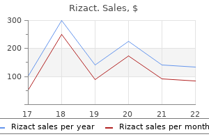
Rizact
Rizact dosages: 10 mg, 5 mg
Rizact packs: 4 pills, 8 pills, 12 pills, 24 pills, 32 pills, 48 pills

Rizact 10mg cheap fast delivery
Recently pain management for dying dog rizact 10 mg discount online, joint ache following a diarrheal illness attributable to pathogenic Escherichia coli has been reported lower back pain treatment videos purchase rizact 5 mg online. Which joints are most commonly concerned in a reactive arthritis following a bowel infection. Sacroiliitis occurs in 20% to 30%; enthesitis (Achilles tendon, plantar fascia attachments) and toe dactylitis happen. The frequency of the triad varies with the causative enteric organism: � Shigella, 85% � Yersinia, 10% � Salmonella, 10% to 15% � Campylobacter, 10% 20. A, Radiograph showing unilateral sacroiliitis (arrows) in a patient with reactive arthritis. B, Radiograph displaying giant, nonmarginal syndesmophytes (arrows) of the backbone in a patient with reactive arthritis. Explain the current theory for the pathogenesis of a postenteritic reactive arthritis. Bacterial lipopolysaccharide antigens (but not viable organisms or nucleotides) from the pathogens (Yersinia, Shigella, Salmonella) causing the infectious gastroenteritis have been shown to be deposited in the joints of patients who develop a postenteritic reactive arthritis. These bacterial cell wall components are thought to incite irritation within the joint. This leads to heavy chains accumulating in the endoplasmic reticulum resulting in an "unfolded protein response," inflicting the release of inflammatory cytokines. Whipple also became a Nobel laureate in physiology in 1934 and was the founding father of the University of Rochester Medical School. W � Wasting and weight loss H � Hyperpigmentation (skin) I � Intestinal ache P � Pleurisy P � Pneumonitis L � Lymphadenopathy E � Encephalopathy S � Steatorrhea D � Diarrhea I � Interstitial nephritis S � Skin rashes E � Eye inflammation A � Arthritis S � Subcutaneous nodules E � Endocarditis 26. Describe the medical traits of the arthritis associated with Whipple illness. Whipple disease happens most commonly in middle-aged white males (male/female ratio, 8:1). More than 70% of patients will develop arthritis at a while during their disease course. Synovial fluid analysis shows an inflammatory fluid with 5000 to 100,000 cells/mm3 (predominantly neutrophils). Whipple disease is caused by a gram-positive actinomycete known as Tropheryma whipplei. These deposits include the rod-shaped free Tropheryma whipplei bacilli seen on electron microscopy. What rheumatic manifestations have been described in sufferers with celiac disease (CeD; gluten-sensitive enteropathy) CeD is an enteropathy ensuing from an autoimmune response to wheat gluten and gliadin by T lymphocytes within the intestine in genetically predisposed people. The most frequent rheumatic manifestations include: � Symmetric polyarthritis (4% to 26%) involving predominantly large joints (knees and ankles more frequently than hips and shoulders) happens. The rheumatic manifestations can respond dramatically to a gluten-free diet but not all the time. In the past, this syndrome occurred in 20% to 80% of patients who had undergone intestinal bypass (jejunoileal or jejunocolic) surgical procedure for morbid weight problems. The arthritis is very painful, inflammatory, oligoarticular, and regularly migratory, affecting both upper and decrease extremity small and large joints. Radiographic findings normally stay regular, regardless of 25% of patients having continual recurring episodes of arthritis. Up to 80% develop dermatologic abnormalities, essentially the most characteristic of which is a maculopapular or vesiculopustular rash. The pathogenesis includes bacterial overgrowth within the blind loop, leading to antigenic stimulation that purportedly causes immune complicated formation (frequently cryoprecipitates containing secretory IgA and bacterial antigens) in the serum that deposits in the joints and skin. Treatment consists of nonsteroidal antiinflammatory medicine and oral antibiotics, which normally enhance signs. Only surgical reanastomosis of the blind loop or improvement in peristalsis can lead to full elimination of signs. What forms of arthritis may be associated with carcinomas of the esophagus and colon The arthritis is usually acute in onset and uneven and predominantly entails lower extremity joints while sparing the small joints of the hands and wrists. Another sort of arthritis related to colorectal malignancy is septic arthritis attributable to Streptococcus bovis. Its clinical manifestations could be remembered by the following mnemonic: P � Pancreatitis A � Arthritis (60%) and arthralgias, normally of the ankles and knees (synovial fluid is typically noninflammatory and creamy in color on account of lipid droplets that stain with Sudan black or oil pink O) N � Nodules that are tender, purple, and normally on extremities (frequently misdiagnosed as erythema nodosum but really are areas of lobular panniculitis with fats necrosis) C � Cancer of the pancreas (a more widespread trigger than pancreatitis) R � Radiologic abnormalities caused by osteolytic bone lesions from bone marrow necrosis (10%) E � Eosinophilia A � Amylase, lipase, and trypsin launched by the diseased pancreas (causes fat necrosis in skin, synovium, and bone marrow) S � Serositis, together with pleuropericarditis, incessantly with fever 33. Outcomes of sufferers with microscopic colitis handled with corticosteroids: a population-based study. At what serum level of bilirubin do adults and infants develop clinically noticeable jaundice Adults develop clinically detectable jaundice when serum ranges of bilirubin reach 2. Hyperbilirubinemia precedes jaundice by a quantity of days as a result of the bilirubin has not but sure to tissue. After serum levels of bilirubin normalize, patients could stay visually jaundiced, because it takes several days for tissue-bound bilirubin to be released. The mucosae of the taste bud and sublingual region are sometimes the first mucocutaneous surfaces to seem yellow in response to hyperbilirubinemia. Bilirubin also has a strong affinity for elastin, which accounts for its early appearance within the sclera of the attention. The pores and skin can also reveal a sallow, refined yellowish hue in sufferers with profound hypothyroidism. Terry nails are characterised by uniform white discoloration of the nail, with the distal 1 to 2 mm remaining pink. The white colour results from abnormalities within the nail bed vasculature and is most commonly seen in patients with liver cirrhosis, heart illness, and diabetes. Muehrcke nails are characterised by double white transverse strains throughout the nails. Kayser-Fleischer rings (brown to green circle of pigment in Descemet membrane of the eye) are pathognomonic of Wilson disease. The pulsation of the central, vertically oriented arteriole in bigger lesions can be visualized with diascopy (observing the lesion through a glass slide firmly pressed on the lesion). Three spider angiomas demonstrating central arteriole and radiating dilated blood vessels. Patients with liver cirrhosis and spider angiomas have elevated plasma levels of vascular endothelial development factor, which may play a job within the growth of spider angiomas. However, the correlation is excessive sufficient that one report means that barmaids in New York used to guess the degree of severity of liver cirrhosis of their clients based on the variety of seen spider angiomas. The number of spider angiomas additionally correlates with the presence of esophageal varices. One study demonstrated that the presence of more than 20 spider angiomas correlated with a 50% likelihood of esophageal bleeding. Approximately 40% of sufferers with hepatic cirrhosis demonstrate reasonable to severe pruritus. The mechanism of pruritus associated with hepatobiliary illness has not been firmly established, however is likely attributable to elevated ranges of bile acids secondary to cholestasis. Serum bile acids are frequently elevated in patients with hepatobiliary illness and pruritus, and bile acid�binding resins relieve the pruritus. Studies on purified bile salts placed on blister bases have proven that each one bile salts produced pruritus, however unconjugated chenodeoxycholate is essentially the most potent.
ROLLED OATS (Oats). Rizact.
- Preventing cancer in the large intestine (colon cancer) when oat bran is used in the diet.
- Are there safety concerns?
- What other names is Oats known by?
- Blocking fat from being absorbed from the gut, preventing fat redistribution syndrome in people with HIV disease, preventing gallstones, treating irritable bowel syndrome (IBS), diverticulosis, inflammatory bowel disease, constipation, anxiety, stress, nerve disorders, bladder weakness, joint and tendon disorders, gout, kidney conditions, opium and nicotine withdrawal, skin diseases, and other conditions.
- Lowering high blood pressure.
- How does Oats work?
- Reducing blood sugar levels in people with diabetes when oat bran is used in the diet.
- Reducing the risk of heart disease, when oat bran is used as part of a diet low in fat and cholesterol.
- Reducing the risk of colon cancer.
Source: http://www.rxlist.com/script/main/art.asp?articlekey=96791
Buy rizact 5 mg amex
Once sufferers depart from their isolation rooms pain treatment history 5 mg rizact discount overnight delivery, these rooms must be cleansed with a 10% bleach solution midsouth pain treatment center germantown tn rizact 5 mg order fast delivery. No Metronidazole 500 mg orally 3�/day* Yes Vancomycin one hundred twenty five mg orally 4�/day* No Good clinical response in 5-7 days Yes Complete 10-14 days of remedy No *If surgical diversion (Hartmann pouch, ileostomy/colostomy) and distal colon is concerned, add vancomycin enemas. Treatment algorithm for Clostridium difficile infection primarily based on disease severity. Subsequent reports identified related findings in different sufferers with persistent diarrhea but without the thickened collagen band. Histologic demonstration of lymphocytic colitis; note the rise in intraepithelial lymphocytes in the surface epithelium and crypts. The most typical medical signs are persistent, nonbloody diarrhea (95%), weight reduction (91%), abdominal pain (40%), urgency (29%), and nocturnal diarrhea (22%). Celiac disease and lactose intolerance also can current with similar signs and ought to be dominated out. Histologic demonstration of collagenous colitis; note the thickened subepithelial collagen band (> 10 microns; notice that the diameter of a standard pink blood cell is 7-8 microns). Barium enemas and colonoscopy typically are normal, however can show subtle mucosal changes. Cases have been recognized within the United States, Europe, Canada, Africa, Asia, Australia, and Latin America, suggesting worldwide distribution. At least a few of this increased incidence is attributed to enhanced scientific consciousness. Most instances may be recognized by biopsies taken inside the vary of versatile sigmoidoscopy; colonoscopy with biopsy of the right colon may be essential to detect 10% of sufferers with isolated right-sided histopathologic findings. Often the disease is insidious, however could have acute onset in up to 40% of patients. Some patients do well on antidiarrheal agents (loperamide) or on cholestyramine alone. These patients responded to retreatment with budesonide and often required gradual subsequent tapers. Bismuth subsalicylate and sulfasalazinemesalamine have proven efficacy in some research. In rare circumstances, sufferers could require surgical procedure, corresponding to diverting ileostomy or colectomy, for severe and refractory disease. Radiation colitis refers to radiation-induced changes in the mucosa of the colon and rectum. Generally, radiation colitis is a chronic, ischemic process attributable to obliterative endarteritis, in distinction to acute inflammation seen in other types of colitis. Radiation damage to the colon happens following therapy of rectal, cervical, uterine, prostate, urinary bladder, and testicular most cancers. Because prostate cancer is the commonest of those cancers, most data has been obtained in this group of patients. The peristaltic movement of the small gut in and out of the sector of radiation decreases the diploma of harm to the small bowel. Brachytherapy, or inside radiotherapy, can deliver high-energy radiation to more centered tissues and subsequently causes much less harm to the colon than external-beam radiation. Tumors within the pelvic area often require greater dosages of radiation and end in higher threat of injury to the colon. The extent of radiation colitis is dependent upon the cumulative radiation dose, fraction dimension, strategy of radiation delivery, amount of tissue exposed, and presence of different treatments similar to surgery or chemotherapy. Radiation injury can be decreased by limiting the dosage and area of publicity whereas shielding adjacent tissues. Additionally, amifostine has been shown to scale back the incidence of radiation colitis by scavenging free radicals produced throughout remedy. Acute radiation injury to the colon usually occurs inside 6 weeks and is manifested by diarrhea, mucus discharge, tenesmus, and infrequently, bleeding. These signs are self-limited and sometimes resolve in 2 to 6 months with out therapy. Chronic signs of radiation colitis and proctitis (or chronic radiation proctopathy) can occur 9 to 12 months following radiation remedy, however could be delayed by a long time after the initial radiation exposure. The primary signs associated with continual damage to the colon and rectum include diarrhea, obstructed defecation, rectal ache, and rectal bleeding. Severe radiation colitis could manifest with bowel necrosis, perforation, fistula development, and uncontrolled rectal bleeding. Colonoscopy could additionally be normal or might present telangiectasias, pallor, and friable mucosa. Early or acute changes include microscopic injury to mucosal and vascular epithelial cells, which can be asymptomatic to the affected person. Late changes generally involve fibrosis with obliterative endarteritis leading to continual ischemia, stricture formation, and bleeding. There is restricted knowledge on the suitable therapy for radiation colitis and proctitis. Medications used to treat radiation colitis and proctitis include oral and rectal sucralfate, steroids, 5-acetylsalicylic acid compounds, hyperbaric oxygen, and antibiotics, similar to metronidazole. Stool softeners are additionally beneficial, as straining may cause telangiectasias to bleed. The major objective of endoscopic therapy is to treat telangiectasias, which are the most common source of rectal bleeding. Colorectal surgeons could apply formaldehyde, also referred to as chemical cautery, to control bleeding. Patients with lengthy or angulated strictures could profit from surgical procedure as these lesions are extra doubtless to perforate with dilating procedures. Fecal microbiota transplantation and emerging purposes, Nat Rev Gastroenterol Hepatol 2011 Dec 20;9(2):88�96. Long-term follow-up of colonoscopic fecal microbiota transplant for recurrent Clostridium difficile infection. Decreased range of the fecal Microbiome in recurrent Clostridium difficileassociated diarrhea. The roles of Clostridium difficile and norovirus amongst gastroenteritis-associated deaths in the United States, 1999�2007. Microscopic colitis: A common and an easily ignored explanation for persistent diarrhoea. Clostridium difficile: emergence of hypervirulence and fluoroquinolone resistance. Clostridium difficile-associated disease: new challenges from a longtime pathogen. Probiotics, antibiotic-associated diarrhoea and Clostridium difficile diarrhoea in people. Alteration of the intestinal microbiome: fecal microbiota transplant and probiotics for Clostridium difficile and past, Expert Rev Gastroenterol Hepatol 2013 Sep;7(7):615�28. Other much less widespread sources include esophagitis, angiodysplasia, Mallory-Weiss tears, most cancers, gastric varices, portal hypertensive gastropathy, Dieulafoy lesions, and aortoenteric fistulas. Mild to moderate blood loss (500-1000 mL) leads to resting tachycardia, whereas lack of a thousand mL will produce orthostatic changes.
5mg rizact fast delivery
Osteoporosis is a frequent complication pain treatment endometriosis discount 10 mg rizact, and studies using pamidronate best treatment for shingles nerve pain 10 mg rizact buy with mastercard, a robust inhibitor of bone absorption, have been proven to enhance signs of ache, tenderness, and swelling considerably. Pain relief, bodily and vocational rehabilitation, and psychological intervention are pillars of an built-in interdisciplinary strategy to affected person care. These patients, their families, and caregivers require ongoing help, schooling, and counseling. MarinusJ,etal: Clinical features and pathophysiology of advanced regional ache syndrome. Trigeminaltrophicsyndrome Interruption of the peripheral or central sensory pathways of the trigeminal nerve might result in a slowly enlarging, unilateral, uninflamed ulcer on ala nasi or adjacent cheek skin. The neck has been reported to be affected in the so-called cervical trophic syndrome, secondary to herpes zoster�associated nerve damage. Biopsy to exclude tumor or a variety of granulomatous or infectious etiologies is often indicated. The trigger is self-inflicted trauma to the anesthetic pores and skin; the suitable therapy is to prevent this by occlusion or with psychotropic. A slough slowly develops, and an indolent necrotic ulcer is left that lasts indefinitely. Whereas the neuropathy renders the ulceration painless and walking continues, plantar ulcers on this situation have a surrounding thick callus. Deeper perforation and secondary infection often result in osteomyelitis of the metatarsal or tarsal bones. Treatment consists of aid of strain on the ulcer through use of a total-contact forged and debridement of the encircling callosity. Removable off-loading gadgets have been discovered to be considerably much less efficient in a scientific review and metaanalysis. Older patients are more prone to injection-induced sciatic nerve damage due to their decreased muscle mass or the presence of debilitating illness. Other common causes of sciatic neuropathy are hip surgery complications, hip fracture and dislocation, and compression by benign and malignant tumors. There is sensory loss and absence of sweating over the distribution of the sciatic nerve branches. Surgical exploration, guided by nerve action potentials, with restore of the sciatic nerve is value it and is most successful if carried out quickly after harm. Syringomyelia Syringomyelia results from cystic cavities contained in the cervical spinal twine brought on by alterations of cerebrospinal fluid circulate. Compression of the lateral spinal tracts produces sensory and trophic changes on the higher extremities, notably in the fingers. The disease begins insidiously and gradually causes muscular weak point, hyperhidrosis, and sensory disturbances, particularly in the thumb and index and middle fingers. The pores and skin modifications are characterised by dissociated anesthesia with lack of pain and temperature sense however retention of tactile sense. Bullae, warts, and trophic ulcerations happen on the fingers and arms, and ultimately contractures and gangrene happen. Other uncommon options embrace hypertrophy of the limbs, arms, or toes and asymmetric scalp hair growth with a sharp midline demarcation. Early surgical therapy allows for enchancment of signs and prevents progression of neurologic deficits. Malperforanspedis Also generally recognized as neuropathic ulceration or perforating ulcer of the foot, mal perforans is a chronic ulcerative disease seen on the sole in circumstances that end in loss of ache sensation at a website of fixed trauma. In most patients, mal perforans begins as a circumscribed hyperkeratosis, often on the ball of the foot. From a dermatologic standpoint, altered ache and temperature sensation, trophic modifications, sweating abnormalities, ulcers of the hands and toes, and in some patients, self-mutilating conduct could also be present. These 5 syndromes and their variants at the moment are known to be secondary to disease-producing mutations in 12 genes. RotthierA,etal: Mechanisms of illness in hereditary sensory and autonomic neuropathies. Atopy is now so common within the inhabitants that the majority people have a family history of atopy. Rather, a dermatologist should infrequently make the diagnosis of grownup "atopic dermatitis" for a dermatitis showing for the primary time after age 30. It is associated with other allergic situations, including meals allergic reactions, asthma, and allergic rhinoconjunctivitis. If one mother or father is atopic, greater than half the kids will develop allergic symptoms by age 2. However, youngsters in Iceland typically have positive pores and skin prick tests to environmental allergens (24%). This leads to a vicious cycle of barrier failure and progressive inflammation, producing a chronic, relapsing, pruritic disorder. Breastfeeding mothers must keep away from the incriminated meals if their infant has been identified with a food allergy. These standards have specificity at or above 90% but have a lot decrease sensitivities (40�100%). Extensively hydrolyzed casein formulation could additionally be used as a supplement or substitute for breast milk during the first four months of life. Aggressive emollient therapy early in life is really helpful to restore any genetic or acquired epidermal barrier defect. The eruption may prolong to the scalp, neck, brow, wrists, extensor extremities, and buttocks. There could also be significant exudate; secondary effects from scratching, rubbing, and an infection embrace crusts, infiltration, and pustules, respectively. The infiltrated plaques eventually take on a characteristic lichenified look. Testing, if carried out, ought to only include foods to which the kid is more likely to be exposed. Double-blind placebocontrolled food challenges are the "gold normal" for diagnosing food allergy. Possible meals allergy detected by testing should be confirmed by clinical history. Higher serum IgE ranges and larger wheal sizes (>8�10 mm) are related to higher chance of reacting to these meals when challenged. Typical morphology and distribution � Flexural lichenification in adults � Facial and extensor involvement in infancy three. Eczema � Typical morphology and age-specific sample � Chronic or relapsing historical past Importantfeatures 1. Early age at onset Atopy Personal and/or household historical past IgE reactivity Xerosis Minorcriteria Must also have three of the next: 1.
Buy discount rizact 5 mg
Young kids best treatment for pain from shingles 10mg rizact effective, due to developmental stage pain medication for dogs tylenol order rizact 10 mg fast delivery, are unable to articulate signs of dysphagia and as an alternative present with signs of feeding difficulties. Additionally, there are a number of potential phenotypes based mostly on the depth of intestinal involvement. Three points concerning the natural history of EoE have become obvious due to broader scientific experiences. First, EoE is a continual disease in which most patients will reply to standard medical therapies. Second, problems associated with EoE embrace food impactions, esophageal narrowing and feeding dysfunction. Finally, there could also be other EoE phenotypes based on whether or not or not sufferers reply to food plan elimination and topical steroids. What is considered an "abnormal" variety of intestinal eosinophils remains unclear and is a matter that must be mentioned between clinicians and pathologists at native establishments. Making the prognosis of EoE is much less complicated as diagnostic tips have been established (Box 43-1). Diagnostic Criteria for Eosinophilic Esophagitis Symptoms are associated to esophageal dysfunction. Esophageal biopsy demonstrates eosinophil-predominant inflammation with a peak worth of 15 eosinophils per high-power subject. Isolated esophageal mucosal eosinophilia persists after a proton pump inhibitor trial. Depending on the precise intestinal organ involved and its accompanying symptoms, there are several approaches. This contains timing of onset, duration, development, aggravating and alleviating elements, related signs. Laboratory Tests � General evaluation: Obtain complete blood rely with differential, complete immunoglobulin E, erythrocyte sedimentation fee, and stool for infectious analysis. In instances in which deeper involvement is suspected, a surgical full-thickness biopsy could additionally be indicated. A, Esophageal biopsy demonstrating EoE, B, Gastric antrum biopsy demonstrating eosinophilic gastroenteritis. Endoscopic view of concentric rings or "trachealization" of the esophagus in eosinophilic esophagitis. They can be used to rule out different etiologic elements such as malrotation and inflammatory bowel illnesses. Upper gastrointestinal contrast examine demonstrating a quantity of rings within the proximal esophagus in eosinophilic esophagitis. Its prevalence is unknown, nevertheless it tends to have an effect on young to middle-aged people with a predilection for males. Table 43-2 illustrates the breadth of those secondary illnesses and the importance of evaluating patients with eosinophils on intestinal biopsy or any form of intestinal inflammation related to peripheral eosinophilia. Current potential administration strategies, based on retrospective studies and case reports, embrace a formal allergy analysis, dietary modification, topical and systemic steroids, and endoscopic dilation or surgical resection. Specific treatment methods must be tailor-made to the individual, considering elements such as extent of disease, severity of signs, price, and compliance. Eosinophilic gastrointestinal diseases-clinically various and histopathologically confounding. A shift in the medical spectrum of eosinophilic gastroenteritis toward the mucosal illness type. The hypereosinophilic syndrome: evaluation of fourteen instances with review of the literature. Quantity and distribution of eosinophils within the gastrointestinal tract of children. World Health Organization-defined eosinophilic issues: 2012 update on prognosis, threat stratification, and administration. A quantitative evaluation of mucosal eosinophils in the pediatric gastrointestinal tract. Update on medical and immunological features of eosinophilic gastrointestinal diseases. Eosinophilic gastroenteritis: a clinicopathological research of sufferers with disease of the mucosa, muscle layer, and subserosal disease. In health, small bowel micro organism resemble oropharyngeal flora with gram-positive, facultative micro organism, which might survive under cardio or anaerobic situations. In overgrowth, micro organism are principally gram-negative, corresponding to Escherichia coli; anaerobic micro organism, including Clostridia and Bacteroides spp. Obstruction to outflow of luminal contents can happen on the web site of surgical anastomosis or with webs, adhesions, or strictures. The jejunoileal bypass, as soon as a popular surgical procedure for morbid weight problems, created a protracted phase of diverted bowel and was typically sophisticated by overgrowth. Diverticula and duplications are incessantly colonized with colonic-type bacteria, leading to overgrowth. The intestinal housekeeper migratory motor complex, when disrupted, is related to bacterial overgrowth. Any situation that may trigger disordered motility, such as diabetes and irritable bowel syndrome or medications, predisposes to overgrowth. Absence or incompetence of the ileocecal valve and enteric fistula can ship colonic bacteria to the small bowel in amounts that exceed clearing capacity. Diarrhea, anorexia, nausea, weight loss, and anemia are cardinal signs, but the nature of the small bowel abnormality influences the presentation. Overgrowth in small intestinal diverticula may current insidiously with metabolic derangements. Bacterial overgrowth leads to small bowel mucosal derangements with brush border defects and bile acid deconjugation commonly resulting in low B12, iron and vitamin deficiencies, and fat malabsorption. Anaerobic bacteria compete with the host for uptake of cobalamin�intrinsic issue complicated, predisposing to B12 deficiency. Whereas luminal micro organism consume cobalamin, folic acid is a product of bacterial substrate fermentation. In addition to iron calcium and cobalamin deficiencies, other micronutrient deficiencies include deficiencies of water-soluble nutritional vitamins. Trace element malabsorption has not been carefully studied in overgrowth syndromes. The gold commonplace for diagnosis is aspiration of small intestinal fluid and culture. Diagnosis of Small Intestinal Bacterial Overgrowth History Prior surgery, older age, medical conditions or medicines related to altered motility, evidence of malabsorption or malnutrition corresponding to metabolic bone illness, night time blindness, easy bruisability, tetany Evidence of systemic disease: weight loss, malnutrition, and malabsorption Hemoglobin (decreased), mean corpuscular volume (increased), vitamin B12 (decreased), folic acid (increased), fecal fat (increased) 14 C-glycocholic acid (increased), 14C-D-xylose (decreased), hydrogen testing with glucose or lactulose, jejunal aspirate for bacterial colony counts and pressure identification Examination Laboratory values Tests 14. There is a danger of potential contamination by oropharyngeal micro organism contaminating the biopsy channel of endoscopes used to acquire small bowel tradition samples.
Rizact 10mg discount without a prescription
Initially neck pain treatment exercise rizact 10 mg generic online, belly angina is often minimal; however pain ischial tuberosity treatment cheap 10mg rizact with visa, it progressively increases in severity over weeks to months. Long-term hypoxia of the small intestinal mucosa could cause villous atrophy resulting in diarrhea, protein-losing enteropathy, steatorrhea, weight reduction, and malnutrition. The risk of intestinal hypoperfusion can improve with shock, severe hypovolemia, decreased cardiac output, and during major thoracic or abdominal surgery as the mesenteric vasculature vasoconstricts. It can be seen in patients taking digoxin or cocaine as these irritate mesenteric vasoconstriction. What ought to I know about mesenteric venous occlusion as a explanation for ischemic bowel illness Mesenteric venous occlusion is a uncommon explanation for ischemic bowel illness, which requires an consciousness of associated danger elements (often a hypercoagulable state) and a high index of suspicion for correct diagnosis. Patients with mesenteric venous occlusion generally present with extreme midabdominal pain out of proportion to the minimal stomach physical examination findings. If infarction is suspected, immediate surgical intervention ought to be undertaken to avoid irreversible ischemia and subsequent bowel resection. This happens by the same pathophysiologic processes that trigger extensive bowel ischemia. The widespread presenting signs of occlusive mesenteric ischemia vary by the purpose for ischemia. These patients may also turn into incontinent of bowel function due to tonic contractions of smooth muscle provoked by ischemia. These contractions cause extreme ache but produce few belly bodily examination findings. Additionally, patients may have diarrhea, steatorrhea, or protein-losing enteropathy, which can further complicate the chronically ischemicinduced atrophy of the small intestine. This happens when large influx of fluid into the bowel wall and lumen causes systemic hypotension and an eventual decrease in arterial move. It must be suspected within the acceptable scientific settings corresponding to abdominal sepsis, hypercoagulability, and the use of oral contraceptive tablets. Again, the physical findings related to mesenteric ischemia vary based on etiologic elements and period of ischemia. Early in the center of the disease process the abdominal examination often consists solely of gentle stomach distension and regular or hypoactive bowel sounds. With progression of ischemic harm, bowel sounds decrease, ileus develops, and abdominal distension worsens. Volume sequestration is manifested by hypotension and tachycardia, whereas fever and peritoneal indicators are indications of transmural harm and likely infarction. Of observe, most patients have evidence of peripheral vascular disease and may also have weight loss. Nonspecific laboratory abnormalities that develop over the course of the illness course of are a result of the consequence of ischemia. Flat and upright stomach plain films should be obtained first in a patient complaining of abdominal pain unless a prognosis of ischemia is obvious. Laparoscopy and enteroscopy may be indicated within the appropriate clinical setting. Additionally, in patients with persistent kidney disease or impaired kidney perform, gadolinium could not trigger the contrast induced nephropathy seen with iodine. It can, nonetheless, uncommonly result in nephrogenic systemic fibrosis, which is an irreversible situation. Describe the position of Doppler ultrasound studies in diagnosis Doppler ultrasound is a noninvasive take a look at that evaluates the patency of and blood flow through the most important mesenteric vessels. It must be carried out whereas the patient is fasting and subsequently meal-stimulated. It is most useful in diagnosing multivessel stenosis in suspected mesenteric angina by demonstrating narrowing or occlusion at a vessel origin and excessively turbulent circulate. Of note, duplex ultrasound has limited capabilities in obese patients as ultrasound waves should pass by way of body tissue previous to producing a diagnostic picture. What is the diagnostic function of endoscopy (sigmoidoscopy, colonoscopy, enteroscopy) and laparoscopy In spite of the truth that a small variety of printed case reports describe diagnostic findings of mesenteric ischemia through enteroscopy, this strategy can be extraordinarily harmful because of the excessive risk of bowel perforation. Lower endoscopy, nevertheless, has been shown to be relatively safe and can aid in figuring out the diagnosis of a patient with suspected ischemic colitis (see Questions 24-28). Laparoscopy, though invasive, has additionally been shown to be a relatively protected technique in assisting with analysis and assessing the diploma of damage to the intestines. Additionally, when intraperitoneal pressure exceeds 20 mm Hg, a stage typically attained after insufflation throughout laparoscopy, splanchnic blood flow decreases. When the diagnosis and therapy of ischemic bowel illness is delayed and peritoneal indicators and acidosis ensue, the mortality fee will increase considerably. Angiography is the gold standard for prognosis of mesenteric arterial occlusion and might help to differentiate between embolic and thrombotic etiologic components. The cutoff of a major artery in the absence of collateral vessel enlargement is indicative of an embolic trigger, whereas vessel narrowing with the development of collaterals signifies thrombosis. Additionally, the venous section of angiography may reveal venous occlusive disease. Angiography can be used as a therapeutic modality by selectively infusing vasodilating medication or thrombolytics, and aiding within the completion of angioplasty, balloon embolectomy, or stent placement. Because of the risks associated with the administration of thrombolytic agents, their use should in all probability be limited to poor surgical candidates without peritoneal signs, to those in whom the ischemic event is taken into account to be reversible or of quick period, and to tertiary care facilities with technical experience. Atherosclerosis commonly includes the femoral artery, which is normally the site of entry for the angiographic catheter. This makes it more durable to access the mesenteric system and can even trigger emboli to distant arteries. Furthermore, iodine distinction will increase the chance of growing renal insufficiency. Management of the underlying disease process, including antiplatelet brokers for vascular disease B. Adequate ache management (avoiding opiates as they lower peristalsis and worsen ischemia) D. Theoretical treatments: small meals, smoking cessation, vasodilators, and suppression of gastric acid secretion to lower mucosal oxygen demand throughout meals 21. What is the position of angioplasty and stenting within the management of ischemic bowel illness Percutaneous transluminal angioplasty with or with out stent placement can be thought of as an alternative to surgical procedure in sufferers with extra distal lesions. Lesions located on the aortic orifices of mesenteric arteries is in all probability not as amenable to dilation and angioplasty because of their fixed diameter. Initial clinical suspicion of acute ischemic bowel illness when other diagnoses have been excluded should prompt angiography. During preliminary surgical procedure (whether or not revascularization has been attempted), some bowel may be left intentionally intact because the standing of its viability will not be clear. Ischemic colitis is the most typical form of non-occlusive intestinal ischemia occurring in older grownup sufferers with impaired cardiac output via a nonocclusive mechanism.

10 mg rizact buy
Melanocytes Melanocytes are derived from the neural crest and by the eighth week of development may be found inside the fetal epidermis lateral knee pain treatment purchase rizact 5mg with mastercard. In normal spine diagnostic pain treatment center rizact 5mg proven, sun-protected trunk epidermis, melanocytes reside in the basal layer at a frequency of about 1 in each 10 basal keratinocytes. Areas such because the face, shins, and genitalia have a larger density of melanocytes, and in closely sun-damaged facial skin, Mart-1 immunostaining can reveal ratios of melanocytes to basal keratinocytes that approach 1: 1. Recognition of the variation in melanocyte/ keratinocyte ratio is critical within the interpretation of biopsies of suspected lentigo maligna (malignant melanoma in situ) on sun-damaged skin. It is the number, dimension, and distribution of the melanosomes or pigment granules within keratinocytes that decide variations in pores and skin colour. Pale skin has fewer melanosomes, and these are smaller and packaged inside membrane-bound complexes. Chronic sun exposure can stimulate melanocytes to produce bigger melanosomes, thereby making the distribution of melanosomes inside keratinocytes resemble the sample seen in dark-skinned people. In histologic sections of skin routinely stained by H&E, the melanocyte appears as a cell with ample amphophilic cytoplasm or as a transparent cell in the basal layer of the epidermis. Keratinocytes also incessantly demonstrate clear spaces but may be differentiated from melanocytes as a outcome of they demonstrate cell-cell junctions and a layer of cytoplasm peripheral to the clear space. Its dendrites prolong for lengthy distances within the dermis, and anyone melanocyte is subsequently involved with a giant number of keratinocytes; collectively they type the so-called epidermal melanin unit. Keratinocytes actively ingest the tips of the melanocytic dendrites, thus imbibing the melanosomes. Melanosomes are synthesized within the Golgi zone of the cell and move via a collection of stages during which the enzyme tyrosinase acts on melanin precursors to produce the densely pigmented granules. Melanocytes in red-haired individuals are most likely to be rounder and to produce extra pheomelanin. Most redheads are compound heterozygotes or homozygotes for a big selection of loss-of-function mutations on this gene. Antimicrobial peptides, including cathelicidin and defensins, are key components of the innate immune system. They shield against infection, are implicated in the pathogenesis of atopic dermatitis, and play a role in charge of pigmentation. The -defensins encompass a class of small, cationic proteins important to each the innate and the adaptive immune system. Within keratinocytes, melanin sometimes varieties a cap over the nucleus, where it presumably capabilities principally in a photoprotective role. Evidence of keratinocyte photodamage within the form of thymidine dimer formation can be assessed utilizing gas chromatography�mass spectrometry or enzyme-linked immunosorbent assays. Areas of leukoderma, or whitening of pores and skin, may be attributable to very different phenomena. In vitiligo, the affected skin turns into white because of destruction of melanocytes. The typical freckle results from a localized improve in manufacturing of pigment by a near-normal number of melanocytes. Black "sunburn" or "ink spot" lentigines demonstrate basilar hyperpigmentation and distinguished melanin throughout the stratum corneum. Melanocytes and keratinocytes categorical neurotrophins (ectodermal nerve progress factors). Langerhanscells Langerhans cells are normally found scattered amongst keratinocytes of the stratum spinosum. The highest density of Langerhans cells within the oral mucosa happens within the vestibular area, and the lowest density is within the sublingual area, suggesting the latter is a comparatively immunologically "privileged" website. At the sunshine microscopic degree, Langerhans cells are troublesome to detect in routinely stained sections. However, they seem as dendritic cells in sections impregnated with gold chloride, a stain specific for Langerhans cells. In their absolutely developed form, the organelles are rod formed with a vacuole at one finish, resembling a tennis racquet. Functionally, Langerhans cells are of the monocytemacrophage lineage and originate in bone marrow. They perform primarily within the afferent limb of the immune response by offering for the popularity, uptake, processing, and presentation of antigens to sensitized T lymphocytes and are essential in the induction of delayed-type sensitivity. Hyaluronan (hyaluronic acid) plays a critical function in Langerhans cell maturation and migration. Macrophages that present antigen in Langerhans cell-depleted pores and skin can induce immune tolerance. ChenJ,etal: Skin permeation habits of elastic liposomes: position of formulation components. ChenY,etal: Biomaterials as novel penetration enhancers for transdermal and dermal drug supply methods. ErnforsP: Cellular origin and developmental mechanisms during the formation of skin melanocytes. HombergM,etal: Beyond expectations: novel insights into epidermal keratin operate and regulation. Iglesias-BartolomeR,etal: Control of the epithelial stem cell epigenome: the shaping of epithelial stem cell id. RobertsN,etal: Developing stratified epithelia: lessons from the dermis and thymus. SakabeJ,etal: Kallikrein-related peptidase 5 functions in proteolytic processing of profilaggrin in cultured human keratinocytes. Much of this regulation takes place through activation of integrins and syndecans. Extracellular matrix protein 1 demonstrates loss-of-function mutations in lipoid proteinosis, leading to reduplication of the basement membrane. MasunagaT: Epidermal basement membrane: its molecular group and blistering problems. Embryologically, they originate as downgrowths from the epidermis and are due to this fact ectodermal in origin. Hedgehog signaling by the transducer generally recognized as smoothened appears crucial for hair growth. Abnormalities in this pathway contribute to the formation of pilar tumors and basal cell carcinoma. In the absence of hedgehog signaling, embryonic hair germs might develop as a substitute into modified sweat gland or mammary epithelium. Although the varied adnexal constructions serve specific capabilities, all can operate as reserve epidermis, in that reepithelialization happens after injury to the surface dermis, principally due to the migration of keratinocytes from the adnexal epithelium to the skin floor. No extra granulation tissue will type, and the contour defect created by the saucerization will persist. The pseudoepitheliomatous hyperplasia famous in infections and inflammatory circumstances consists nearly solely of adnexal epithelium.
Syndromes
- Spinal tap to check for infection around the spinal column
- Cisplatin
- Respiratory distress
- Sores (abrasions) on the skin from scratching and digging
- Death
- Intestinal blockage
Buy 10 mg rizact amex
Sulfonamide antibiotics pain management utilization purchase rizact 5mg on line, associated hypoglycemic agents medial knee pain treatment 5 mg rizact free shipping, and the sulfonylurea diuretics could all be related to photosensitivity reactions. In addition, sufferers may tolerate one of the medications from this group, however when further members of the group are added, clinical photosensitivity occurs. The typical pattern is erythema, scale, and in persistent instances, lichenification and hyperpigmentation. Fluoroquinolone antibiotics are incessantly related to photosensitivity reactions. Sparfloxacin is very photosensitizing; enoxacin, ciprofloxacin, and sitafloxacin are mildly photosensitizing; and levofloxacin rarely, if ever, causes photosensitivity. Histologically, photodistributed lichenoid reactions are sometimes indistinguishable from idiopathic lichen planus. The lichenoid nature of the eruption may not be clinically apparent, and histology is required to affirm the diagnosis. Voriconazole, a second-generation triazole, has been related to an unusual mixture of photosensitive phenomena. Photosensitivity happens in 1�2% or more of sufferers taking voriconazole for more than 12 weeks. Usually, the photosensitivity is gentle, and with the utilization of solar protection and topical remedy, voriconazole could be continued. Pseudoporphyria (with foot erosions as well), eruptive lentigines and atypical nevi, premature growing older, and even the event of extremely aggressive and doubtlessly fatal squamous cell carcinomas in sun-exposed sites have been reported. Photodistributed telangiectasia is a uncommon complication of calcium channel blockers (nifedipine, felodipine, amlodipine). Pseudoporphyria is a photodistributed bullous response clinically and histologically resembling porphyria cutanea tarda. Patients present with blistering on sun-exposed skin of the face and palms and pores and skin fragility. The blistering usually resolves steadily as soon as the offending medicine is stopped. Tanning booth (sunbed) publicity and even extreme sun publicity can produce pseudoporphyria. Patients on dialysis could develop pseudoporphyria, and N-acetylcysteine in doses up to 600 mg twice daily may lead to enchancment in these circumstances. Anticoagulant-inducedskinnecrosis Both warfarin and heparin induce lesions of cutaneous necrosis, although by totally different mechanisms. Obese, postmenopausal girls are predisposed, and lesions are most likely to occur in areas with plentiful subcutaneous fat, such because the breast, stomach, thigh, or buttocks. The scientific look overlaps with calciphylaxis, and sufferers with warfarin-induced calciphylaxis have been described. Patients with a means more delayed onset (up to 15 years) are ascribed to noncompliance, drug-drug interactions, or liver dysfunction. Lesions begin as purple, painful plaques that develop petechiae, then type a big bulla. Warfarin-induced venous limb necrosis is most frequently seen in cancer sufferers, but in addition in the setting of heparin-induced thrombocytopenia and antiphospholipid syndrome. Early in warfarin treatment, the serum ranges of the vitamin K�dependent antithrombotic protein C fall. This is extra prone to occur if the degrees of protein C are already low, if other antithrombotic proteins are poor, or if the affected person has an related hypercoagulable state. Histologically, noninflammatory thrombosis with fibrin in the subcutaneous and dermal vessels is seen. Administration of purified protein C can rapidly reverse the syndrome, in addition to related priapism. Dabigatran etexilate has been instructed for prevention of warfarin-induced pores and skin necrosis within the patient with protein C deficiency. Heparin induces necrosis each on the sites of native injections and in a widespread sample when infused intravenously or given by native injection. Heparin can even induce local allergic reactions at injection websites, that are distinct from the necrosis syndrome. Independent of its method of supply, heparin-induced pores and skin necrosis lesions current as tender pink plaques that bear necrosis, often 6�12 days after the heparin remedies are started. This antibody causes both the thrombocytopenia and the aggregation of platelets in vessels, leading to thrombosis (white clot syndrome). The antibody may appear up to 3 weeks after the heparin has been discontinued, so the onset of the syndrome may be delayed. Histologically, fibrin thrombi are less reproducibly found in affected tissues, as a outcome of the vascular thrombosis is the result of platelet aggregation, not protein deposition. The course of may not only produce infarcts in the skin, but also trigger arterial thrombosis of the limbs, coronary heart, lung, and brain, resulting in important morbidity or mortality. Bilateral adrenal necrosis brought on by hemorrhagic infarction can occur and, if not detected early, may lead to demise from acute addisonian crisis. The syndrome have to be recognized instantly in any patient receiving heparin with late-developing thrombocytopenia. The therapy is to cease the heparin and give a direct thrombin inhibitor and vitamin K. After the platelet count has returned to normal, warfarin therapy is typically given for 3�6 months. HafijiJ,etal: Warfarin-induced calciphylaxis successfully treated with sodium thiosulphate. HermansC,etal: Dabigatran etexilate (Pradaxa) for stopping warfarin-induced pores and skin necrosis in a patient with extreme protein C deficiency. MartinelliI,etal: Anticoagulant therapy with rivaroxaban in extreme protein S deficiency. The lesions are pruritic red patches or plaques that might be deep seated, involving the dermis and subcutaneous tissue. Plaques on the hip are inclined to progress across the waist and down the thigh, forming a "cowboy gunbelt and holster" sample. Small, generalized eczematous papules may happen on other skin sites in severe reactions. These reactions often persist for 1�3 weeks, but may persist for a lot longer, or resolve only to recur spontaneously. On testing, sufferers with this pattern of reaction are positive on intradermal testing to the pure vitamin K1. Subcutaneous sclerosis with or without fasciitis seems at the injection web site many months after vitamin K treatment. The addition of vitamin K1 to cosmetics has led to allergic contact dermatitis from the vitamin K, confirmed by patch testing. Injectionsitereactions In addition to allergic reactions, as described with vitamin K, cutaneous necrosis could occur at websites of medication injections. These embrace chemotherapeutic brokers, calcium salts, radiocontrast materials, and nafcillin.
Rizact 5mg best
The eruption is often seen on the face treatment of cancer pain guidelines discount rizact 5 mg online, especially the eyelids back pain treatment uk rizact 10mg buy visa, carried there by hands which have watered or cared for the plant. English ivy follows philodendron in frequency of instances of occult contact dermatitis. Primrose dermatitis affects the fingers, eyelids, and neck with a punctate or diffuse erythema and edema. Primin, a quinone, is the causative oleoresin abounding within the glandular hairs of the plant Primula obconica. The well-liked minimize flower, the Peruvian lily, is the commonest explanation for allergic contact dermatitis in florists. When handling flowers of the genus Alstroemeria, the florist uses the thumb and second and third digits of the dominant hand. Chrysanthemums frequently trigger dermatitis, with the hands and eyelids of florists most often affected. The -methylene portion of the sesquiterpene lactone molecule is Fruit and vegetables Many greens may trigger contact dermatitis, including asparagus, carrot, celery, cow-parsnip, cucumber, garlic, Indian bean, mushroom, onion, parsley, tomato, and turnip. Onion and celery, amongst other vegetables, have been incriminated within the manufacturing of contact urticaria and even anaphylaxis. Several crops, including celery, fig, lime, and parsley, can cause a phototoxic dermatitis because of the presence of psoralens. Trees Trees with timber and sawdust that will produce contact dermatitis embrace ash, birch, cedar, cocobolo, elm, Kentucky espresso tree, koa, mahogany, mango, maple, mesquite, milo, myrtle, pine, and teak. Melaleuca oil (tea tree oil), which may be applied to the pores and skin to deal with quite lots of maladies, could cause allergic contact dermatitis, primarily through the allergen D-limonene. The unique woods, especially cocobolo and rosewood, and tea tree oil are prominent among allergens that may produce erythema multiforme after cutaneous publicity. Toxicodendron, various medicaments, and a selection of other allergens could induce this response. Plant-associated dermatitis Phototoxic contact dermatitis from plants is mentioned in Chapter 3. Randox (2-chloro-N,N-diallyl-acetamide) has been reported as the cause of hemorrhagic bullae on the ft of farmers. Lawn care firms spray herbicides and fungicides throughout the spring, summer season, and fall. Dryene, thiuram, carbamates, and chlorothalonil are potential sensitizers in these workers, whose clothes frequently turns into wetted whereas spraying. Barbs, bristles, spines, thorns, spicules, and cactus needles are a few of the mechanical accessories of plants that will produce dermatitis. It additionally occurs in persons handling Indian figs in Israel, the place the situation is seen from July to November. Agave americana is a low-growing plant used for ornamental functions in lots of southwestern U. Trimming during landscaping can induce an irritant dermatitis caused by calcium oxalate crystals. The stinging nettle is a common weed that bears tiny spines with biologically active substances such as histamine that produce itching and urticaria inside minutes of contact. Tree-associated crops Foresters and lumber employees may be exposed to allergenic plants apart from timber. Foresters and wood choppers exposed to these lichens growing on trees could develop severe allergic contact dermatitis. Exposure to the lichens may also occur from firewood, funeral wreaths, and also fragrances added to aftershave lotions (oak moss and tree moss). Sensitization is produced by D-usnic acid and other lichen acids contained in lichens. The leafy liverwort (Frullania nisquallensis), a forest epiphyte rising on tree trunks, has produced allergic dermatitis in forest employees. The protein fraction causes the respiratory symptoms of bronchial asthma and hay fever, and the oil-soluble portion causes contact dermatitis. Ragweed oil dermatitis is a seasonal disturbance seen primarily through the ragweed growing season from spring to fall. Contact with the plant or with wind-blown fragments of the dried plant produces the everyday dermatitis. The oil causes swelling and redness of the lids and whole face, and a pink blotchy eruption on the forearms that, after several attacks, might turn into generalized, with lichenification. It intently resembles persistent atopic dermatitis, with lichenification of the face, neck, and major flexures, and severe pruritus. The distribution additionally mimics that of photodermatitis, with ragweed dermatitis differentiated by its involvement of the higher eyelids and the retroauricular and submental areas. Chronic instances could continue into the winter, although signs and signs are most extreme on the top of the season. Coexistent sensitization to pyrethrum may account for prolongation of ragweed dermatitis. Men outnumber ladies in hypersensitivity reactions; farmers outnumber sufferers of all other occupations. Plant derivatives Marine vegetation 98 Numerous aquatic crops are toxic or produce contact dermatitis. The onset is inside a few minutes of leaving the ocean, with severe itching and burning, followed by dermatitis, blisters, and deep, painful desquamation that impacts the areas coated by the washing suit, particularly the scrotum, perineum, and perianal areas and occasionally the breasts in women). Bathing in recent water inside 10 or 15 min of leaving the ocean could stop the dermatitis. Trawler fishermen within the Dogger Bank area of the North Sea develop allergic dermatitis after contact with Alcyonidium hirsutum. These could also be localized in sure elements of the plant, corresponding to in the peel of citrus fruits, leaves of the eucalyptus tree, and bark of the cinnamon tree. Aromatherapy, an increasingly well-liked remedy for relief of stress, includes both inhaling or massaging with important oils; this will likely trigger allergic contact dermatitis in therapists or clients. Exposure to botanical extracts by way of many cosmetics and homeopathic treatments has resulted in an rising variety of reports of allergic contact sensitivity to particular person elements, especially tea tree oil. It can also be used as a flavor for lipstick, bitters, alcoholic and nonalcoholic beverages, toothpaste, and chewing gum. Eugenol, clove oil, and eucalyptus oil are utilized by dentists, who may purchase contact dermatitis from them. Lemon oil from lemon peel or lemon wooden might cause sensitization within the varied handlers of these substances. Myroxylon pereirae incorporates numerous substances, together with essential oils just like the oil of lemon peel.
Generic 5 mg rizact fast delivery
These are (1) congestive coronary heart failure; (2) constrictive pericarditis; (3) hypoalbuminemia; (4) renal failure; (5) portal venous congestion or portal hypertension; (6) hepatic venoocclusive disease; (7) continual cholecystitis; (8) acquired immune deficiency syndrome�related cholangitis; (9) adenomyomatosis; (10) major sclerosing cholangitis; (11) leukemic infiltration; and (12) inflammation from hepatitis pain treatment center pasadena drive lexington ky generic 10 mg rizact mastercard, pancreatitis pain treatment center hazard ky rizact 10 mg buy cheap line, and colitis. Gallbladder carcinoma also causes wall thickening however is normally differentiated from different causes by a masslike appearance, adenopathy, and liver metastases. Describe the differential imaging options seen in the common causes of biliary obstruction. Dilatation of the pancreatic duct sometimes localizes the obstruction to the pancreatic or ampullary stage. It must be suspected when abrupt biliary obstruction is current but no mass or stone is recognized. Sonographic image demonstrates markedly dilated widespread bile duct (calipers) with obstructing echogenic stone (arrow). Choledolcholithiasis in an 83-year-old lady with fever, elevated white blood cell count, elevated total bilirubin and prior cholecystectomy. A, Coronal T2-weighted magnetic resonance picture demonstrates quite a few gallstones (arrows) within the widespread bile duct. B, Magnetic resonance cholangiopancreatography depicts choledolcholithiasis (arrows) in similar patient. Doppler can readily differentiate biliary ducts from vasculature within the portal triad. A, Sonographic picture demonstrates the "double duct" signal (arrow) according to intrahepatic ductal dilatation. B, Contrast-enhanced axial computed tomography picture in a 41-year-old woman with a gastrointestinal bleed demonstrates nonenhancing dilated ducts (arrows). In extra severe circumstances of pancreatis, the pancreas may appear enlarged and hypoechoic. Occasionally, the pancreas could seem enlarged and slightly heterogeneous, with increased attenuation in the peripancreatic fats ("soiled fats") attributable to irritation. In more severe illness, intraglandular intravasation of pancreatic fluid causes intrapancreatic fluid collections. Fluid extending into the pararenal area can lead to the Grey Turner signal (flank ecchymosis) and fluid extending into the gastrohepatic and falciform ligaments may end up in Cullen sign (periumbilical ecchymosis). Posterior leakage of fluid can present with a pleural effusion, classically on the left. The gland dimension is variable, however focal enlargement caused by a persistent inflammatory mass could necessitate biopsy to exclude carcinoma. The pancreatic duct may be dilated (>3 mm) to the level of the papilla and will seem beaded, irregular, or clean. Extensive stranding in the peripancreatic fats (arrows), a nonenhancing pseudocyst anterior to the pancreatic physique (*) and a typical bile duct stent (dotted arrow) are noted. Ten percent to 20% of sufferers with acute pancreatitis and fluid collections develop pseudocysts after four to 6 weeks. Drainage may be indicated for pseudocysts (1) failing to resolve after 6 weeks; (2) remaining bigger than 5 cm in diameter; or (3) causing pain, an infection, hemorrhage, bowel obstruction, or fistula. Necrosis is recognized by a scarcity of contrast enhancement throughout the pancreatic tissue. Abscesses end result from liquefactive necrosis with subsequent infection and normally occur four weeks after the onset of acute pancreatitis. The distinction between abscess and contaminated necrosis is troublesome, however important as a outcome of a pancreatic abscess usually requires extra aggressive remedy. Enzymatic breakdown of the arterial wall may end up in a pseudoaneurysm, mostly within the (1) splenic, (2) gastroduodenal, or (3) pancreaticoduodenal arteries. Up to 10% of pseudoaneurysms rupture, normally right into a pseudocyst, but sometimes into the retroperitoneum, peritoneum, pancreatic duct, or bowel. Pancreatic enlargement is normally focal and finest appreciated within the pancreatic physique and tail. Enlargement and distortion of the pancreatic contour or form are essentially the most frequent findings of pancreatic cancer. Pancreatic duct dilatation (>2-3 mm in diameter) could be the only indirect evidence of a small neoplasm. Biliary tract dilatation is more generally seen with neoplasms in the head of the pancreas. Isolated intrahepatic biliary ductal dilatation may be seen with pancreatic cancer that has unfold to the porta hepatis. Local invasion is most commonly into the peripancreatic fat, but often into the porta hepatis, stomach, spleen, and adjoining bowel loops. Regional lymph node enlargement occurs, together with nodes in the porta hepatis, paraaortic region, and space across the celiac and superior mesenteric artery axis. Liver metastasis happens as pancreatic metastases that often are low-density lesions. What are the characteristic options of the 4 main cystic pancreatic neoplasms They produce giant quantities of mucin, which may finish up in ductal dilatation brought on by mucin plugs. Findings related to malignancy embrace (1) ductal dilatation larger than 10 mm, (2) giant mural nodules, (3) intraductal calcifications, (4) bulging duodenal papilla, and (5) diffuse or multifocal involvement. Intraductal papillary nodules and a distinguished duodenal papilla can distinguish this tumor from chronic pancreatitis. Solid pseudopapillary tumor of the pancreas is most frequently seen in youthful (25-year-old) black or Asian ladies and is characteristically current within the pancreatic tail. Ascites is "free-flowing" and situated in the dependent portions of the abdomen and pelvis. Bowel appears to "float" within the fluid, normally within the heart of the abdomen, if giant amounts of ascites are present. Noncontrast computed tomography picture demonstrates marked ascites (*) with elevation of the omental fats (arrow). Loculated ascites is typically (1) nondependent, (2) stable when patient changes place, and (3) may displace adjoining bowel loops. Aspiration could additionally be required to decide whether or not a set is simple or complex. Ascites is positioned anterior and pleural fluid posterior to the diaphragmatic crus. Unlike pleural fluid, ascites spares the naked space of the liver, which lies alongside the posterior border of the right hepatic lobe. Abscesses differ in look however generally are irregularly marginated and hypoechoic, with internal areas of increased echogenicity. The abscess wall usually enhances, increased density in the adjacent fat is frequent, and mass effect on the surrounding structures may be seen. Axial computed tomography image demonstrates an abscess (arrows) likely resulting from diverticulitis.
Buy cheap rizact 5 mg on-line
Approximately 10% of patients listed for liver transplantation in the United States die each year awaiting an appropriate donor organ oceanview pain treatment medical center 10mg rizact purchase otc. In addition pain treatment hypnosis generic 5mg rizact with mastercard, an equal quantity are faraway from the record as "too sick to transplant. Congenital and metabolic liver disease (8%) � Hemochromatosis � Wilson illness � 1 Antitrypsin deficiency � Cystic fibrosis � Amyloidosis D. Patients usually current with progressive lethargy and jaundice over several days. The most typical causes of acute liver failure in the United States, in descending order, are acetaminophen (46%), indeterminate (14%), drug-induced (11%), hepatitis B (6%), autoimmune hepatitis (6%), ischemia (4%), hepatitis A (3%), and different (9%). Acute ingestion of acetaminophen may trigger severe hepatic harm via the poisonous metabolite, N-acetyl-p-benzoquinone imine, a metabolite of the cytochrome P450 system. Chronic alcohol ingestion might result in induction of the cytochrome P450 system and a reduction within the quantity of acetaminophen required to trigger hepatotoxicity. Without treatment, an acetaminophen stage of more than 300 mcg/mL at four hours or greater than forty five mcg/mL at 15 hours is related to a 90% danger of hepatotoxicity. If patients current within 4 hours of ingestion, activated charcoal can cut back acetaminophen absorption. Careful recipient and donor selection are essential in optimizing outcomes on this inhabitants. In most instances, cholangiocarcinoma stays a relative contraindication for liver transplantation; however, some transplant centers have reported acceptable outcomes in selected individuals. Liver transplants are more commonly carried out in unresectable circumstances of early stage perihilar cholangiocarcinoma (tumor size <3 cm, no metastases) by which protocols involving neoadjuvant chemotherapy adopted by liver transplantation are associated with recurrence-free survival charges of 68%. In instances of intrahepatic cholangiocarcinoma, liver transplantation is generally not performed due to very excessive recurrence charges. The decision to carry out a liver transplant in a specific affected person is predicated on the judgment and expertise of the physicians on the transplant middle. Providing a kidney transplant in chosen liver transplant recipients can have a serious effect on posttransplant survival and high quality of life. Recognition of alcoholism by the affected person and relations is particularly necessary, and patients demonstrate this through adherence to an alcohol rehabilitation program. Features associated with a low rate of recidivism include absence of comorbid substance abuse, good social operate, and absence of household historical past of alcohol abuse. Which components measured in the recipient previous to transplant correlate with decreased postoperative survival Pretransplant recipient traits related to an elevated danger of liver-related demise beyond 1 year after transplant embrace requirement for retransplantation, renal insufficiency, and diabetes. Current immunosuppressive therapy normally entails two or three agents to forestall allograft rejection in the quick postoperative interval. Corticosteroids are used as first-line remedy in immunosuppression at many facilities. Therefore most liver transplant recipients are weaned utterly off of corticosteroids inside a couple of months after surgery. As a end result, posttransplant immunosuppressive regimens are configured to minimize nephrotoxicity. Many facilities have launched using rabbit antithymocyte globulin as induction remedy. A liver transplant affected person has just sustained a grand mal seizure 36 hours posttransplant. The neurologic unwanted facet effects are often reversible with a discount in dosage or discontinuation of the drug. Medications that induce P450-3A4 decrease ranges and improve the danger of rejection or require greater doses of the immunosuppressant. What are the histologic findings of acute rejection versus posttransplant hepatitis C on liver biopsy The differentiation between recurrent hepatitis C and acute cellular rejection is amongst the most problematic areas in medical transplantation. In many instances, the histologic findings on the liver biopsy are inconclusive in differentiating these two issues. The histologic options of acute mobile rejection embrace: � Mixed mobile infiltrate (including eosinophils) in the portal triad � Inflammation of the bile ducts presenting as either apoptosis or intraepithelial lymphocytes � Endotheliitis of the central or portal veins Recurrent hepatitis C could be difficult to distinguish from rejection. The histologic findings may show a predominant lymphocytic infiltrate within the portal areas rather than the mixed mobile infiltrate of rejection. In distinction, bile duct inflammation and venous endothelial irritation are extra distinguished options in rejection. Describe the other posttransplant complications manifested by elevated liver enzymes. The medical presentation may be variable, however is usually related to elevated aminotransferases. Cessation of hepatic artery blood circulate preferentially causes ischemic harm to the biliary tree, leading to breakdown of the biliary tree and growth of bilomas, bile leaks, and ultimately strictures. In hepatic artery thrombosis, retransplantation is often required for profitable long-term outcomes. In the early posttransplant period, portal vein thrombosis might current with signs of graft dysfunction and require immediate revascularization or retransplantation. Late thrombosis may be nicely tolerated or lead to graft dysfunction or portal hypertension. Balloon angioplasty, stent placement, and thrombolytic infusion have been used to reestablish the portal circulation. Biliary leaks or strictures may be asymptomatic however also can lead to jaundice, bacteremia, or sepsis. Biliary leaks can occur on the biliary anastomosis and inside the liver because of bile duct destruction. A hepatocellular pattern could occur with azathioprine, nonsteroidal antiinflammatory medication, and a few antibiotics. Tissue invasive illness may trigger life-threatening problems when the liver, lungs, or gastrointestinal tract are concerned. Chronic allograft rejection is mostly characterized by an insidious but progressive rise in alkaline phosphatase and bilirubin. Patients are normally asymptomatic and synthetic operate remains intact until the late stages. The pathogenesis of this syndrome remains unclear, however the evidence favors lack of bile ducts and the event of obliterative arteriopathy in the small hepatic arteries. Histologic findings embrace a normal-appearing parenchyma with few mononuclear infiltrates in the portal areas but absence of bile ducts in almost all of the portal triads. Later in the course, patients develop strictures and dilations within the bigger bile ducts resembling major sclerosing cholangitis. In these cases, the scientific course could additionally be complicated by recurrent assaults of biliary sepsis. The course of regularly progresses to graft failure, but latest reviews indicate that 20 to 30 p.c of patients could reply to further immunosuppressive remedy. Patients with progressive liver failure caused by continual rejection might require analysis for retransplantation. How typically is it essential to carry out a second liver transplant, and for what causes are retransplantations carried out Fewer than 10% of the liver transplants carried out in the United States are retransplants. Early retransplants are usually performed for primary nonfunction and hepatic artery thrombosis.