
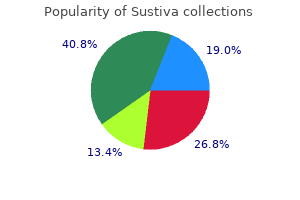
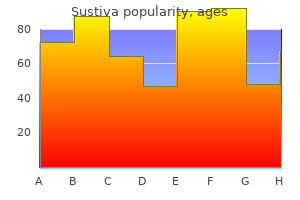
Sustiva
Sustiva dosages: 600 mg, 200 mg
Sustiva packs: 10 pills, 20 pills, 30 pills, 60 pills, 90 pills, 120 pills

Sustiva 200mg effective
The impact of regular childbirth on eyes with abnormalities predisposing to rhegmatogenous retinal detachment treatment wax 200 mg sustiva buy free shipping. Influence of pregnancy on the evolution of background retinopathy: preliminary results of a potential fluorescein angiography research medications beta blockers 200 mg sustiva buy with mastercard. Safety of indocyanine green angiography during being pregnant: a survey of the retina, vitreous, and macula societies. Choroidal vascular lesions in serous retinal detachment considered with indocyanine green angiography. Exposure to verteporfin and bevacizumab therapy for choroidal neovascularization secondary to punctate inner choroidopathy during being pregnant. First case of postconception Verteporfin exposure: pregnancy and neonatal outcome. Accidental being pregnant publicity to verteporfin: obstetrical and neonatal outcomes: a case report. Effect of anti-vascular endothelial development factor antibody throughout early fetal improvement in rats. Intravitreal bevacizumab for a subfoveal myopic choroidal neovascularization within the first trimester of pregnancy. Intravitreal administration of ranibizumab for idiopathic choroidal neovascularization in a pregnant woman. Inadvertent use of bevacizumab to treat choroidal neovascularisation throughout being pregnant: a case report. Hypoplastic optic discs are sometimes underdiagnosed and should vary in degree of growth, leading to variable ranges of visible acuity and visual area defects. Papilledema Papilledema is elevation of the optic nerve due to elevated intracranial strain. Thus for a diagnosis of papilledema the intracranial pressure must be measured, usually by lumbar puncture opening pressure in the lateral decubitus position. An elevated disc or nerve fiber layer swelling with out elevated intracranial strain is optic disc edema not papilledema. Pseudopapilledema Pseudopapilledema is disc elevation in the absence of nerve fiber layer edema. Congestion of the Optic Nerve Head Most causes of pseudopapilledema are acquired and relate to an underlying ocular or systemic disease. The most typical congenital cause of congestion on the optic nerve head is optic disc drusen. Megalopapilla Megalopapilla is a uncommon anomaly of the optic disc, involving thinning of the nerve fiber across a big optic nerve head, and is often associated with massive refractive errors and with midline congenital deformities. Slusher and coworkers9 described a household of 35 members spanning five generations with an autosomal dominant sample of congenital optic disc abnormalities. Remarkably, a myriad of morphologic variations of phenotype were expressed, including optic disc pits, morning glory syndrome, and coloboma of the optic nerve. One gene defect may find yourself in a wide selection of optic disc abnormalities; the standard classification schemes that describe varieties of cavitary optic disc anomalies ought to therefore be reconsidered. Not surprisingly, anomalies of structure at this important juncture typically lead to marked physiologic penalties. This was adopted in 1868 by Liebrich,14 who described the fundus look of optic disc drusen and made the hyperlink to the histopathology findings of M�ller. Retrospective studies of family members of individuals with optic disc drusen found that solely certainly one of 27 relatives of seven probands with optic disc drusen additionally had optic disc drusen. Vascular maldevelopment has been theorized to trigger elevated transudate launch into the intercellular area which in flip acts as a nidus for the formation of optic disc drusen. True disc edema is doubtlessly a life threatening course of, in distinction to the extra benign nature of optic disc drusen. These axons project to varied primary visual nuclei in the brain,10 represent a fiber tract rather than a nerve, and, as such, have histologic and useful similarities to mind tissue. The optic nerve is enclosed by three meningeal sheaths which are contiguous with the meningeal coverings of the brain. Fibers from the retina gather on the optic disc, move down through the lamina cribrosa, become myelinated, and type fascicles. The first is the end result of direct compression or displacement of the fibers by the drusen and is often arcuate in nature, usually in the inferior nasal quadrant. The development, nevertheless, is gradual and sufferers will not be aware of the sphere defect with out formal visible subject testing. The reported frequency of visible area defects varies extensively, from 24% to 87%, with the very best occurrence in eyes with superficial optic disc drusen. This can cause transient visible loss, or in some cases anterior ischemic optic neuropathy can happen. Detection the appearance of the optic nerve head with optic disc drusen can be fairly variable, with some discs showing superficial drusen which are readily seen. The use of B-scan ultrasound to detect optic disc drusen was described way back to the 1970s. The method can be helpful for detecting buried drusen, but the echogenicity of drusen relies on the calcium content. These gadgets provide an unprecedented capacity to evaluate optic disc drusen to the depth of the lamina cribrosa and to evaluate the interplay with structures of the optic nerve. Associated Retinal Changes the retinal vasculature in eyes with optic disc drusen is often anomalous with elevated tortuosity, vascular loops, and abnormal branching patterns together with optociliary shunt vessels7,forty one There can be an elevated incidence of cilioretinal arteries in patients with optic disc drusen with as a lot as 40% of sufferers having the vascular aberration versus 15% within the normal inhabitants. The first description of a vascular disturbance related to optic disc drusen was in 1895 when Gifford described an 11-year-old woman with a central retinal artery occlusion. The presence of drusen alters the flow dynamics of the central retinal vein by altering the path and probably constricting the vascular diameter. Typically in a juxtapapillary location, the neovascularization extends towards the macula but not often involves it. Once the prognosis is established, nonetheless, common examinations are important to ensure that treatable problems of optic disc drusen are appropriately managed. The presence of optic disc drusen can predispose the affected person to glaucomatous optic nerve injury at lower intraocular pressures. Assessment for glaucoma may additionally be complicated by the presence of the optic disc drusen as a small crowded nerve can mask the appearance of cupping. Glaucoma may coexist with optic disc drusen47 and determining which portion of the sphere loss is as a end result of of glaucoma and which is as a result of of optic disc drusen is tough. The use of pressure-lowering medications should contemplate the dangers and advantages, tailor-made to every particular person affected person. The presence of a neovascular membrane was traditionally handled with photocoagulation depending on the situation. Most optic disc pits are nonfamilial; however, there are a few stories with an autosomal dominant sample of inheritance.
Diseases
- Fibrochondrogenesis
- Ausems Wittebol Post Hennekam syndrome
- Freeman Sheldon syndrome
- Succinyl-CoA acetoacetate transferase deficiency
- Xeroderma pigmentosum
- Congenital facial diplegia
- Retroperitoneal liposarcoma
- Leukemia, T-Cell, chronic
- Bulimia nervosa
- Atresia of small intestine

Discount sustiva 600 mg with amex
Effect of intensive diabetes administration on macrovascular events and risk elements within the Diabetes Control and Complications Trial medicine game sustiva 200mg buy visa. The relationship of glycemic publicity (HbA1c) to the risk of improvement and progression of retinopathy within the Diabetes Control and Complications Trial treatment xeroderma pigmentosum order 600 mg sustiva overnight delivery. Influence of intensive diabetes treatment on quality-of-life outcomes within the Diabetes Control and Complications Trial. The absence of a glycemic threshold for the event of long-term complications: the attitude of the Diabetes Control and Complications Trial. Lifetime benefits and costs of intensive remedy as practiced in the Diabetes Control and Complications Trial. Effect of intensive therapy on residual -cell operate in sufferers with sort I diabetes in the Diabetes Control and Complications Trial. Design, implementation, and preliminary results of a long-term follow-up of the Diabetes Control and Complications Trial cohort. Diabetes Control and Complications Trial/Epidemiology of Diabetes Interventions and Complications Research Group. Retinopathy and nephropathy in patients with type I diabetes four years after a trial of intensive therapy. Writing Team for the Diabetes Control and Complications Trial/Epidemiology of Diabetes Interventions and Complications Research Group. Prolonged impact of intensive remedy on the danger of retinopathy issues in sufferers with kind 1 diabetes mellitus. Comparison of digital and movie grading of diabetic retinopathy severity within the Diabetes Control and Complications Trial/ Epidemiology of Diabetes Interventions and Complications Study. Effect of glycemic publicity on the risk of microvascular problems within the Diabetes Control and Compllications Trial - revisited. The long-term results of type 1 diabetes therapy and issues on health-related high quality of life. United Kingdom Prospective Diabetes Study, 30: Diabetic retinopathy at diagnosis of non-insulin-dependent diabetes mellitus and associated danger components. A phase 2 randomized scientific trial of intravitreal bevacizumab for diabetic macular edema. A randomized trial comparing intravitreal triamcinolone acetonide and focal/grid photocoagulation for diabetic macular edema. Agreement between clinician and studying heart gradings of diabetic retinopathy severity degree at baseline in a part 2 examine of intravitreal bevacizumab for diabetic macular edema. Three-year comply with up of a randomized trial comparing focal/grid photocoagulation and intravitreal triamcinolone for diabetic macular edema. The Diabetic Retinopathy Clinical Research Network Laser-Ranibizumab-Triamcinolone Clinical Trials. Effect of focal/grid photocoagulation on visual acuity and retinal thickening in eyes with non-center-involved diabetic macular edema. Factors related to enchancment and worsening of visual acuity 2 years after focal/grid photocoagulation for diabetic macular edema. Frequency of intraocular strain increase inside days after intravitreal triamcinolone injections in the Diabetic Retinopathy Clinical Research Network. Randomized trial evaluating short-term results of intravitreal ranibizumab or triamcinolone acetonide on macular edema after focal/grid laser for diabetic macular edema in eyes additionally receiving panretinal photocoagulation. Cost-effectiveness evaluation of ranibizumab plus immediate or deferred laser or triamcinolone plus immediate laser for diabetic macular edema. Evaluation of masking study individuals to intravitreal injections in a randomized scientific trial. Randomized scientific trial evaluating intravitreal ranibizumab or saline for vitreous hemorrhage from proliferative diabetic retinopathy. Exploratory evaluation of the effect of intravitreal ranibizumab or triamcinolone on worsening of diabetic retinopathy in a randomized clinical trial. Evaluation of outcomes 1 year following use of short-term ranibizumab for vitreous hemorrhage due to proliferative diabetic retinopathy. Ranibizumab plus prompt or deferred laser for diabetic macular edema in eyes with vitrectomy before anti-vascular endothelial growth issue therapy. Lack of association between thiazolidinediones and macular edema in sort 2 diabetes. Comparison of standardized clinical classification with fundus photograph grading for the evaluation of diabetic retinopathy and diabetic macular edema severity. The results of medical management on the development of diabetic retinopathy in individuals with sort 2 diabetes. Rationale and design of the AdRem examine: Evaluating the effects of blood pressure decreasing and intensive glucose management on vascular retinal issues in patients with type 2 diabetes mellitus. Retinal vascular lesions in sufferers with Caucasian and Asian origin with type 2 diabetes. Effects of blood stress lowering and intensive glucose control on the incidence and progression of retinopathy in patients with sort 2 diabetes mellitus: a randomised managed trial. A randomized trial of sorbinil, an aldose reductase inhibitor, in diabetic retinopathy. Randomized comparison of krypton versus argon scatter photocoagulation for diabetic disc neovascularization. Randomized controlled research of an intervitreous dexamethasone drug delivery system in patients with diabetic macular edema. Retinal microaneurysm rely predicts progression and regression of diabetic retinopathy. Sustained supply fluocinolone acetamide vitreous inserts present profit for a minimal of 3 years in patients with diabetic macular edema. Ranibizumab monotherapy or combined with laser versus laser monotherapy for diabetic macular edema. Patient-reported visible function outcomes enhance after ranibizumab remedy in patients with vision impairment as a end result of diabetic macular edema. Three-year outcomes of individualized ranibizumab therapy of sufferers with diabetic macular edema. A part 2/3, multicenter, randomized, double-masked, 2-year trial of pegaptanib sodium for the therapy of diabetic macular edema. Changes in vision- and health-related high quality of life in sufferers with diabetic macular edema handled with pegaptanib sodium or sham. The effect of ruboxistaurin on visible loss in patients with moderately severe to very severe nonproliferative diabetic retinopathy. Effect of ruboxistaurin on visual loss in sufferers with diabetic macular retinopathy.
Cheap 200 mg sustiva with amex
Retinal pigment epithelial modifications in persistent Vogt�Koyanagi�Harada illness: fundus autofluorescence and spectral domain-optical coherence tomography findings medicine zanaflex order sustiva 600 mg free shipping. Ultrawide-field green-light (532-nm) autofluorescence imaging in continual Vogt�Koyanagi�Harada illness 911 treatment buy sustiva 600 mg overnight delivery. Evaluation of the effect on outcomes of the route of administration of corticosteroids in acute Vogt�Koyanagi�Harada disease. Prognostic elements for clinical outcomes in patients with Vogt�Koyanagi� Harada illness treated with high-dose corticosteroids. Chronic noninfectious uveitis related to Vogt�Koyanagi�Harada disease treated with lowdose weekly systemic methotrexate. Photodynamic therapy for subfoveal choroidal neovascularisation in Vogt�Koyanagi�Harada illness. Intravitreal bevacizumab for choroidal neovascularization secondary to Vogt�Koyanagi�Harada syndrome. Correlation between peripapillary atrophy and corticosteroid therapy in patients with Vogt� Koyanagi�Harada disease. They often present a diagnostic and therapeutic problem for clinicians and researchers. These embody granulomatous ailments, such as sarcoid, tuberculosis, and sympathetic ophthalmia; masquerade syndromes like syphilis and intraocular lymphoma; infectious etiologies together with toxoplasmosis and pneumocystis choroidopathy; and other entities corresponding to presumed ocular histoplasmosis and Beh�et disease. Blurred vision, photopsias, visual area adjustments, floaters, and modifications in distinction sensitivity can occur. Inflammatory multifocal chorioretinopathies may actually be a more descriptive term. It has additionally been suggested that several of those entities may be "associated" or even represent a spectrum of the identical process. These ailments are not often overlapping, and clear distinctions can be made with multimodal imaging strategies. These lesions are scattered across the optic disc and radiate to the equator in a "shotgun" pattern. Shah and colleagues did an intensive review of English literature in 2005 and reported the following epidemiologic traits. It is necessary to acknowledge that signs can precede the onset of the traditional fundus findings by several years. These birdshot lesions may be oval or round in form, sometimes about one-half or one-quarter disc diameter in size. They tend to cluster close to the nerve and mostly nasal and inferior to the disc. The anterior segment typically has minimal irritation with out distinguished keratic precipitates. Finally, exclusion criteria embrace presence of serious keratic precipitates, posterior synechiae, or prognosis of infectious, neoplastic, or inflammatory illness which will trigger multifocal choroidal lesions. Again, signs of blurred imaginative and prescient, color deficiency, contrast sensitivity points, and visible subject problems could additionally be current for years previous to the onset of fundus lesions. It remains a poorly understood disease, and no consensus on management and therapy has been found. However, in a latest analysis of forty six sufferers, method of remedy was correlated with visual outcome. They found that prognosis for central visual acuity and maintenance of visible fields was improved in sufferers on long-term immunosuppression in comparison with short-term therapy. The diffuse hyperfluorescence seen might represent a deep inflammatory focus that accumulates fluorescein. Theories to explain this early hypofluorescence include choroidal ischemia versus blockage from inflammatory infiltrates. In the acute phases, the inflammatory infiltrates could additionally be denser and block fluorescence. The lesions can turn into more isofluorescent on this part of the disease, or they could stay hypofluorescent. They recommend utilizing this protocol and looking out on the ellipsoid zone to monitor disease progression. They discovered that some of these hypofluorescent lesions might be placoid in nature and contain the macula. In this stage, there could additionally be a greater decrease in b-wave amplitude versus a-wave amplitude. It means that the M�ller and bipolar cells are extra affected than the photoreceptor�retinal pigment epithelium advanced. Rod dysfunction could occur earlier than cone dysfunction: the rod b-wave could additionally be affected prior to photopic b-wave and flicker response in most patients. The 30 Hz flicker implicit time is irregular in 70% of patients at baseline and is a vital marker to observe. Local steroid injections enhance the risk of cataract and trigger glaucoma with repeated dosing. Implantation of a fluocinolone acetonide sustained-release device has been shown to remove the necessity for systemic remedy; nevertheless, it also has a excessive threat of cataract development and glaucoma. Cyclosporine has been used as it inhibits T lymphocytes and prevents S-Ag-induced experimental uveitis. Side-effects similar to bone-marrow suppression and growth of malignancies must be weighed for this class. Other biologics, corresponding to infliximab and adalimumab, have additionally been used with evidence supporting efficacy. Of 102 sufferers, 16 had hypertension, 5 had coronary artery disease, 2 had a history of cerebrovascular accident, and a pair of had a central retinal vein occlusion. Some lymphocytes have been found across the retinal blood vessels and in the optic disc. However, entities such as pars planitis, intraocular B-cell lymphoma, syphilitic chorioretinitis, sarcoidosis, sympathetic ophthalmia, and different white spot illnesses, especially multifocal choroiditis and panuveitis syndrome, should be considered. Indolent nonprogressive multifocal choroidal lesions additionally give a similar image and ought to be thought-about when unilateral. Symptoms embody blurred imaginative and prescient, photopsias, scotomas, nyctalopia, and poor distinction sensitivity. The course of is related to a low-grade inflammation of the anterior and posterior segments. Management/Treatment In the evaluate of the literature by Shah and colleagues,10 most instances had been treated; nonetheless, no definitive tips for the initiation of remedy were given. Oral, sub-Tenon, intraocular, and most recently sustained release fluocinolone acetonide55 have been used.
Purchase 600 mg sustiva visa
Flat irregular retinal pigment epithelium detachments in chronic central serous chorioretinopathy and choroidal neovascularization treatment kidney cancer symptoms discount 600 mg sustiva with amex. Subretinal dot-like precipitates and yellow material in central serous chorioretinopathy medicine dropper generic 200 mg sustiva. Fundus autofluorescence of elongated photoreceptor outer segments in central serous chorioretinopathy. Optical coherence tomography options of acute central serous chorioretinopathy versus neovascular age-related macular degeneration. Morphologic changes in the outer layer of the detached retina in rhegmatogenous retinal detachment and central serous chorioretinopathy. Near-infrared and shortwavelength autofluorescence imaging in central serous chorioretinopathy. Central serous chorioretinopathy: current findings and new physiopathology hypothesis. Toward a selected classification of polypoidal choroidal vasculopathy: idiopathic illness or subtype of age-related macular degeneration. Polypoidal choroidal vasculopathy pattern in age-related macular degeneration: a clinicopathologic correlation. Clinicopathological correlation of polypoidal choroidal vasculopathy revealed by ultrastructural study. Improved visualization of polypoidal choroidal vasculopathy lesions using spectral-domain optical coherence tomography. Time-lag between subretinal fluid and pigment epithelial detachment reduction after polypoidal choroidal vasculopathy remedy. Evaluation of pulse corticosteroid therapy for Vogt�Koyanagi�Harada illness assessed by optical coherence tomography. Retinal fluorescein and indocyanine green angiography and optical coherence tomography in successive stages of Vogt�Koyanagi�Harada illness. Bartonella and intraocular irritation: a series of circumstances and evaluation of literature. Characterization of macular edema from various etiologies by optical coherence tomography. Optical coherence tomography features in the course of the evolution of serous retinal detachment in patients with diabetic macular edema. A systematic correlation between morphology and useful alterations in diabetic macular edema. Aqueous vascular endothelial growth factor ranges are related to serous macular detachment secondary to branch retinal vein occlusion. Bullous retinal detachment and multiple retinal pigment epithelial detachments in patients receiving hemodialysis. Secondary retinal adjustments associated with choroidal naevi and melanomas documented by optical coherence tomography. Comparison of serum glycosylated hemoglobin levels in sufferers with diabetic cystoid macular edema with and without serous macular detachment. Association of pathomorphology, photoreceptor status, and retinal thickness with visible acuity in diabetic retinopathy. A permeability defect of the retinal pigment epithelium: prevalence in early streptozocin diabetes. For example, presently a minimum of 256 genes are recognized that can trigger one or another form of retinal illness,1 and over 12,000 mutations have been reported, in total, in these genes. However, real families are sometimes extra complicated, particularly for late-onset, progressive forms of retinal illness. The distinction between late onset and no onset could simply be the age of the affected person when examined. Whatever the terminology, the underlying finding is that dominant retinal disease mutations could have highly variable penalties, confounding prognosis. An further rare but confounding chance has been observed in giant, multigenerational households with inherited retinal illness: mutations in multiple gene could also be segregating independently within the household. This occurs as a end result of families with late onset, nonlethal ailments are prone to meet and socialize with comparable families. Descendants of those families are vulnerable to inheriting mutations in more than one gene. An affected individual can be either homozygous for a single mutation or heterozygous for two distinct mutations. An particular person with two distinct recessive mutations can also be called a compound heterozygote. Recessive mutations must be in trans to trigger disease Examples of autosomal recessive retinopathies embrace Leber congenital amaurosis and Usher syndrome. Approximately one-fourth of kids of provider parents are affected and one-half of children are carriers. Affected people may happen in a couple of era and in more than one department of those households. Autosomal Dominant Inheritance Autosomal dominant inheritance occurs when a single copy of a mutation on an autosomal chromosome is sufficient to trigger illness. Males are as more likely to be affected as females, and roughly 50% of kids of an affected particular person might be affected. Forms of retinal illness which are typically autosomal dominant include maculopathies such as Best disease. Two phenomena that may confuse the picture of autosomal dominant illness are variable expression and incomplete penetrance. Variable scientific expression means that people with the same mutation might differ in onset, progression, or severity of illness or, in some instances, might have distinctly totally different scientific findings. Since females have two Xs, this suggests that the majority X-linked mutations might be recessive in Genetic Mechanisms of Retinal Disease 713 females. For a truly recessive X-linked mutation, one-half of the sons of a carrier feminine are affected, one-half of her daughters are unaffected carriers, and not considered one of the sons of an affected male are affected. Although females have two Xs, one of the Xs, chosen at random in each cell, is inactivated in most tissues. This is X-inactivation or Lyonization, named for Mary Lyon, who first described the phenomenon. Isolated Cases Isolated circumstances deserve an entry of their own because the mode of inheritance is commonly unclear. A practical definition of an isolated case is an affected individual with no affected firstdegree family members (parents, sibs, and children) and no stories of extra distant affected relations. One instant concern is that there may be different affected members of the family however the individual describing the household is unaware of the disease standing in these individuals. Clinical examination of first-degree relations is often informative in these circumstances. The more than likely prospect is that this is an autosomal recessive case and the dad and mom are carriers. Another possibility is autosomal dominant or X-linked inheritance with nonpenetrance in prior generations.
Primula veris (Cowslip). Sustiva.
- What is Cowslip?
- Inflamed nasal passages or sinusitis when taken with gentian root, European elder flower, verbena, and cowslip flower (SinuComp, Sinupret).
- Are there safety concerns?
- Dosing considerations for Cowslip.
- How does Cowslip work?
Source: http://www.rxlist.com/script/main/art.asp?articlekey=96202

Sustiva 600mg cheap fast delivery
Subretinal membranes may trigger visual distortion by preventing proper flattening of the retina or by disturbing the contour of the overlying retina medications 5 rights order 600 mg sustiva mastercard. Clinically medicine university sustiva 200 mg discount fast delivery, subretinal membranes differ in nature to those seen on the retinal surface, and this can be partly defined by the different expression of proteins on their surface. Immunocytochemical labeling and confocal imaging studies demonstrate their distinctive nature. In felines, M�ller cell processes within the retina that preferentially categorical vimentin within the outer portion of the cell develop into the subretinal area. Microvilli usually prolong from the apical surface of the M�ller cells, just past the outer limiting membrane. Rod synaptic terminals showed transforming with extension of rod bipolar cell dendrites and horizontal cell processes into the outer nuclear layer. Animal fashions have given us a clearer understanding of mobile recovery following retinal reattachment. In the feline mannequin, retinal reattachment inside 1�3 days may be very efficient at reversing the cellular adjustments induced by retinal detachment. For example, a normal balance between disc addition and disc shedding must be restored if the outer segments are going to attain regular size. Clinical evidence indicates that this course of might occur over months or even years. In monkey retinas detached for 1 week, rod and cone outer segments regain approximately 30% of their regular mean length within 7 days of reattachment, 60% of their size after 30 days, and one hundred pc by one hundred fifty days. Disc shedding, then again, seems to interact after the first reattachment week. In cat retinas detached for periods longer than 7 days, many outer segments remained shorter than normal several months after reattachment,sixty four implying that defects in the meeting or shedding phases (or both) of the renewal course of could persist nicely past 30 days of reattachment in retinas indifferent for longer durations. Neurites were invariably observed in all membranes examined but only in regions containing glial cells. The green in (D) represents labeling of the photoreceptor synaptic terminals with an antibody to synaptophysin. In the feline model, reattachment induces development into the vitreous of M�ller cell processes, that type epiretinal membranes. These M�ller cell processes then act as a substrate for ganglion cell neurite growth. At present it appears affordable to conclude that a return in course of completely normal retinal morphology happens steadily over a timespan of months or years, even after temporary episodes of detachment. Cellular remodelling in mammalian retina: results from research of experimental retinal detachment. Histological evaluation of retinas sampled during translocation surgical procedure: a comparability with normal and transplantation retinas. Glial remodelling and neuronal plasticity in human retinal detachment with proliferative vitreoretinopathy. Histopathological study of the impact of retinal detachment surgery on forty nine eyes obtained post mortem. The extracellular matrix of human retinal pigment epithelial cells in vivo and its synthesis in vitro. The relationship of the retinal pigment epithelium to photoreceptor outer segments within the human retina. Observations on the retinal pigment epithelium and retinal macrophages in experimental retinal detachment. Evidence from normal and degenerating photoreceptors that two outer section integral membrane proteins have separate transport pathways. A survey of molecular expression by photoreceptors after experimental retinal detachment. Limiting photoreceptor death and deconstruction throughout experimental retinal detachment: the worth of oxygen supplementation. Inhibition of retinal detachment-induced apoptosis in photoreceptors by a small peptide inhibitor of the fas receptor. The efficacy of delayed oxygen therapy in the therapy of experimental retinal detachment. Experimental retinal detachment in the cone-dominant ground squirrel retina: morphology and primary immunocytochemistry. M�ller cell outgrowth after retinal detachment: affiliation with cone photoreceptors. Retinal detachment repair by vitrectomy: simplified formulae to estimate the chance of failure. Long-term visual recovery after scleral buckling for macular-off retinal detachments. Fate of biotinylated basic fibroblast progress factor in the retina following intravitreal injection. Basic fibroblast progress factor: a potential regulator of proliferation and intermediate filament expression in the retina. Immunocytochemical localization of two retinoid-binding proteins in vertebrate retina. Animal fashions of retinal detachment and reattachment: identifying cellular occasions that may have an result on visible restoration. Recovery of photoreceptor outer segment size and analysis of membrane meeting rates in regenerating primate photoreceptor outer segments. Identification of ganglion cell neurites in human subretinal and epiretinal membranes. Retinal reattachment of the primate macula: photoreceptor restoration after short-term detachment. Microglial cell activation following retinal detachment: a comparison between species. Retraction and transforming of rod spherules are early occasions following experimental retinal detachment: an ultrastructural examine using serial sections. Mueller cell and neuronal remodelling in retinal detachment and reattachment and their potential consequences for visible restoration: a evaluate and reconsideration of current knowledge. Upregulation of glial fibrillary acidic protein in response to retinal injury: its potential function in glial remodelling and a comparison of vimentin expression. Expression profiles of nestin and synemin in reactive astrocytes and M�ller cells following retinal damage: a comparison with glial fibrillar acidic protein and vimentin. The impact of alkylphosphocholines on intraretinal proliferation initiated by experimental retinal detachment. Ranibizumab is a potential prophylaxis for proliferative vitreoretinopathy, a nonangiogenic blinding illness. Effects of the neurotrophin brain-derived neurotrophic consider an experimental mannequin of retinal detachment. Basic fibroblast progress factor and local damage defend photoreceptors from mild injury in the rat. The term "central" refers to the type of the illness causing visible signs as a outcome of the presence of serous detachments in the macular area. Various mechanisms to accomplish its tasks are used including membrane pumps, transporters, and channels, transcytosis, metabolic alteration of solutes in transit, and passive but selective diffusion.
Discount sustiva 600mg with amex
This is believed to happen as a end result of ciliary body swelling symptoms gallstones generic sustiva 200 mg without a prescription, choroidal effusion medications after stroke cheap 600 mg sustiva with visa, or each, or with swelling of the lens itself with subsequent forward rotation of the lens�iris diaphragm. The folds usually resolve following discontinuation of the offending treatment. It ought to be noted, nevertheless, that every so often, topiramate might result in growth of angle closure glaucoma. It is used as a food-coloring agent, for pores and skin pigmentation within the treatment of vitiligo, and for the treatment of photosensitivity problems corresponding to erythropoietic protoporphyria, psoriasis, and photosensitive eczema. It additionally has been used Drug Toxicity of the Posterior Segment 1735 92 A B 200 �m 1000 Trickness [�m] 800 600 four hundred 200 zero zero. Photograph (A) exhibiting a blunted foveal reflex in a affected person on paclitaxel for treatment of metastatic breast carcinoma. Photograph (A) shows perifoveal retinal folds related to chlorthalidone remedy, which resolve within 2 weeks following discontinuation of the drug (B). Fundus photograph demonstrating macular and extramacular striae in a patient on topiramate. Characteristic yellow�white macular crystals are seen in a perifoveal distribution. The crystals seem extra prominently in eyes with preexisting retinal illness and with concurrent use of beta-carotene. Canthaxanthin crystals are localized to the spongy degeneration of the internal neuropil and are related to atrophy of the M�ller cells. MiscellaneousAgents A single case of crystalline retinopathy following 19 years of nitrofurantoin (Macrodantin) use has been reported. Prominent perifoveal punctate yellow deposits in a doughnut-shaped ring surrounding the macula. Drug Toxicity of the Posterior Segment 1739 therapy, with each intravenous and intravitreous (20 �g) routes of administration, has been related to an anterior uveitis, hypotony, and visual loss. Cidofovir has been shown experimentally and clinically to trigger a direct toxic effect to the ciliary body, with a resulting iritis and intraocular strain decrease. The degree of systemic acidosis correlates well with the extent of visual dysfunction. Hydroxychloroquine retinopathy regardless of regular ophthalmologic evaluation: a consecutive collection. Detection of early hydroxychloroquine retinal toxicity enhanced by ring ratio analysis of multifocal electroretinography. The incidence of irreversible retinal toxicity in sufferers handled with hydroxychloroquine: a reappraisal. Rates and predictors of hydroxychloroquine retinal toxicity in patients with rheumatoid arthritis and systemic lupus erythematosus. Ophthalmological monitoring of sufferers taking antimalarials: preferred apply patterns. Ophthalmological monitoring for hydroxychloroquine toxicity: a scientific evaluate of obtainable knowledge. Revised suggestions on screening for chloroquine and hydroxychloroquine retinopathy. Retinal toxicity related to hydroxychloroquine and chloroquine: risk components, screening, and progression despite cessation of therapy. Multifocal electroretinography evaluation for early detection of retinal dysfunction in patients taking hydroxychloroquine. Multifocal electroretinographic adjustments in sufferers receiving hydroxychloroquine therapy. Visual subject and multifocal electroretinography and their correlations in sufferers on hydroxychloroquine therapy. Highspeed ultra-high-resolution optical coherence tomography findings in hydroxychloroquine retinopathy. Spectral-domain optical coherence tomography and adaptive optics might detect hydroxychloroquine retinal toxicity earlier than symptomatic vision loss. Selective thinning of the perifoveal inner retina as an early sign of hydroxychloroquine retinal toxicity. Comparison of hydroxychloroquine and chloroquine use and the development of retinal toxicity. A decade of chloroquine upkeep therapy: rate of administration governs incidence of retinotoxicity. Chloroquine toxicity in the human eye: histopathologic observations by electron microscopy. Chloroquine retinopathy: lipofuscin- and melanin-related fundus autofluorescence, optical coherence tomography and multifocal electroretinography. Chloroquine causes lysosomal dysfunction in neural retina and implications for retinopathy. Ultrastructural alterations in rat and cat retina and pigment epithelium induced by chloroquine. Incidence of hydroxychloroquine retinopathy in 1,207 sufferers in a big multicenter outpatient follow. Recommendations on Screening for Chloroquine and Hydroxychloroquine Retinopathy (2016 Revision). Progressive severe visible loss after long-term withdrawal from thioridazine remedy. Pharmacologic research on the structure-activity relationship of hydroxyindole alkylamines. Pathogenesis of degenerative retinopathies induced by thioridazine and other antipsychotics: a dopamine hypothesis. In vivo observations of chlorpromazine ocular deposits in a patient on long-term chlorpromazine remedy. The impact of quinine on the electroretinograms of youngsters with pediatric cerebral malaria. Ocular toxicity of desferrioxamine: mild microscopic histochemical and ultrastructural findings. Retinal toxic effects following inadvertent intraocular injection of Celestone Soluspan. Maculopathy caused by intra-arterially administered cisplatin and intravenously administered carmustine. Gentamicin retinal toxicity after cataract surgery in a watch that underwent vitrectomy. The affect of aphakia and vitrectomy on experimental retinal toxicity of aminoglycoside antibiotics. Retinal toxicity of gentamicin after subconjunctival injection carried out adjacent to thinned sclera. Immediate pars plana vitrectomy within the management of inadvertent intracameral injection of gentamicin. Decreased postoperative endophthalmitis rate after establishment of intracameral antibiotics in a Northern California eye department.
Sustiva 600 mg buy low price
The most refined and earliest change could be the temporal enlargement of the foveal pit medication 3 checks sustiva 600mg order with mastercard, leading to an uneven pit with the temporal area being thinner than the nasal medicine dictionary pill identification buy cheap sustiva 200mg. With development, there might be hyporeflective cavities in the inner retina, and these could clinically be described as "pseudolamellar macular holes. The foci of retinal pigment hyperplasia manifest as hyperreflective intraretinal lesions that appear to migrate into the inside retinal layers, with associated posterior shadowing. Adaptive Optics Imaging Adaptive optics imaging, a technique that corrects for the optical aberrations of the attention and often utilizes a scanning laser ophthalmoscope, allows evaluation of the cone photoreceptor mosaic. However, this was not demonstrated in one other study that was performed using a flood-illumination adaptive optics camera. These studies highlight the necessity for multimodal imaging in the future to totally consider the pure course of this condition in regards to the well being of the cones within the so-called "mac tel area. Even within the absence of any fluorescein leakage or any other signs, particularly in patients with uneven mac tel sort 2,7 that is diagnostic of the ocular situation. Stereoscopic angiography has demonstrated that the deeper vasculature is concerned however extra superficial capillaries may also contribute to the fluorescein leakage. The commonest threat elements related to lower visual acuity on this cohort have been traits discovered in additional superior illness, namely retinal pigment hyperplasia and the right-angle venules. The abnormally deep vessels are largely masked from view by the overlying retinal vessels. Note how the retinal vessels seem to be drawn towards a locus in the temporal macula, at the website where the right-angle vein enters the deeper retina (arrow). Note the readability of the visualization of the vessels offered by the volume-rendering process. However, mac tel kind 2 is quite distinct from different conditions the place retinal telangiectasia is a distinguished function. Neovascular mac tel kind 2 could masquerade as neovascular age-related macular degeneration, and vice versa. Late stages of neovascular mac tel sort 2 could, nevertheless, be indistinguishable from a disciform scar with chorioretinal anastomosis in age-related macular degeneration. Another entity to contemplate in the differential prognosis could be tamoxifen retinopathy. Nevertheless, the authors had outstanding clinical acumen and developed a scale that has been part of this landmark examine. With additional research into the pure historical past of mac tel type 2, another scale incorporating newer applied sciences could additionally be of value. A major challenge for finding out therapies for mac tel kind 2 is the event of reproducible and clinically meaningful end result measurements. When structural and useful correlations may be made, such consequence measurements might turn out to be extra evident. A vertical transmission sample would also counsel an autosomal dominant pattern, however there may be variable expressivity or penetrance with monozygotic twins showing totally different illness severities. Instead, pericyte degeneration and lipid accumulation throughout the capillary partitions, in addition to multilaminated basement membrane, have been found52 � all much like these seen in diabetics and prediabetics. Another histopathologic study53 demonstrated dilation and proliferation of retinal capillaries into the outer retinal, subretinal, and preretinal spaces. A sharp demarcation between the edematous and nonedematous retina was current and concerned all layers of the retina, including the nerve fiber layer and the ganglion cell layer. The most detailed histopathologic examine to date examined the eye of a 65-year-old affected person with mac tel sort 2 and located decreased expression of M�ller cell-specific markers within the fovea, which correlated with macroscopically visible pigment depletion in this space. Baseline characteristics of individuals within the natural historical past study of macular telangiectasia (MacTel) MacTel Project Report No. Morphologic options of group 2A idiopathic juxtafoveolar retinal telangiectasis in three-dimensional optical coherence tomography. Macular full-thickness and lamellar holes in affiliation with sort 2 idiopathic macular telangiectasia. Evolution and administration of macular hole secondary to type 2 idiopathic macular telangiectasia. Volume-rendering optical coherence tomography angiography of macular telangiectasia type 2. High-resolution photoreceptor imaging in idiopathic macular telangiectasia sort 2 using adaptive optics scanning laser ophthalmoscopy. Assessing photoreceptor construction associated with ellipsoid zone disruptions visualized with optical coherence tomography. The National Eye Institute visible perform questionnaire within the Macular Telangiectasia (MacTel) project. The longitudinal influence of macular telangiectasia (MacTel) kind 2 on vision-related quality of life. Correlation of macular perform with retinal thickness in nonproliferative kind 2 idiopathic macular telangiectasia. There are anecdotal case reports, but no randomized controlled clinical trials performed on this ocular situation. Antiangiogenic brokers appear to be equally ineffective in most reported collection despite angiographic and tomographic results,58,59 though individual patients could expertise a functional benefit60 However, antiangiogenic remedy has been reported to lead to probably deleterious results in eyes with the non-neovascular form of the disease. Most individuals progress from an asymptomatic, however clinically identifiable state, via a wellcharacterized pathogenic sequence ultimately to develop important visible incapacity, despite pharmacologic, laser, and surgical makes an attempt at treatment. Future analysis will hopefully unravel the molecular foundation and identify more specific targets for a remedy. A fluorescein angiographic examine of macular dysfunction secondary to retinal vascular disease. The prevalence of kind 2 idiopathic macular telangiectasia in two African populations. Early morphological modifications and useful abnormalities in group 2A idiopathic juxtafoveolar retinal telangiectasis using spectral domain optical coherence tomography and microperimetry. Fundus autofluorescence in type 2 idiopathic macular telangiectasia: correlation with optical coherence tomography and microperimetry. Medical characteristics of patients with macular telangiectasia type 2 (MacTel Type 2) MacTel Project Report quantity three. A case of idiopathic perifoveal telangiectasia preceded by options of cone dystrophy. Abnormal macular pigment distribution in kind 2 idiopathic macular telangiectasia. Grid laser photocoagulation for macular edema in bilateral juxtafoveal telangiectasis. Lack of obvious short-term good factor about photodynamic remedy in bilateral, acquired, parafoveal telangiectasis with out subretinal neovascularization. Monthly ranibizumab for nonproliferative macular telangiectasia type 2: a 12-month potential research. Thirty-month follow-up after intravitreal bevacizumab in progressive idiopathic macular telangiectasia sort 2. Poor long-term outcome of anti-vascular endothelial development factor remedy in nonproliferative macular telangiectasia kind 2.
600mg sustiva order overnight delivery
Two years after radiotherapy treatment gastritis sustiva 200mg order otc, and 15 months after diagnosis of the radiation retinopathy medications j-tube 200mg sustiva purchase free shipping, subhyaloidal and vitreous hemorrhages as a end result of optic disc neovascularization and widespread retinal nonperfusion (D) had been observed. Radiation retinopathy was recognized, with macular edema and numerous cotton-wool spots and hemorrhages within the posterior pole (A). The radiation retinopathy remained delicate in the course of the 27 months follow-up, and visible acuity was 20/32 eventually visit (G). Analogous to proliferative diabetic retinopathy, it suggests profound ischemia and carries a worse prognosis for long-term visible acuity. If left untreated, it could lead to neovascular glaucoma, vitreous hemorrhage, and traction retinal detachment caused by fibrovascular proliferation similar to that seen in diabetic retinopathy. The main affected person factor is concomitant vascular disease corresponding to diabetes mellitus. Destruction of those two cell sorts leaves little cellular structural help for capillaries and thus leads to capillary closure, aneurysms, vessel leakage, and hemorrhage. Concomitant diabetes mellitus can additionally be a poor prognostic indicator for visual acuity, increasing the chance of visual loss by almost 300%. Finger and Kurli proposed a four-stage, prognosis-related classification that uses ophthalmoscopic and fluorescein angiographic findings to classify macular and extramacular changes. These are radiation sort, treatment Radiation Retinopathy 1223 modality (external beam vs. High-dose, single-fraction treatment regimens are related to a more speedy onset. However, neither of these radiation types is as highly related to severe sight-limiting issues (such as rubeosis and neovascular glaucoma) as gamma knife treatment, which can lead to full loss of vision in almost 50%. However, a dilated ophthalmic examination combined with a careful historical past, together with questioning of the patient and a radical evaluate of the remedy data to determine whether or not the eyes were included in the field of radiation, will usually result in the prognosis. The prognosis must be thought of following cephalic radiation for any reason, including treatment for orbital inflammatory illness similar to thyroid disease67 and orbital pseudotumor,sixty eight sinus malignancies, and periocular cutaneous lesions. Although the prognosis can usually be made clinically, additional evaluation must be thought of. Fluorescein and indocyanine green angiography may be helpful to outline the extent of retinal ischemia and vascular anomalies. A newer retrospective review means that the incidence may be significantly lowered by hyperfractionated (twice-daily doses of 1. Treatment has until now been based mostly on its scientific, histopathologic, and angiographic similarities with diabetic retinopathy and retinal vein occlusion, ailments for which giant randomized managed research have already been efficiently performed. Although macular ischemia is currently not treatable, the frequently related macular edema may respond to photocoagulation. Further, sufferers should be completely informed of the potential penalties of radiation therapy, particularly in cases in which the therapy is being administered for nonlethal pathology corresponding to Graves orbitopathy. In the primary state of affairs, sufferers could have been handled with plaque brachytherapy. They might develop capillary nonperfusion in the retinal sector containing the plaque, in addition to exudative lesions that can threaten the macula. Patients should thus be adopted up intently and potentially treated prophylactically with sector laser if indicated. The second state of affairs involves sufferers who could have been handled by nonophthalmologists for extraocular tumors with techniques in addition to brachytherapy. A nonophthalmologist is more likely to take action solely when the affected person complains of visual loss, which often implies that the retinopathy has progressed to a sophisticated stage. Indeed, an ophthalmologist may not be consulted until imaginative and prescient loss has been detected by sufferers. The ophthalmologist should contemplate this prognosis when confronted with a affected person with retinovascular disease with cotton-wool spots, microaneurysms, exudative adjustments, and capillary dropout that looks like extra widespread conditions, significantly diabetic retinopathy, but with a medical historical past that features radiation therapy which can have damaged the posterior section of the eye. Radiant vitality as (a) a pathogenic and (b) a therapeutic agent in ophthalmic disorders. Stereotactic radiotherapy for remedy of juxtapapillary choroidal melanoma: 3-year follow-up. Incidence of radiation retinopathy after high-dosage single-fraction gamma knife radiosurgery for choroidal melanoma. If the perifoveal capillary internet is concerned, the prognosis for central vision is grave, as a outcome of both retinal atrophy or persistent macular edema, as properly as from neovascularization, which occurs on the interface of perfused and Radiation Retinopathy 6. Heavy-charged-particle radiosurgery of the pituitary gland: scientific outcomes of 840 patients. Radiation remedy for orbital tumors: ideas, current use, and ophthalmic radiation side effects. Plaque radiotherapy for large posterior uveal melanomas (> or = eight mm thick) in 354 consecutive patients. Radiation retinopathy: clinical, histopathological, ultrastructural and experimental correlations. Posterior ocular abnormalities after irradiation for retinoblastoma: a histopathological examine. Radiation retinopathy as an experimental model for ischemic proliferative retinopathy and rubeosis iridis. Radiation retinopathy � scientific, histopathological, ultrastructural and experimental correlations. Radiation retinopathy: an experimental mannequin for the ischemic-proliferative retinopathies. Late retinal problems of radiation therapy for nasal and paranasal malignancies: relationship between irradiated-dose area and severity. High-resolution optical coherence tomography correlates in ischemic radiation retinopathy. The use of optical coherence tomography to determine the severity of radiation retinopathy. Clinical investigation of radiation retinopathy fundus and fluorescein angiographic features. Early macular morphological changes following plaque radiotherapy for uveal melanoma. Noninvasive grading of radiation retinopathy: using optical coherence tomography angiography. Estimates of ocular and visual retention following therapy of extra-large uveal melanomas by proton beam radiotherapy. External beam irradiation for choroid metastases: identification of things predisposing to long-term sequelae. Frequency and risk factors for intraocular pressure elevation after posterior sub-Tenon capsule triamcinolone acetonide injection. Intravitreal triamcinolone for refractory diabetic macular edema: two-year outcomes of a double-masked, placebo-controlled, randomized scientific trial.
Discount sustiva 600mg visa
Subthreshold grid laser treatment of macular edema secondary to department retinal vein occlusion with micropulse infrared (810 nanometer) diode laser symptoms 4dp5dt 600 mg sustiva purchase with mastercard. Prospective randomised controlled trial evaluating sub-threshold micropulse diode laser photocoagulation and standard green laser for clinically significant diabetic macular oedema symptoms mononucleosis cheap 600mg sustiva overnight delivery. Non-damaging retinal phototherapy: dynamic vary of warmth shock protein expression. Long-term safety, highresolution imaging, and tissue temperature modeling of subvisible diode micropulse photocoagulation for retinovascular macular edema. Nondamaging photothermal remedy for the retina: preliminary scientific expertise with persistent central serous retinopathy. Effect of pulse period on size and character of the lesion in retinal photocoagulation. The Impact of pulse length and burn grade on measurement of retinal photocoagulation lesion: implications for sample density. Retinal Laser Therapy: Biophysical Basis and Applications detachment: three case stories. Selective concentrating on of the retinal pigment epithelium in rabbit eyes with a scanning laser beam. Noninvasive optoacoustic temperature dedication at the fundus of the attention throughout laser irradiation. Pennesi Introduction Early History Typical Retinitis Pigmentosa Complicated Retinitis Pigmentosa Differential Diagnosis � Phenocopies of Retinitis Pigmentosa Differential Diagnosis: Pseudoretinitis Pigmentosa Basic Science Genetic Consultation Support Services Treatment Future Management congenital deafness) is estimated to be from 1: 60,000 to 1: 30,000. Numerous scholarly critiques have been written, a lot of that are cited in earlier versions of this chapter. Because the sphere is altering so rapidly, we advocate current evaluations via major digital journals as the best technique of acquiring the most recent info. In some sufferers, particularly these residing in an city setting, night time vision problems will not be obvious until ocular disease is at a sophisticated stage. Deteriorating evening imaginative and prescient can be a distinguished function of other ocular disease, similar to excessive myopia. In some patients, especially these with extreme disease beginning in childhood, this can be manifested as a progressive contraction of the visible field (see also Perimetry, beneath Psychophysical findings). This is a random occasion determining which genes of the two X chromosomes (the regular or mutant copy) are expressed in a specific cell. This leads not only to unusual, irregular patterns of visible area loss in individual eyes but in addition to fairly striking differences in subject loss between the two eyes. A patient might not notice what could additionally be a striking loss of peripheral visual area if the central field remains clear. As the visible field reaches the stage of "tunnel imaginative and prescient," the affected person usually turns into conscious about subsequent change with time. This often leads the patient to the conclusion that the speed of degeneration is accelerating. Legal blindness happens when fields turn out to be "very contracted" � a stage at which acuity loss has also usually occurred. These are reported as occurring within the midperipheral field of regard, often adjacent to areas of relative or absolute scotomas. These photopsias are described as tiny, blinking or shimmering lights or as a coarse, sparkling graininess to vision. The phenomenon is much like that reported by patients with ophthalmic migraine besides that, although retinal disease involvement expands over time, the photopsias are usually stationary inside the area. Also, in distinction to ophthalmic migraine, the photopsias could also be continuous rather than episodic. As the scotomas turn into denser over the years, the photopsias decrease and at last disappear. Since eight patients in this collection had retinal detachments, the symptom of light flashes have to be thought-about a sign for careful fundus examination. Color vision might fail early in cases where central cones seem to be irregular from the start. In such circumstances, pericentral scotomas encroach very near fixation early in the disease. Notethe midperipheral pigmentary adjustments, retinal pigment epithelium mottling,vascularattenuation,andopticnervepallor. This process occurs most prominently at the junctions of vessels producing perivascular pigmentary cuffing and spicule-shaped deposits. The lack of pigment throughout the pigment epithelial cells often produces an total grey, desaturated look to the retina with larger visibility of underlying choroidal vessels by way of the extra transparent pigment epithelium. Eventually, the conventional salmon-pink color of the complete mid- and much peripheral fundus is replaced by dense bone spicule pigmentary formations. In some sufferers abnormal pigmentation and atrophy are confined to one area of the retina. Notethethread-likeretinalvessels, pale, waxy optic discs, epiretinal membrane and mottling of retinal pigmentepithelium. No histopathologic evaluation has but identified the source of this tapetal-like reflex. Cideciyan and Jacobson46 advised that abnormal cones may be the sources of the tapetal reflex. Note that the disc is almost normal in the son (B) however reveals severe waxy pallor within the father(C). These have been mistaken for astrocytic hamartomas60 and are consistent with disc drusen as a result of aberrant axoplasmic transport. Vitreous hemorrhage associated to preretinal neovascularization has additionally been described in uncommon instances. These midperipheral depressions of retinal sensitivity are readily documented as decreased retinal thresholds on static perimetry. The peripheral islands remnants of visible subject are often lost before the central subject contracts sufficiently to qualify the person as legally blind. Kinetic visible subject testing has historically been carried out with the handbook Goldmann perimeter. Most facilities perform kinetic visible fields utilizing a Goldmann perimeter for clinical evaluation and for assessment for driving and for incapacity. Periodic assessment of full-field kinetic perimetry helps to present the affected person with information of his or her visible limitations and when to restrict and ultimately cease driving. Subgroup D (13 patients in 4 families) had diffuse lack of rod perform and night time blindness earlier than the age of 10. Subgroup R (28 sufferers in 13 families) had regional lack of rod perform, and most of those patients were unaware of night blindness till after the age of 20. It must be emphasised, however, that a big proportion of patients (27 of the 44 households studied by Lyness et al.