Tadacip
Tadacip dosages: 20 mg
Tadacip packs: 10 pills, 30 pills, 60 pills, 90 pills, 120 pills, 180 pills, 270 pills, 360 pills
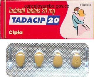
Tadacip 20 mg discount without prescription
Pape and colleagues have defined borderline sufferers as patients with polytrauma and � An damage severity score of >40 factors in the absence of thoracic damage � An damage severity rating of >20 factors with thoracic damage � Polytrauma with an abdominal and pelvic trauma (Moore grade >3) and hemodynamic shock with blood strain < 90 � A chest radiograph displaying bilateral lung contusions � An initial mean pulmonary artery pressure of >24 mm Hg � An increase in pulmonary artery strain of >6 mm Hg during nailing erectile dysfunction pills that work 20 mg tadacip with visa. Among different elements icd 9 erectile dysfunction nos cheap 20 mg tadacip, thoracic trauma appears to play a vital role on this predisposition. However, whether femoral fractures in patients with chest trauma should be treated with definitive stabilization or ought to be stabilized with a brief external fixator remains a subject of debate. The scientific situation, together with the presence or absence of a criterion indicating borderline standing and components associated with a high risk of antagonistic outcomes, ought to determine how the patient is handled. Markers of Inflammation Inflammatory markers might maintain the important thing to identifying patients in danger for the event of post-traumatic problems such as a number of organ dysfunction syndrome. Genetic Predisposition and Adverse Outcomes Biological variation and genetic predisposition are more and more talked about as explanations of why critical post-traumatic problems develop in some sufferers and never in others. Some individuals may be "preprogrammed" to have a hyper-reaction to a given traumatic insult. In addition to the second hit, which leads to a further systemic inflammatory response, embolic fat from use of instrumentation within the medullary canal will worsen the pulmonary status. Patients with a chest harm (an abbreviated injury score of >2 points) are most susceptible to deterioration after an intramedullary nailing process. Lactic acid is a byproduct of anaerobic metabolism, which happens throughout local tissue hypoxia. Serum lactate measurements have been proven to correlate with tissue perfusion and reversal of the shock state. Several studies have revealed that occult hypoperfusion, outlined as elevated blood lactate levels without indicators of scientific shock or serum lactate levels equal to or greater than 2. Early intramedullary fixation of femoral fractures in polytrauma sufferers, who had occult hypoperfusion, was related to a high incidence of postoperative issues. Geriatric Trauma Elderly trauma patients require particular evaluation and remedy because of their larger mortality price following trauma, even comparatively much less major trauma. During this period, marked immune reactions are ongoing and increased generalized edema is noticed. A latest potential research demonstrated that multiply injured patients subjected to secondary definitive surgery between days 2 and 4 had a considerably increased inflammatory response compared with that in sufferers operated on between days 6 and 8 (p < 0. Evidence Suggesting Efficacy of Damage Control in Orthopedics Study published from Hanover, eleven Germany, a retrospective analysis, studied trauma patients throughout three different timeperiods (Flow chart 1). In the early total care period, the protocol for the remedy of a femoral shaft fracture was early definitive stabilization (within lower than 24 hours). In the intermediate period, the standard protocol for treating a femoral shaft fracture in a multiply injured affected person at risk for post-traumatic problems modified from early definitive stabilization to early momentary fixation. Practical Considerations for Damage Control Orthopedics the use of spanning external fixation carries the chance of pin-track infection. Self-drilling pins, which can be manually inserted, could be utilized rapidly with a restricted need for fluoroscopy. Operating time could be decreased by multiple operating groups engaged on reverse ends of the identical limb or on completely different extremities. External fixation systems that employ snapand-click clamps could be assembled quickly. In addition, a system that enables flexibility in pin placement is preferable in order that areas of future incisions may be prevented. Biochemical changes after trauma and skeletal surgery of the lower extremity: quantification of the operative burden. Stimulation of the inflammatory system by reamed and unreamed nailing of femoral fractures. Damage control: an approach for improved survival in exsanguinating penetrating belly damage. Changes within the management of femoral shaft fractures in polytrauma sufferers: from early whole care to harm management orthopedic surgical procedure. Major secondary surgical procedure in blunt trauma patients and perioperative cytokine liberation: determination of the clinical relevance of biochemical markers. Optimal timing for secondary surgery in polytrauma patients: an evaluation of four,314 seriousinjury circumstances. The timing of fracture treatment in polytrauma patients: relevance of injury management orthopedic surgical procedure. A longitudinal, prospective and observational research of the procedure-related impact on cardio pulmonary and inflammatory responses. It is most likely going that we shall be able to distinguish people who discover themselves prone to produce severe response and will have the flexibility to manage them more aggressively. Overview Damage control orthopedic surgery requires an understanding of the physiological condition of the patient, their response to the first hit of damage and utility of orthopedic stabilization for the lengthy bone injuries based mostly on the necessity to reduce the second hit or surgical insult, and thus improve the physiological response of the patient to their accidents. Hence in lower thoracic trauma both diaphragm or abdominal organs are presumed to be injured until proved otherwise and vice versa. Similarly in lower belly and perineal trauma pelvic organs are likely to be involved. Additionally abdomen is crowded with viscera with variable structural patterns a few of them when injured both bleed profusely or contaminate the peritoneal cavity with extremely pathologenic organisms. All the major vessels run vertically in the midline and therefore midline penetrating injuries are extra critical than these within the flanks. There are sure peculiar pathological processes following belly trauma like "abdominal compartment syndrome" which leads rapidly to metabolic disturbances and a quantity of organ dysfunction or failure. Unrecognized damage to intra-abdominal organs stays a distressingly preventable explanation for demise. Geography and Demography Geographical distribution of blunt and penetrating accidents is variable. Penetrating injuries mainly because of stabs and gunshots are more frequent in city areas and regions infested with terrorist, whereas blunt accidents mainly because of falls and blows are more common in rural areas. However with speedy industrialization, urbanization, elevated variety of vehicles, straightforward availability of crude weapons and various conflicts in societies, ethnic teams and nations any sort of damage can happen anyplace in nation and medical persons must remain alert and educated to present pressing remedy. Regional hospitals must be outfitted to deal mass causalities and be familiar with rules of triage and rapid transportation. Prevention Public training, governmental infrastructure insurance policies and authorized provisions play a considerable role in preventing some accidents together with belly ones. Prehospital triage scheme of trauma sufferers from the American College of Surgeons Committee on trauma based mostly on the AbdominAl TrAumA presence of physiologic derangement, specific anatomical injuries, mechanism of damage and comorbid conditions along with the assets and services available must be followed. The distance to special middle and methods of transport available also wants to be thought of. The second mechanism is as a outcome of of penetrating objects like bomb fragments, bolts, nuts and steel pellets included in bomb inflicting indiscriminate harm to a number of organs. The tertiary mechanism is due to the sufferer being propelled towards an object by blast wind and/or native burn. Classification of Injuries and Mechanisms Abdominal trauma is classed in accordance with the sort and mechanism of damage: � Blunt trauma � Penetrating trauma � Blast injury � Iatrogenic harm. Blunt trauma is as a end result of of speedy deceleration and exhausting influence over the belly wall disrupting the intra-abdominal tissues via the results of crushing, bursting or shearing. Noncompliant parenchymatous organs like spleen, liver and kidneys are more vulnerable to injury then follow viscera, typically leading to massive bleeding and shock.
Tadacip 20 mg purchase line
The heart of the femoral head is positioned where the distance to the medial border of the femoral head is identical as half of the longitudinal diameter erectile dysfunction can cause pregnancy tadacip 20 mg discount with mastercard. The mid-width of the talus and the mid-width of the ankle measured clinically yield the identical point (Source: Modified from Moreland et al erectile dysfunction at age 20 purchase tadacip 20 mg with visa. Connect two factors at either finish of the ankle plafond line; (B) Ankle joint orientation line, sagittal airplane. Connect two points from anterior to posterior lip of joint; (C) Proximal tibial knee joint orientation line, frontal plane. Connect two factors on the concave side of the tibial plateau subchondral line; (d) distal femoral knee joint orientation line, frontal plane. Connect the two anterior and posterior factors the place the condyle meets the metaphysic. For kids, this is drawn the place the expansion plate exits anteriorly and posteriorly; (g) Hip joint orientation line, frontal airplane. For instance, sagittal plane orientation angles usually refer to the anatomic axis as a end result of mechanical axis strains are rarely used within the sagittal plane. The joint edge ratio (JeRs) for the sagittal aircraft (Source: � Springer-Verlage Berline, Heidelberg 2003). The intersection of the anatomic axis with the joint line is pretty constant and is important in understanding normal alignment and in planning for deformity correction. The distance from the intersection point of anatomic axis lines with the joint line may be described relative to the center of the joint line or to considered one of its edges. Mechanical Axis and Mechanical Axis deviation3 Alignment refers to the collinearity of the hip, knee, and ankle. Orientation refers to the position of each articular surface relative to the axes of the person limb segments (tibia and femur). Although normal alignment is commonly depicted with the mechanical axis passing by way of the middle of the knee, a line drawn from the middle of the femoral head to the center of the knee. Normal axial alignment of the decrease extremity and load-bearing distribution on the knee. For standing radiographs, the radiography technologists are usually taught to place the patient with the feet collectively within the "stand at consideration" posture. The appropriate method is to orient the patella ahead, regardless of the foot place. To orient the patella ahead, really feel the patella with the index finger and thumb of 1 hand and rotate the foot till the patella is pointing ahead. The radiograph confirms the right place, exhibiting the patella centered between the femoral condyles. In full extension, the patella is often centered on the femoral condyles, even in sufferers with patellar instability. However, patients with giant amounts of distal femoral valgus usually have true lateral patellar subluxation in full knee extension. Because the frontal aircraft of the knee forward place is kind of the identical as the plate of the knee flexion-extension axis, the latter can be used to position the limb in the frontal airplane. The airplane of the knee flexion-extension axis is approximately 3� externally rotated to the frontal aircraft. The limb is positioned in order that the x-ray beam is perpendicular to the flexion-extension axis of the knee. The knee joint axis is parallel to the x-ray movie cassette CorreCtions of Deformity of Limbs orientation angles. Alternatively, two separate films can be obtained: one of the tibia alone and the other of the femur alone. To make certain that the beam will cowl the complete bone length, it could be necessary to angle the beam generator diagonally. Long radiographs ought to be obtained with the radiography tube at a distance of 10 ft (305 cm) from the movie. Magnification on a 51 in (130 cm) cassette at 10 ft is roughly 4�5%, in contrast with 10�20% for radiographs obtained on 17 in (43 cm) cassettes at a more in-depth distance. A magnification marker positioned in the mid-sagittal axis of the limb can be utilized to measure the precise magnification factor. These compensatory mechanisms trigger uneven loading of the limbs and should alter the alignment and leg length measurement on the radiograph. When separate radiographs of the femur or tibia is obtained, it could be very important specify where to middle the beam. To higher assess the joint orientation of the proximal tibia or the distal femur, the radiographs should be centered on the knee. To see the proximal femur, the pelvis is rotated posteriorly 30�45� without rotating the knee on the research side 143. Angular deformity of the femur or tibia includes angulation not only of the bone additionally of its axes. Where there was one axis line to symbolize the bone, there are actually two axis strains: proximal and distal. This step is labeled step 0 as a reminder that it comes before any step within the preoperative planning course of. It is performed before tibial and femoral mechanical and anatomic axis planning of frontal airplane deformities. If one side is taken into account normal, its angles and distance can be utilized as templates for the deformed aspect. The distal tibial mechanical axis line is drawn as a line extending proximally from the middle of the ankle at the template angle to the ankle joint line. The distal tibial mechanical axis line is drawn from the center of the ankle at an angle 90� to the ankle joint line. Magnitude of angulation (Mag) is measured between the proximal and distal axis lines. The knee and ankle are normally oriented to the proximal and distal axis traces, respectively. In the previous case, draw a 3rd line corresponding to the mechanical axis of the mid-tibia. Start on the distal axis line on the degree of the plain apex, and draw the third line parallel to the tibia. Measure the magnitude of angulation of the angle between the plafond axis line and the distal tibial mechanical axis B. This axis line is drawn starting with a degree on the obvious apex on the axis line that passes at the degree of the obvious apex. A third line is drawn on the template angle from the middle of the ankle joint line. This rule applies to the lower and higher extremities from the humeral and femoral necks distally. CorreCtions of Deformity of Limbs this might be mentioned separately in reference to slipped capital femoral epiphysis deformities. Translation of bone ends leads to lack of bone contact and delicate tissue disruption. In distinction, angulation results in stretching of soft tissues with maintenance of bone contact.
Syndromes
- The quadriceps tendon (where the thigh muscles attach to the top of the kneecap)
- Fluids
- Does it go away without self care?
- Hematoma (blood accumulating under the skin)
- CT or MRI of the brain
- The baby begins to store fat.
- Dizziness
- Abnormally foul-smelling stools
Tadacip 20 mg buy generic
Further follow-up after 20 weeks revealed ache on strenuous work cheap erectile dysfunction pills uk purchase tadacip 20 mg mastercard, but there was no avascular necrosis of the lunate erectile dysfunction causes high blood pressure proven tadacip 20 mg. He discovered the hitherto unknown phenomenon in nature, namely, distraction histogenesis, 2. He developed the versatile ring fixator-this discovery constitutes one of the exceptional advances in the historical past of musculoskeletal analysis. The conditions by which he labored had been very primitive and he lacked many necessary medicines and devices. The new institute of orthopedics in Kurgan has turn out to be the biggest orthopedic middle on the earth, with 1200 beds, 350 orthopedic surgeons, 60 scientists with PhD degrees, and a staff of 1500 nurses, therapists, and ancillary personnel. Ilizarov Type1,three the second kind, the ring fixator (Ilizarov type) has clean, skinny (1. Cross Kirschner wires are used for circumferential multilevel, multiplanar, and multidirectional transosseous osteosynthesistransfixion of fractures, limb-lengthening, deformity correction, and so on. In most purposes, the half-pins are attached to the body work on one facet only. Ideal external fixator ought to have stability of fragments of the bone in alignment and on the same time should permit axial micromotion by its elasticity. Cantilever Type the conventional sort of external fixators with giant diameter (4�6 mm) stiff pins, generally threaded, fixed to bone in one airplane (uniplanar) act as cantilevers. Their advantages are as follows: � Minimal transfixion of sentimental tissue � Less bulky � Ease of assembling. They may turn into jammed � the forces, performing on the surface of bone are in a single plane as cantilever, causes angulations and deformity, on the fracture or nonunion or distraction web site. Cantilever loading creates increasing moments under larger masses that result in uncontrollable forces within the osteogenic zone. Counter forces are manually tough and mechanically unimaginable to effect Biomechanics of ilizarov ring fixator Thus, the fixator should have stability and elasticity. The Ilizarov technique obtains the stability and elasticity with this elastic materials by: � the wires are tensioned-the higher the tension on the wire, the larger is its solidity, on the expense of elasticity. In this manner, a linear traction pressure is replaced by a traction pressure performing on a plane, with the advantage of a much greater and extra even distribution of forces. This chance is peculiar to the Ilizarov methodology, in which the skinny Kirschner wires as fixators allow the installation of two wires on virtually equivalent planes. The disadvantages are as follows: � As the pin passes from one side of the limb to the other it transfixes the delicate tissues � Apparatus is cumbersome and time consuming to assemble and, � Steep learning curve. Thus, biomechanically ideas of two techniques of external fixators are different (Table 1). Pin passes from skin one aspect to other and transfixes the delicate tissue, thereby causes pain, joint stiffness subluxation or frank dislocation, and joint contractures 2. It is troublesome to assemble and takes lot of time, due to this fact preconstruction is by the patients. They are simple to look after in the course of the postoperative essential to save operating time. This function has simplified bodily remedy for the sufferers, elevated ambulatory capacity, and lowered the requirements of analgesic pain medication substantially 4. Forces acting on the surface of bone are in a single airplane as cantilever causes angulations and deformity on the fracture nonunions or distraction site. Cantilever loading creates growing moments under giant hundreds that lead to uncontrollable forces within the osteogenic zone. Counter forces are manually difficult and mechanically unimaginable to have an result on Does not allow immediate weight bearing and performance Advantages 1. They are elastic sort of exterior fixator and permit axial micromotion which are conducive to therapeutic of fractures and regenerate 2. Axial distraction or compression angular and translational corrections are all attainable using gradual mechanical technique Circular fixator is a stable fixator and elastic. These fixators permit quick weight bearing As the wires are skinny the holes are small Circular fixator can have capacity for three-dimensional correction. The Ilizarov system is prepared to management shear at the fracture site whereas allowing axial and bending dynamization producing an ideal environment for bone therapeutic. Wire stoppers add shear rigidity to the system Circular fixators are higher for sufferers with osteoporosis using wires Circular fixators can work in a septic environment allowing correction of limb length discrepancy because of aseptic pseudoarthrosis or following the removing of a secure implant 3. Another significant deficiency of monolateral fixators is their limited capability for correction of angular or rotational deformities and lack of bone substance. These frames are difficult to use when lengthening in the presence of a history of sepsis. Stuart Green has used titanium half pins with virtually negligible pin tract an infection. Patient acceptance is poor 1062 textBook of orthopedics and trauma Increasing the wire rigidity from ninety kg to one hundred thirty kg will increase the bending and axial stiffness however lowers the torsional stiffness. All the uni- and biplanar fixators show very excessive axial stiffness to axial loading. Multiplanar positioning of wires on all sides of the rings or introduction of extra wires further aside with the posts or accent rings/half rings will increase the soundness of the meeting. These wires act as small springs within the extra inflexible system of rings and threaded connecting rods. For any fixator system, there are two elementary interrelated issues, stability and rigidity. For hundreds to 100 N, the Ilizarov body is much less stiff in axial loading than other frames. This nonlinear habits of the Ilizarov body is attributable to increasing wire rigidity under loading. The Ilizarov frame is less stiff in bending, significantly within the lateral-medial course, but its values for lateralmedial and anteroposterior bending are related, bending stiffness will increase with rising axial loading. Large distances between rings can result in buckling or elevated torsional displacement of the frame. The dimension and configuration of the half pin techniques are essential factors to preserve the body in an enough stability. Biomechanics of the Wire4 Tensioning of the Wire the tensioned wires are thought-about nonlinear, self-stiffening pins. However, they preserve the elasticity and low axial stiffness to axial loading, 75% decrease than typical fixators. It is attention-grabbing to notice that the rise in stiffness is nonlinear (rate of improve decreases with growing tension). Recently with the arrival of the new calibrated wire tensioning device (dynamometer), particular pressure can be achieved. The Lecco group routinely uses one hundred thirty kg pressure for full rings and ninety kg for half rings. While theoretically one might count on an increased wire fracture fee, this has not proved to be the case clinically. While increased tension provided increased stability, by growing the wire stiffness, it also decreases the axial excursion of the wire on loading.
Cheap 20 mg tadacip free shipping
The blocks required depend on the positioning of the surgical procedure and the necessity for a tourniquet erectile dysfunction tea tadacip 20 mg buy on-line. Three-in-one blocks (femoral plexus block) could also be used for knee arthroscopy offered that prolonged tourniquet occasions could be avoided erectile dysfunction treatment nj tadacip 20 mg generic mastercard. Tourniquet may be deflated solely after a minimum period of 45 minutes, and the anticipated surgical period should be inside 1�1. Risk of native anesthetic toxicity is significant, especially throughout injection (leak underneath the cuff) and after launch of the tourniquet, as a end result of a potentially poisonous dose is intentionally positioned intravenously. Epidural and subarachnoid (spinal) blocks are major regional anesthetic strategies for surgical procedure involving the decrease half of the physique. Regional anesthesia (spinal and epidural) provides a number of benefits over common anesthesia. However, intraoperative anticoagulation with heparin seems relatively protected if epidural catheters are inserted 2�3 hours prior to anticoagulation. The common peroneal nerve could be anesthetized because it programs superficially under the pinnacle of the fibula, or both branches of the sciatic nerve could be blocked in the popliteal fossa. In any surgical procedure involving the medial facet of the foot, the saphenous department of the femoral nerve must be anesthetized either on the level of the ankle or maybe larger up. For ankle blocks, Esmarch bandages utilized instantly above the ankle allow a minimum of 2 hours of surgery to be carried out with out tourniquet ache. Because hypotensive anesthesia reduces blood loss intraoperatively, it reduces the necessities for blood transfusion. Preoperative autologous blood donation and cell-saver methods may scale back transfusion necessities. This creates a possible V/Q mismatch with resultant hypoxemia, an issue that appears most frequently in sufferers with underlying lung illness. The lateral decubitus place can create neurovascular problems as well as because the dependent shoulder presses on the axillary artery and brachial plexus, and the anterior stabilizing post compresses the femoral triangle. These issues can be minimized by placing an axillary roll beneath the upper thorax and by careful positioning of the anterior stabilizing submit on the dependent groin. Hypotensive anesthesia has been shown radiographically to enhance the standard of cement-bone fixation, as it reduces bleeding from bone. Although rare, this could occur from traumatic needle insertion, infection, epidural hematoma, spinal cord or cerebral ischemia. Headaches after spinal blocks are more widespread in younger female patients and with the use of giant gauge needles. The headache is completely relieved by epidural injection of 10�15 mL of freshly drawn autologous blood. This technique is gaining popularity and is the tactic of choice in all surgical interventions of the decrease extremities. G), increased incidence of postspinal headache, transient neurological impairment and cauda equina syndrome. In extended procedures, the discomfort of the conscious patient in an immobilized place is prevented. Regional anesthesia reduces the amount of general anesthesia needed and thereby reducing the excessive cardiac depressant effect of general anesthetic. Postoperative regional analgesia is offered utilizing opiates with or without native anesthetic. Intraoperative Hypotension Profound hypotension instantly following insertion of cemented femoral prostheses has resulted in cardiac arrest and dying. Therefore, it seems doubtless that hypotension is said ultimately to the utilization of cement. Attempts to reduce this complication have included (1) using a plug in the femoral shaft to limit the distal spread of cement in the femur, (2) venting of entrapped air, and (3) ready for cement to turn out to be extra viscous earlier than its insertion. Two possible explanations are that (1) it could be brought on by direct vasodilatation and/or cardiac despair from methyl methacrylate, or (2) it may be due to the compelled entry of air, fat, or bone marrow into the venous system with resultant pulmonary emboli. Large echogenic emboli have been described following insertion of femoral prostheses; this helps the concept that the circulatory collapse is embolic quite than from a poisonous impact of the methyl methacrylate. The emboli could induce a launch of vasoactive substances from the lung, which may contribute to circulatory collapse. Hypoxia has been described immediately following insertion of a cemented femoral prosthesis and for as much as 5 days into the postoperative period. In the event of hypoxemia, one should first ascertain whether or not it has a selected cause such as atelectasis of the dependent lung, hypoventilation or fluid overload. Complex procedures corresponding to those involving acetabular bone grafting, insertion of a long-stem femoral prosthesis, removing of a prosthesis, revision surgical procedure, or surgical procedure in patients with acetabular protrusion (which entails a threat of coming into the pelvic cavity and/ or the iliac vessels) complicate the administration of the anesthetic. Fluid administration should be carefully managed throughout this type of in depth surgery. AnesthesiA in OrthOpedics surgical procedure and is believed to be secondary to the embolic results of femoral shaft cement or fats embolism. Postoperative management should embody nasal oxygen and pulse oximetry (if necessary for several days), considered use of narcotics to provide analgesia and but avoid hypoventilation or airway obstruction and appropriate fluid management. Hypoxia and fluid overload could additional improve pulmonary pressures and thus improve the probability of pulmonary edema or right heart failure. Because of the added surgical stress, invasive hemodynamic monitoring lasting 24�48 hours must be thought-about for sufferers (particularly aged or infirm patients) undergoing bilateral procedures. Bonecement:When acrylic cement is utilized to the cavities of the tibia, femur, and patella, acute hemodynamic responses seldom comply with. Such responses do occur, however, when long-stem femoral prostheses are inserted following in depth femoral reaming. Lesser levels of femoral reaming may scale back the incidence of embolic events, but the significance of those events is unclear. Patients present process bilateral procedures are at further risk of turning into hypovolemic through the first few hours after the operation. Preoperative autologous blood donation can minimize homologous transfusions on this setting. Patients with congenital scoliosis may have congenital heart illness, airway abnormalities and preexisting neurological deficits. Patients with neuromuscular disease similar to muscular dystrophy, poliomyelitis, dysautonomia, spinal twine harm, and neurofibromatosis may develop scoliosis. Perioperative issues embrace intraoperative positioning, spinal wire monitoring, minimization of blood loss, prevention of postoperative hyponatremia and postoperative respiratory care. Nowadays, many of these patients bear both anterior and posterior procedures, which may be staged or performed underneath one anesthetic and which regularly involve a thoracotomy. Particular attention should be focused on positioning of the neck, arms, and eyes to defend pressure factors adequately, particularly if hypotensive anesthesia is to be used.
Cheap 20 mg tadacip amex
The reported complication rate of megaprosthesis stays 5�10 times higher than the rates observed in routine complete joint arthroplasties erectile dysfunction tadacip 20 mg cheap mastercard. These are largely due to the extended bone and gentle tissue defects necessitating massive dimension implants resulting in abnormal biomechanical loading of the complex reconstruction erectile dysfunction radiation treatment 20 mg tadacip for sale. Complications include mechanical failures such as breakage or fracture of the implant and aseptic loosening. Wound infection and gentle tissue breakdown requiring additional treatment and sometimes revision of prosthesis are additionally not unusual. Custom-made prostheses are individually manufactured for every patient to give an accurate match; nonetheless, as they should be specifically ordered and manufactured, delays can occur. In kids or in places where the anatomy is distorted, personalized implants have the advantage to modify for smaller or abnormal bone dimension and to enable expansion (expandable prosthesis). Modular methods are available off the shelf and permit variable resection because of their multicomponent design. Modularity allows intraoperative flexibility based on the ultimate amount of tissue 100 Chapter Role of Allografts and Bone Substitutes Rajesh Malhotra, Ravijot Singh Introduction There are numerous methods by which the defect left behind after surgical excision of bone tumors may be reconstructed. They can be used as fillers, to present mechanical stability and assist improve reattachment of biological host tissue. Depending upon the type, measurement and the placement of defect, allografts can be used within the following forms: � Osteoarticular allografts � Intercalary allografts � Allograft-prosthesis composites � Morselized allografts � Allograft struts. Tables 1 and 2 outline the indications of use of huge allograft replacement after resection in higher and decrease limbs respectively. Intercalary Allografts � Intercalary segmental allografts: Tumors confined to metaphyseal or diaphyseal areas of bone could additionally be resected with broad margins, but preserving the joint. In such instances, intercalary segmental allografts can be fastened to the periarticular host bone, thus attaining the limb size and stability and preserving the joint function. This technique avoids the complications related to osteoarticular allografts, similar to chondrolysis and joint instability. Allograft-host junction is ready with a transverse or stepcut osteotomy and intramedullary nail, or, plate and screws can be used to achieve fixation. Impregnation of the medullary canal of the allograft with cement and/or reverse cortical allograft strut could also be used to present more steady fixation when using plates. A higher price of nonunion for diaphyseal junctions (15%) than for metaphyseal junctions (2%) has been reported. Thus, the structural advantages of the allograft are coupled with vascular and osteogenic capabilities of the vascularized fibular graft in order to obtain decrease charges of infection, fracture and nonunion. Satisfactory allograft incorporation with no local recurrence has been reported when utilized in sufferers with low-grade malignant tumors. The allografts are then fastened to the host bone using an acceptable fixation technique relying upon the location. The delicate tissue across the allografts is used to attach the host tendons and ligaments. Meticulous reconstruction of the ligaments, tendons, and capsule should be accomplished because the longevity of these grafts relies upon upon the steadiness of the joint to a big extent. Malalignment must be avoided because it subjects the allograft cartilage to greater mechanical stresses. These embrace resorption of the graft, delayed union or nonunion on the host-graft junction, chondrolysis, bone graft fractures, joint or junction instability and anatomic mismatch between allograft and the host. Most of these issues can be prevented to a big extent by reaching enough joint stability, anatomic matching of the articular surfaces, maintaining joint alignment and steady fixation of allograft to host bone. Histological research present that extra superior degenerative modifications occur within the allograft articular cartilage if the joint is unstable. In addition, anatomic mismatch between graft and the host articular surfaces can result in altered joint kinematics and irregular loading of the joint, thus giving rise to elevated fee of joint degeneration and bone resorption. Therefore, chemotherapy may delay union because of its motion towards the osteoblasts. As within the case of whole condylar allografts, anatomic match and gentle tissue reconstruction stay important. In addition, the surgical method is sort of demanding, as improper placement of the graft would lead to inappropriate loading of the joint and deformity. Furthermore, the ligament balancing is tougher, as the ligaments on one side of the joint are regular. In case of lengthy bones, allograft is prepared to the required length on a separate sterile table at the time of tumor resection and a step-cut osteotomy is common at the graft-host junction. The allograft is ready in order to accommodate the suitable size of the prosthesis into it. It is then docked onto the host bone and the ligaments and muscle attachments secured. Augmentation of the fixation with a plate, cables or allograft struts may be accomplished (if required). If successful, the useful outcomes are higher than a flail hip or an arthrodesis. However, due to scarcity of data out there and high complication charges, together with mechanical failure and an infection, their use remains to be debatable. Additionally, they noticed, that the gentle tissue adherent sleeve formation around an allograft is very important in stopping late allograft an infection, which is more frequent in metallic implants. The use of impacted morselized autograft in a cage has been described in animal model for reconstructing segmental diaphyseal defects along with intramedullary nail fixation. Nevertheless, allograft struts have been used to augment the fixation of intercalary segmental allografts and allograft-prosthesis composites to the host bone. Complications of Reconstructions utilizing Allografts in Musculoskeletal Tumors Infection An an infection rate of 8. Failure of implant following main reconstructions in 70% circumstances and amputation in 15% instances has been reported to end result from infection. Meticulous bone banking, which incorporates techniques of harvesting, preservation and sterilization, is understood to reduce the incidence of infections. Note that the lesion has healed and the allografts have included reconstructions, reduces the fracture fee remarkably. Significantly higher charges have been reported in chemotherapy handled patients (32%) in comparison to nonchemotherapy patients (12%). Recently, the deficiency of vascular endothelial growth issue and receptor activator of nuclear factor kappa-B ligand within the allografts has been recognized to be a explanation for this nonunion. Allograft sterilization using gamma- 688 TexTbook of orThopedics and Trauma are cheap to be used in benign tumors. They reviewed the prevailing literature to facilitate the decision-making concerning their use in orthopedic apply. The authors found scientific data obtainable for under 22 merchandise (37%) and nonetheless fewer had Level I proof. Hence, the need for thus many different merchandise, particularly with limited printed clinical evidence for their efficacy was questioned.
Generic tadacip 20 mg otc
Alternatively erectile dysfunction drug related tadacip 20 mg discount fast delivery, measurement could additionally be taken with a tape from anterior superior iliac spine to medial joint line of knee what causes erectile dysfunction in 30s tadacip 20 mg buy discount line, medial malleolus and plantar surface of heel. This simple method has a quantity of advantages: First, because it encompasses the entire limb, the orthoroentgenogram allows assessment of angular deformity and bony pathology which may in any other case be missed. Teleradiograph: Teleradiograph is a single exposure of each legs on a protracted film 14 � 36 inches, taken from 6 toes distance with sufferers standing. Arthrogram-radiograph: In arthrogram-radiograph, on single long film three successive exposures are made, centered exactly over the hips, knees, and ankles. The target-to-film distance is 6 ft, each exposure including about one-third of the whole lower limb. The advantages are: (A) True length of the every bone could be measured, (B) the complete size of each lower limbs from the iliac crests to the soles of the feet, with excellent detail of bone and delicate tissue throughout, (C) technically, the procedure is straightforward, (D) radiations to the affected person is minimal. Disadvantages are: It may be very cumbersome and wishes a quantity of exposures causing risk of errors if affected person strikes. Certain precautions must be taken: (A) the tube should be centered over the articular ends of the long bones. This technique is straightforward, accurate, visualizes the whole pelvis and lower limb, and the scans are straightforward to retailer. Currently, three techniques are used: (1) orthoroentgenogram, (2) the scanogram, and (3) computerized digital scanogram. Motivation is important as a result of leg lengthening is demanding on the patient and the dad and mom. It is important to give realistic expectations of surgical procedure and clarify the problems that can happen. Tupman (1962) observed the growth in British boys and girls between the age of 8 and 14 years. Patient have to be explained all of the complications, ache and ordeal that he or she has to bear. In a normal lower limb, between the age of four years and maturity, the femur usually increases its total size by 2 cm per year, whereas the common fee of development of the tibia is 1. Growth of the whole limb is as follows: Distal femoral physis-35% Proximal femoral physis-15% Proximal tibial physis-30% Distal tibial physis-20% Thigh length: 70% contribution by distal, 30% by proximal epiphyseal plate. Table 4: Ready reckoner for the growth of femur and tibia in girls and boys eight Girl-years Femur Tibia Boy-years Femur Tibia 7. They have additionally supplied a ready reckoner for the remaining progress at varied ages in boys and girls (Table 4). The fundamental insurance policies of the graph are: (A) the growth of the legs can be represented by a straight line by suitable manipulation of the abscissa; (B) the size of the longer extremity is represented by a straight line because of the strategy of plotting factors; (C) the growth of the short limb can be represented by a straight line which lies beneath the road of the longer limb and may have a different slope; (D) the discrepancy is represented by the vertical distance between the two lines; (E) the proportion inhibition of growth of the short limb is represented by the difference in slopes of the 2 traces, designating the conventional slope as 100%. The Menelaus rule of thumb (Australian) method: the tactic predicts development of 10 mm per yr (three-eight inch) at the distal femoral physis and 6 mm per 12 months at the proximal tibial physis, with growth terminating at age of 14 years for women and sixteen years for boys. The Menelaus method is straightforward, easy to calculate, and supplies a rough estimate as to the timing of epiphysiodesis. The method of computerized calculations: the skeletal age, limb length and calendar date are wanted to enter in the computer. The application-based technique is an easy, easy-to-use method for sophisticated calculations, which had been sometimes performed using hand with the assistance of assorted formulation. This software reduces the time needed for these tedious calculations and likewise considerably reduces human errors. Treatment of Limb Length Discrepancy General Principles It is typically advisable to appropriate coexisting deformities before correcting leg length discrepancy as a outcome of the correction of some deformities modifications the remedy goal. This graph shows the amount of development potential remaining in the growth plates of the distal femur and proximal tibia of girls and boys as capabilities of skeletal age. It is beneficial in determining the amount of shortening that may end result from epiphysiodesis the selection of therapy technique is determined by the magnitude of predicted discrepancy at maturity than on etiology. There are four strategies of correction of limb length inequality: (1) stimulation development of the shorter limb, (2) retarding the growth of the lower limb, (3) operative shortening of the longer limb, and (4) operative lengthening of the shorter limb. The first two methods act by influencing the growth at physis of the bones of the shorter or longer limbs and therefore to be efficient, want enough time before closure of physis. Various strategies were tried like lumbar sympathectomy, periosteal stripping, insertion of overseas materials like ivory peg or metal near the epiphysis and even two different metals to produce electrical energy. Though some lengthening was claimed by each technique, useful elongation was by no means achieved, to not speak of managed or predictable gain in size. Retardation of Growth Arrest of progress could additionally be temporary by epiphyseal stapling or permanent by epiphysiodesis. Growth arrest by stapling relies on "Hueter-Volkmann regulation" (1862, 1869) which states that elevated pressure together with long axis will inhibit, and diminished stress will accelerate bone development. This was further corroborated by Hass (1945, 1948) and Arkin and Kartz (1956) each in animal experiments and likewise clinically. The baby later developed bow leg because the dad and mom refused a second arrest by staples. Stimulation of Bone Growth the primary written account of growth stimulation is by Pare who used mild venous congestion. Therefore, repeated assessment each 3�6 months for 2�3 years immediately preceding the contemplated operation is mandatory. For epiphysiodesis, an oblong piece of cortex is faraway from metaphysio-epiphyseal region on both sides taking extra of metaphysis. Percutaneous epiphysiodesis has been successfully carried out and leaves a smaller scar. Except for the scar it has no benefit over conventional operation, but needs subtle equipment. The removed piece of bone is replaced on both sides Staples are removed after the desired shortening is obtained- whether or not complete equalization or little wanting it. Arrest could also be everlasting if the staples are retained for too long (more than three years), or due to subperiosteal insertion or improper handling throughout insertion or removal. Blount demonstrated radiological thickening of epiphysis after removing of staples. Complications common to both the operations are undercorrection, overcorrection and angular deformities like varum, valgum, and recurvatum. In stapling, further problems are breakage, migration and widening of the staples. One of the most illustrative papers is that by Green and Anderson who performed each the operations with high diploma of success. They concluded that both the operations are good and efficient, epiphysiodesis is particular and ultimate, but if prediction is wrong or progress irregular, the result may be awful. Stapling has a little higher rate of complications, however these are of no nice consequence. Resumption of development after removal of staples is the massive advantage of stapling operation. Complications are little extra frequent and outcomes little much less predictable after upper tibial stapling than after lower femur. With the appearance of eight plate epiphysiodesis described by Peter Stevens in 2007, the difficulties of contralateral epiphysiodesis confronted due to the staples have decreased significantly.
Hydrolysed Collagen (Gelatin). Tadacip.
- A kind of arthritis called osteoarthritis, osteoporosis (brittle bones), strengthening bones and joints, strengthening fingernails, improving hair quality, weight loss, shortening recovery after exercise and sports-related injury, and other conditions.
- What is Gelatin?
- How does Gelatin work?
- Are there safety concerns?
- Dosing considerations for Gelatin.
Source: http://www.rxlist.com/script/main/art.asp?articlekey=97003
Tadacip 20 mg buy with amex
The bodily examination should consist of remark of stance and gait erectile dysfunction in teens proven 20 mg tadacip, any deformity erectile dysfunction treatment bangalore generic 20 mg tadacip free shipping, dysplasia of the joints of the limb, range of movement and stability must be noted. Neurovascular examination must be fastidiously conducted for muscle strength, reflexes, sensation, and peripheral circulation. Children with decrease limb inequality should be assessed clinically and radiologically. Clinical assessment is finest accomplished by placing picket blocks of gradually increasing peak beneath the shorter leg till pelvis becomes sq.. This offers an correct idea of shortening underneath regular strain of physique weight and takes into Limb LengTh discrepancy account the peak of the foot additionally. Eight plates supply a less morbid and completely reversible technique of contralateral epiphysiodesis. Age of arrest is set by the projected quantity of discrepancy at maturity and the speed of lengthening of the shorter (usually abnormal) limb. With the subtrochanteric operation, no plaster immobilization is critical, and knee could be mobilized quickly. The patient is allowed on crutches after a few days and partial weight bearing may be allowed after few weeks. With the assistance of interlocking system, diaphyseal shortening with nail fixation can keep away from problems like nonunion and rotational deformity and can be safely done where necessary instrumentation and image intensifier is available. There are varied methods of femoral shortening similar to oblique osteotomy, step-cut osteotomy, overriding osteotomy, subtrochanteric or supracondylar osteotomy. Kuntscher (1965) instructed the thought of an intramedullary saw to resect a section of femoral diaphysis. Like Limb LengTh discrepancy a normal closed intramedullary nailing, reaming is done from above and a special intramedullary noticed is launched. Then the main fragments are fixed by nail, ideally, interlocking nail, the split pieces behave as grafts. This distinction in girths is especially noticeable, because the originally shorter side was already narrow (commonly as a end result of poliomyelitis), and the relatively cumbersome regular facet is made additional broad by the operative shortening. If the inequality is an excessive amount of for correction by femoral shortening alone, then tibial shortening can also need to be accomplished. The increased bulk because of bunching, whether or not in the thigh or leg is normally taken up in 1 or 2 years. The maximum shortening that might be carried out is 5�7 cm in femur and 2�4 cm in tibia relying on the peak of the individual. Therefore, one can pass drill bits through the medullary canal to make 4�5 holes in the posterior cortex. If the lengthening is to be carried out greater than 6�8 cm, then two ranges lengthening is recommended as in a case of achondroplasia. The progression of histogenesis is managed by mechanical factors (stability on the site of the bony separation and the rhythm of distraction and biological elements (local osteogenic potential and vascularity of the bone). Orthofix Device for Limb Lengthening In many centers the orthopedic surgeons prefer the orthofix for physiological, biomechanical, and technical causes. Bone formation achieved with the orthofix seems to equal to that of the ringed gadgets employing the method of Ilizarov. Advantages of the orthofix compared with ring fixators include ease of application, decreased threat of neurovascular damage, minimal muscle transfixion, ease of radiographic evaluation, workplace removing, and patient acceptance. Segmental bone transport and correction of angular and rotation deformities related to leg length discrepancies can be handled with orthofix. The normal articulated system may be used for lengthening that requires simultaneous correction of angular deformity. A slotted lengthening system for bilevel lengthening and segmental bone transport. Dynamization may be achieved by releasing the locking screw of the telescopic physique. According to Price, axial loading of bone facilitates bone therapeutic and reduces the stresses at the pin bone interface. This lower in pin-bone stress may have theoretic advantages with regard to decreased pin loosening and decreased danger of late fracture through the screw holes after gadget removing. Six pins, three into proximal fragment and three within the distal fragment present a superb stability. Technique (Price): the extra suitable straight lengthener without ball joints ought to be used every time attainable. If any angular or rotational correction is necessary, the articulated gadget may be employed, but it is strongly recommended that methyl methacrylate be used to stabilize the ball joints to prevent angulation of the fixator. Slightly convergent screw placement can be used to forestall valgus in the femur and varus in the tibia. Ilizarov contended that the marrow blood supply is critically essential for regenerate maturation and ossification. For this purpose, he was cautious to avoid transection of the medullary vessels when he carried out a cortical osteotomy. For this cause, a selection of authorities have began to lengthen limbs with a medullary nail in place. The nail serves a monitor to help in stabilizing the lengthening bone and forestall translational deviation or angulation of the separating fragments (Ilizarov, himself, has on occasion used a big diameter wire for the same purpose). In this manner, the patient want not to wear an external fixator during the "impartial fixation" phase of regenerate bone ossification. To stop pin site sepsis from contaminating the intramedullary nail, Paley has developed a special alignment jig that permits a surgeon to place external fixator, half-pins behind an intramedullary nail within the trochanteric region of the femur flares outward. There is normally sufficient room for the insertion of halfpins a couple of millimeters anterior or posterior to the intramedullary nail on this area. The osteotomy for lengthening can be completed with the nail in place or by withdrawing the nail for the procedure. Indeed, intramedullary saw (originally designed for closed femoral shortening) can be utilized for the osteotomy. The rate and rhythm of distraction following insertion of an intramedullary nail and concomitant utility of an external fixator on the identical bone follows the identical old each 6 hour protocol designed by Ilizarov. When the desired size has been achieved, the affected person is taken again to the operating room the place the distal locking screws are inserted into the femur (under fluoroscopic control) and the frame removed. To stop contamination of the surgical site, throughout distal interlocking, Paley and his group now advocate inserting the distal transverse locking screws from the medial, quite than the lateral facet of the limb. It is opinion of the Price that the DeBastiani method is the procedure of choice for limb lengthening within the pediatric age group. In this manner, the surgeon will find a portion of the proximal end of the femur posterior to the intramedullary nail wide enough to allow insertion of 6 mm threaded half-pins. The pins could be inserted by first placing a guidewire within the correct location, and then drilling over this guidewire with a cannulated drill-bit. A drill sleeve should be used to protect the gentle tissues from the spinning drill.
20 mg tadacip purchase otc
An oblique radiograph obtained in the plane of the indirect plane part will show solely the anatomic airplane element erectile dysfunction treatment cream tadacip 20 mg purchase free shipping. An oblique aircraft radiograph obtained perpendicular to the indirect aircraft deformity will show the utmost deformity for that component of lesser magnitude to that measured within the anatomic aircraft young erectile dysfunction treatment generic 20 mg tadacip amex. Because both dreformities are in an indirect aircraft and because the oblique planes are less than 90� aside, 4 completely different oblique radiographs can be essential to present the utmost and minimum angulation and translation components, respectively. The oblique radiograph that exhibits the maximum translation would also show some angular deformity of lesser magnitude to the actual oblique aircraft angulation. The oblique plane radiograph obtained orthogonal to the earlier one shows the absence of translation deformity however the presence of angulation. The same is true for translation deformity on the radiographs obtained in and perpendicular to the oblique plane of angular deformation. Osteotomy Correction of Angulation-translational Deformities Correction of angulation and translation once they occur concurrently depends on the magnitude and aircraft of each deformity and its significance in that aircraft. For example, translation and angulation in the sagittal aircraft are much better tolerated than in the frontal aircraft and subsequently may not have to be corrected. The graph depicts the aircraft of angulation and translation, both are in the same anatomic aircraft. Closing or opening wedge correction at this level will concurrently correct both the angulation and the translation by means of a single bone reduce. Osteotomy by way of the purpose of most translation entails sequential correction of the angulation after which translation or translation and then angulation. The bone at the previous fracture degree is usually sclerotic, hypovascular, beforehand contaminated from an open fracture, and/or underneath poor soft tissue protection. The a-t point is normally a safer stage for osteotomy by way of a beforehand unhurt degree with good soft tissue protection and an open medullary canal. Opening, closing, or impartial wedge angular corrections can be performed at this degree. Straight or focal dome osteotomies at different ranges require angulation with translation of the osteotomy web site. When angulation and translation are in the same indirect aircraft, the correction may be carried out via the a-t level in the same manner as for anatomic aircraft deformities. The main disadvantage of corrections via the a-t level is the residual bump on the malunited previous fracture website. In the tibia, if the bump is on the subcutaneous medial border, it could be bothersome and esthetically displeasing. When the bump is on the medial subcutaneous border of the tibia, it is very obvious. With gradual correction, the reverse is preferred; (B) Closing wedge osteotomy-angulation first, then translation; (C) Opening wedge osteotomy-angulation first, then translation; (d) Opening wedge osteotomy-translation first, then angulation CorreCtions of Deformity of Limbs Both angulation and translation are corrected at this stage. This kind of correction lends itself to intramedullary fixation as a result of the medullary canal may be realigned. If this translation is significant, it ought to be corrected by translation of the osteotomy. In the sagittal aircraft, the limiting issue for this kind of correction is the bone-to-bone contact at the osteotomy website. The angulation is corrected in its plane, and the interpretation is corrected in its airplane. Because the correction is thru the original fracture area, realignment is associated with good bone to bone apposition. An understanding of the relationship between angulation and translation is important to the reduction of those deformities. Insignificant could refer to the magnitude of angulation and/or translation relative to the airplane during which they happen. The osteotomy is carried out to correct probably the most important component(s) of the deformity whereas accepting the much less important component(s). One aircraft of angulation correction is the frontal plane, and the opposite is the sagittal plane. In considering all bypass choices (strategies 1, 2, three and 5), one must take into accounts that a bump may stay regardless of accurate realignment. Strategies 2 and three the a-t point within the frontal (strategy 2) or sagittal (strategy 3) aircraft is chosen as the first deformity apex for the correction of angulation. For most bowing deformities, only a Multilevel fracture deformities follow the same planning steps as with multiapical or uniapical solutions. This leaves residual translation is corrected by translating the osteotomy in its aircraft. This leaves two bumps on the bone, one on the original fracture stage and one at the osteotomy website. This is probably the most sensible solution; (C) Angulation is corrected first, then translation; (d) Fracture reduction follows the identical technique as that offered in (B), translation first, then angulation. The correction will be carried out utilizing two osteotomies because the two apices are far other than each other. Angulation on the former osteotomy is performed for frontal airplane correction solely and at the latter osteotomy for sagittal aircraft correction only. There can also be a very gentle distal femoral valgus and a leg-length discrepancy; (B) the osteotomies were performed within the proximal metaphysis and in the mid diaphysis. The distal osteotomy is for the correction of the varus and the procurvatum deformities; the proximal osteotomy is for the correction of the juxta-articular valgus deformity. Notice the sample of the olive wires, which provide the necessary fulcrums and distraction points for this correction; (C) the equipment is proven in the quick postoperative period. In milder circumstances, the deformity could be resolved right into a single apex using the mechanical axis technique outlined. For multiapical deformities, osteotomy rule three is utilized as described within the section osteotomy concerns. This occurs when one of the deformities is diaphyseal and the other is juxta-articular. Usually a varus or valgus diaphyseal deformity exists with a compensatory juxta-articular angular deformity on the level of the proximal tibial physis. Notice that the bowing within the femur is diffusely distributed throughout the size of the femur, as is the bowing within the tibia. Notice additionally the lateral compartment joint laxity within the knee, which contributes the varus deformity; (C) the apparatus is shown throughout development within the operating room. The two gadgets need to be coordinated to enable at least 90� of free flexion of the knee. These are known as compensatory bowing defor mities as a end result of one deformity compensates for the opposite. Whether because of remodeling or ongoing a number of stress fractures, these deformities reveal no single or double apex.
Tadacip 20 mg discount line
In the backbone erectile dysfunction from stress buy tadacip 20 mg otc, eosinophilic granuloma causes a focal harmful lesion that sometimes includes the vertebral physique and pedicle erectile dysfunction caused by hydrochlorothiazide tadacip 20 mg buy mastercard. The spectrum of vertebral involvement ranges from a purely lytic lesion without collapse to partial collapse and even complete collapse. The other causes of vertebra plana are spinal tuberculosis, fungal infections, osteoporotic collapse and malignancy. Treatment of eosinophilic granuloma consists of statement, oral analgesics and the utilization of bracing for symptomatic relief. The situation is usually self-limited and the vertebral physique often reconstitutes over a period of 6�8 months. However, the vertebral physique collapse not often can lead to painful kyphotic deformity and neurological compromise and this is able to require decompressive surgical procedure with stabilization. Eosinophilic Granuloma Eosinophilic granuloma is a benign, self-limited situation of kids and younger adults. The different two members of the group are Hand-Schuler-Christian disease and Letterer-Siwe disease. MalignantTumorsofSpine Introduction Primary malignant bone tumors of the vertebral column are very rare. They are seen within the center to aged age group sufferers and infrequently present late in the course of the illness. The objective of treatment for sufferers with malignant main spine tumors is to present one of the best likelihood of native control mixed with acceptable systemic therapy. The extension of the tumor past the bone into the spinal canal and the important vascular tissues should be recognized. Oncological ideas of malignant tumor surgical procedure would necessitate extensive and radical excision even for spinal tumors. This includes elimination of the vertebral physique, anterior and posterior longitudinal ligaments and the intervertebral disc to keep away from leaving residual tumor behind. Theoretically, to get hold of a clear surgical margin in main malignancies, neural, muscular, and some vascular buildings may be sacrificed. However, such an aggressive approach results in significant morbidity and mortality although it offers one of the best chance for both native management and treatment of the illness. Surgical outcomes for malignant main spine tumors rely upon the kind of surgical process carried out and the surgical margin obtained. The 5-year survival rate the place only curettage has been carried out for primary malignant spine tumors is 0%. With complete resection, the 5-year survival fee has been quoted to be as high as 75%. Type B lesions begin in the concerned zone, but lengthen beyond the boundaries of the cortical bone. The finest surgical strategy is decided by the zones concerned and the extent of the native tumor spread. Osteosarcoma Approximately 2% of all major osteogenic sarcomas of the bone arise in the backbone. It expands rapidly throughout the bone and beyond the cortical margins into the adjacent soft tissues. Limited tumor excision and radiotherapy has a poor median survival of only a few months. A extra aggressive surgical strategy can outcome in longer survival times, of course with important morbidity. Improvements in adjuvant radiotherapy and chemotherapy protocols have offered improved survival charges. Patients often present in the second or third decade of life with axial pain in the low again area. These tumors normally arise from the vertebral body, but extension into the posterior components and canal can occur. If metastasis and lymphoreticluar malignancies are excluded, chordoma is the most typical tumor occurring in the backbone. The tumor has options of low-grade malignancy and is characterized by gradual, relentless local unfold and potential for distant metastases. Axial pain in the sacral area is the early symptom and neural dysfunction within the form of bladder and bowel involvement happens as the tumor expands. The tumor arises from the anterior sacrum and therefore chordomas can reach appreciable dimension earlier than stress symptoms of constipation, urinary frequency, or nerve root compression happen. Radiographs can reveal sacral destruction however the presence of overlying bowel shadows can simply impede the analysis. Surgical wide excision of the tumor with a cuff of normal tissue is the one healing process because the tumor is resistant to radiotherapy and chemotherapy. Adequate tumor resection ought to be the objective and will have priority over saving neural elements. If all sacral nerve roots could be preserved no much less than on one aspect, the patient can have regular bowel, bladder and sexual function. If nerve root resection is required bilaterally, preservation of the S2 roots might preserve partial urinary and fecal continence in some patients. Preservation of at least one S3 nerve root is required for preservation of bowel and bladder function in most patients. The kind of surgical strategy and the need for fixation of bone is dependent upon the extent of sacral involvement. Tumors that contain only the distal portion of the sacrum (S3 and below) can be treated with a single process from a posterior surgical strategy. Tumors that contain the S1 and S2 segments or people who involve the entire sacrum require a mixed anterior and posterior resection. Sacropelvic reconstruction after extensive resection could require multiple stages to complete, and morbidity is sort of significant. Therefore, a diverting colostomy and ureterostomy must be performed and clearly, success of this complex surgical procedure requires the help of a multidisciplinary staff. Radiographically, chondrosarcoma reveals massive areas of bone destruction and an associated delicate tissue mass with flocculent calcifications inside it. Complete surgical excision offers the only hope of tumor treatment and stays as a problem in vertebral lesions. En-bloc excision with removing of the overlying soft-tissue capsule, muscle, pleura or peritoneum, with a cuff of normal bone is crucial for a disease-free survival. Stabilization of collapsed vertebrae by way of both kyphoplasty or vertebroplasty supplies instant pain reduction for patients. SpinalMetastasis the spinal column is the commonest site of skeletal metastases. At least 5�10% of all most cancers patients develop backbone metastases during the course of their illness. Within the spinal column, the vertebral bodies are affected probably the most (85%) however metastatic deposits also can happen in the paravertebral region (10%), the epidural area (<5%) and the intradural space (<1%).
20 mg tadacip cheap free shipping
Now-a-days erectile dysfunction doctors in cleveland 20 mg tadacip buy otc, bioabsorbable implants show no difference within the stiffness pomegranate juice impotence order 20 mg tadacip with visa, linear load and failure mode when compared with metallic units. In the shoulder rotator cuff tears, shoulder instability, and biceps lesions that require labrum repair or biceps tendon tenodesis can be managed with these implants. Bioresorbable implants have been used as interbody spacers in lumbar interbody fusion; although the overseas body reactions and power currently nuclear for bioabsorbable cages, the early appearance of osteolysis associated with use of poly (L-lactideco-D, L-lactide) cages raises questions concerning their value on this situation. Bioabsorbable anterior cervical plates have been used and studied as alternatives to metallic plates when a graft containment gadget is required. Bioresorbable material use in pediatric conditions was perhaps the earliest recorded use in orthopedic literature. These have been used as self-reinforced absorbable rods for fixation of physeal fractures, in pediatric olecranon and elbow fractures, and as screws for fixation of subtalar extra-articular arthrodesis. Ankle fracture fixation is another area where self-reinforced absorbable rods have been efficiently employed. There are bioabsorbable implants now out there for use in humeral condyle, distal radius and ulna, radial head and different metaphyseal areas. Bioabsorbable implants are also variously used in cranio-maxillofacial surgery and dental surgical procedure. Degradation Crystalline polymers have regular inside structure and because of the orderly arrangement are gradual to degrade. Amorphous polymers have a random structure and are fully and extra simply degraded. Semi-crystalline polymers have crystalline and amorphous (random structure) regions. Hydrolysis begins on the amorphous space leaving the more slowly degrading crystalline particles. Some earlier biodegradable implants have had problems with degradation time and tissue reactions. This homopolymer of L-lactide is very crystalline because of the ordered sample of the polymer chain and has been documented to take greater than 5 years to take in. The newer generation of implants remains predominantly amorphous after manufacturing due to managed manufacturing processes of copolymers. D-lactide when copolymerized with L-lactide increases the amorphous nature of these implants. The best materials is maybe one that has "medium" degradation time of around 2 years, as by that point the aim for which the implant was put has been served. Advantages the largest advantage is that since these implants have the potential for being fully absorbed, the necessity for a second operation for removal is overcome and long-term interference with tendons, nerves and the rising skeleton is prevented. Additionally, the risk of implant-association stress shielding, peri-implant osteoporosis and infections is lowered. Current Uses Biodegradable implants are available for stabilization of fractures, osteotomies, bone grafts and fusions significantly in cancellous bones, as well as for reattachment of ligaments, tendons, meniscal tears and different soft tissue buildings. Arthroscopic surgery is the latest orthopedic self-discipline to embrace biodegradable implant know-how. Osteochondral fractures could be properly fixed by using arthroscopic strategies and biodegradable pins. There are fairly a few problems that need to be addressed with the utilization of these devices. Primarily, the insufficient stiffness of the system and weakness in comparability with metal implant can pose implantation difficulties like screw breakage throughout insertion and likewise make early mobilization precarious. The other potential disadvantages are an inflammatory response described with bioabsorbable implants, rapid lack of initial implant power and higher re-fracture charges. This results in a foreign physique reaction, which nevertheless, has solely been recorded within the medical scenario. No experimental study has been capable of document this, nor have the precise mechanisms and causes identified. Many manufacturers are introducing coloured implants, as sometimes visualization inside the joint could additionally be an issue with non-colored units. This is certainly simpler to implant (personal experience), but the literature information significantly higher charges of inflammatory response with the usage of coloured implants. The evolution of clinical applications of biodegradable implants in arthroscopic surgical procedure. Tissue reactivity and degradation patterns of absorbable vascular ligating clips implanted in peritoneum and rectus fascia. Self-reinforced absorbable screws in the fixation of displaced ankle fractures: a potential clinical examine of 152 sufferers. Biodergradable implants in fracture fixation: early outcomes of treatment of fractures of the ankle. A preliminary study of the osteogenic potential of a biodegradable alloplastic osteoinductive alloimplant. Bioabsorbable fixation devices in trauma and bone surgical procedure: present medical standing. Covalent linking of compounds to plates represents a novel methodology for delivering concentrated ranges of progress factors to a specific web site and doubtlessly extending their half-life. An area for future development must concentrate on growing implants that degrade at the "medium time period" Since the. In vitro studies have shown promising results of antibiotic elution from bioabsorbable microspheres and beads. Animal in vivo checks have proven that antibiotic impregnated polymers can successfully and beads. Animal in vivo checks have shown that antibiotic impregnated polymers can efficiently treat induced osteomyelitis in rabbits and dogs. Recent improvement and research have shown many components in the strategy of elementary healing of fracture. Secondly, distinction additionally should be made between the therapeutic of diaphyseal fractures and cancellous bone fractures of the intra-articular and metaphyseal area, where compression of the fragment is helpful. Thirdly, many biochemical messenger substances are identified which stimulate therapeutic of fractured bones. Bone matrix also incorporates a selection of cytokines, including progress elements that stimulate bone formation. The function of the surgeon in treating simple fractures of this kind could additionally be that described by Voltaire, "to amuse the affected person while nature heals the injury" Fourthly utilizing new methods of inside fixation. The greater the vitality, the extra is comminution, the more is the variety of bones fractured. Healing of fracture is a posh, molecular biological process, which is influenced by a quantity of components, corresponding to delicate tissue accidents, fracture fixation and function. Secondary healing: the primary and the frequent sort is formation of a callus consisting of cartilaginous and fibrous tissue. This alternative of fibrocartilage by bone is named endochondral, oblique bone formation or secondary healing.


