
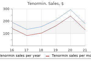
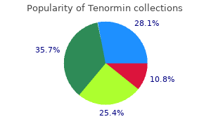
Tenormin
Tenormin dosages: 100 mg, 50 mg
Tenormin packs: 60 pills, 90 pills, 120 pills, 180 pills, 270 pills, 360 pills, 240 pills

Order tenormin 100 mg free shipping
The chylomicrons are too large to cross intercellular junctions linking to capillary epithelial cells atrial fibrillation tenormin 50 mg generic amex. Lipoprotein lipase is sure to the capillary endothelial cells in muscle blood pressure exercise 50 mg tenormin best, adipose, and breast tissues. In addition, within the postpartum state, breast lipoprotein lipase activity will increase 10-fold to promote milk manufacturing. The intracellular pool of ldl cholesterol and cholesterol esters in the hepatocyte is dynamic. These patients might have attribute physical findings (see Plates 7-6 and 7-7). Familial faulty apo B100 is a dysfunction caused by mutations in the gene encoding apo B100. Polygenic hypercholesterolemia refers to mixtures of multiple genetic and environmental factors that contribute to hypercholesterolemia. Polygenic hypercholesterolemia is diagnosed by exclusion of other major genetic causes, absence of tendon xanthomas, and documentation that hypercholesterolemia is current in fewer than 10% of first-degree relatives. Hypercholesterolemia can predispose to atherosclerosis and enhance the chance for vascular disease. Familial hyperapobetalipoproteinemia (with overproduction of apo B100) and familial combined hyperlipidemia are both inherited in an autosomal dominant trend. These cutaneous protuberances characterize the accumulation of enormous (10�20 m in diameter), cholesterol-filled macrophages. Plain and tuberous xanthomas are most regularly discovered over the elbows, knees, and buttocks, presumably associated to steady irritation by garments. In the primary part, atherosclerotic lesions consist of cushionlike elevations of lipid-filled macrophages (foam cells) beneath the intima. The atheroma of the arterial intima is probably the most dangerous function of familial hypercholesterolemic xanthomatosis due to its frequent incidence within the coronary vessels, which may cause angina and myocardial infarction at an early age. To date, most affected sufferers have the identical single level mutation at nucleotide quantity 3500. Some sufferers may have hepatic steatosis and cirrhosis, which may outcome from therapy with medium-chain triglycerides. In one affected person who underwent liver transplantation for hepatic cirrhosis, the serum lipoprotein profile normalized however gastrointestinal fat malabsorption continued. Tangier disease was originally described and named on the premise Lymph nodes, Tonsils enlarged; irregular colour liver, and spleen enlarged Tonsils eliminated H & E stain Foam cells Abetalipoproteinemia Fat stain Malnutrition Retinal lesions (periphery) Lordosis Acanthocytosis Ataxic neuropathy of a kindred living on Tangier Island in Chesapeake Bay. Findings on bodily examination include orange tonsils (caused by ldl cholesterol deposits), corneal opacities, hepatosplenomegaly, and peripheral neuropathy. Serum triglyceride concentrations higher than 199 mg/dL are termed hypertriglyceridemia and are associated with an elevated risk of heart problems. Hypertriglyceridemia results from the buildup of triglyceride-rich lipoproteins. Severe hypertriglyceridemia results because the clearance of triglyceride-rich lipoproteins from plasma is blocked. Familial dysbetalipoproteinemia happens when people are homozygous for the E2 allele. However, serum triglyceride concentrations higher than 1000 mg/dL might result in chylomicronemia syndrome. Signs and signs related to chylomicronemia syndrome embrace abdominal ache, pancreatitis, eruptive xanthoma, flushing with alcohol intake, memory loss, and lipemia retinalis. The acute pancreatitis could be life threatening, and the sufferers most commonly affected are those with poorly managed diabetes mellitus or alcoholism. At markedly increased levels, the serum may be milky because of hyperchylomicronemia. Chylomicronemia syndrome outcomes when there are massive accumulations of those lipoproteins in the blood. Manifestations of chylomicronemia syndrome include recurrent belly pain, pancreatitis, hepatosplenomegaly brought on by the accumulation of triglycerides in reticuloendothelial cells, eruptive xanthomas, lipemia retinalis, lipemic plasma, neurologic manifestations, dyspnea, and severe hypertriglyceridemia (>2000 mg/dL). Eruptive xanthomas are normally present in this setting, particularly when serum triglyceride concentrations are greater than 2000 mg/dL. The therapeutic objective is to keep serum triglyceride concentrations at lower than one thousand mg/dL. The diploma of hypertriglyceridemia is often lower than one thousand mg/dL except aggravated by alcohol use, orally administered estrogen, or hypothyroidism. Treatment of individuals with familial hypertriglyceridemia includes avoidance of alcohol and orally administered estrogens, as well as implementation of a few of the nonpharmacologic and pharmacologic approaches outlined in the following text. Nonpharmacologic treatment options embody weight reduction in overweight sufferers, a daily isotonic exercise program, improved glycemic management in patients with diabetes mellitus, limitation of alcohol intake, and avoidance of free carbohydrates in the diet. Pharmacologic therapy is indicated when hypertriglyceridemia persists despite nonpharmacologic interventions. When the serum triglyceride focus is very excessive (500 mg/dL), the primary goal is to avoid pancreatitis. The sites of atherosclerosis are typically these elements of the arterial vascular tree associated with elevated turbulent blood circulate (bifurcations and curvatures). Typical areas for symptomatic atherosclerotic lesions are the proximal left anterior descending coronary artery, proximal renal arteries, and carotid bifurcations. These sites have an upregulation of proinflammatory adhesion molecules for inflammatory cells. The normal arterial wall is composed of the endothelial cell layer, intima and subendothelial house, inner elastic lamina, media (muscularis layer formed by clean muscle cells), and adventitia (loose connective tissue). The activated macrophage releases mitogens and chemoattractants, which recruit extra macrophages and smooth muscle cells. Smooth muscle cells turn out to be the principle cell sort, lying in parallel layers with proteoglycan and basement membrane in between. Continued irritation ends in the recruitment of increased numbers of macrophages and lymphocytes that release proteolytic enzymes, cytokines, chemokines, and growth factors. Focal necrosis develops, and free cholesterol types the central lipid core of the fibrous plaque. Cycles of accumulation of mononuclear cells, migration and proliferation of smooth muscle cells, and formation of fibrous tissue result in a steady restructuring of the atherosclerotic lesion. The microvessels present a portal of entry for monocytes and lymphocytes into the growing plaque. The microvessels are fragile and are prone to rupture, resulting in small focal hemorrhages inside the plaque. The plaque can progress to an advanced lesion, where the floor endothelial cells may be lost, and the fibrous cap ruptures to expose the subendothelial area. Plaque rupture is liable for roughly 75% of deadly coronary thrombi; these plaques tend to have thin fibrous caps, elevated macrophage content, and enormous lipid cores.
100 mg tenormin cheap otc
Cytologic analysis of the fluid obtained is of little value due to unacceptably high falsepositive and false-negative results blood pressure chart xls tenormin 100 mg buy discount on-line. A centrifugal nodule with sharply circumscribed blood pressure quit smoking purchase tenormin 50 mg with visa, fleshy, and homogeneous character, usually spherical or ovoid in form, characterizes them. Malignant change is extraordinarily uncommon and usually takes the type of fibrosarcoma occurring in the large myxoma. After menopause, fibroadenomas are inclined to regress and turn out to be hyalinized however might remain unchanged or develop with estrogen alternative remedy. The treatment is simple excision, which confirms the prognosis and suffices for the remedy. They are about one-half as common as fibroadenomas and are usually found at or close to the menopause, within the central zone of the breast. The tumors are not often of enormous measurement; they range in diameter from 1 to a number of centimeters. On palpation, the benign papilloma is freely movable, soft, and either tense (cystic) or fluctuant. Smaller papillomas may be discovered within the neighboring ducts or by way of the ramifications of a bunch of ducts a lengthy way from the primary tumor. Microscopically, the arborescent epithelial outgrowths relaxation upon a fibrous stalk with an intact basement membrane. The period of the growth extends over a interval of 6 or 7 years, with rapid growth towards the end of this era, when these tumors can considerably increase in size in just a few weeks. The benign character of the expansion is indicated by the absence of invasion of the skin or of the regional lymph nodes. Because most of these tumors are benign, the name could also be deceptive, leading to the popular terminology of phyllodes tumor or giant myxoma. Dense fibrous tissue in whorls is separated by clefts from polypoid, fibrous, and epithelial masses projecting into cystic cavities. Under the microscope, the predominant part is myxomatous connective tissue with intervening dense fibrous strands. The majority of the growths are benign, however some could be the seat of sarcomatous change in about 10% of cases, particularly when the tumor has existed for many years. Roughly 30% of sufferers with malignant phyllodes tumors will die from their illness. Many kinds of sarcomas, similar to osteogenic, lympho-, myo-, lipo-, and myelosarcomas, have been described. Microscopic view exhibiting numerous crowded spindle cells with abnormal hyperchromatic nuclei Tumor ulcerated by way of pores and skin Giant myxoma Section of breast tissue containing tumor Cyst containing myxoid mass Clinical presentation of tumor in proper breast Giant myxoma. The tremendous dimension and the absence of axillary node involvement distinguish these growths from mammary carcinomas. Grossly, the tumors are solid, fleshy growths, which can invade the pectoralis fascia. The lungs are the most common metastatic site, followed by bone, coronary heart, and liver. Worldwide, breast cancer is the second most common kind of most cancers after lung cancer (10. Approximately one-third of all types of female carcinoma arise within the breast, and more than threequarters of those are the infiltrating scirrhous type or lobular carcinoma. The peak incidence is above 40 years of age; with 85% occurring after forty and 75% after 50. The signs that bring the affected person underneath examination are the discovery of the lump (55% to 65% of cases), its rising dimension, occasional fleeting pains or tenderness, and modifications in the skin or nipple. Approximately 60% of palpable tumors are located within the upper outer quadrant of the breast. The main medical findings on examination are the presence of a single lump in a breast otherwise regular to palpation in a patient greater than 35 years old; the exhausting and irregular feeling of the tumor; the apparent nearness of the tumor to the analyzing fingers due to atrophy of overlying fat; the restricted mobility of the mass; and flattening or retraction of the skin or nipple on the affected side when arms or breast are manipulated. Excisional biopsy with or without radiographic control supplies the one definitive diagnosis. Microscopically, the tumor cells are of average size, with outstanding hyperchromatic nuclei. The cells grow in small nests or in cords with outstanding intervening fibrous tissue. The most typical breast cancer histology is the infiltrating ductal carcinoma, accounting for about 75% of breast cancers. Proliferation of duct cells with hyperchromatic nuclei in strong sheets and no glandular architecture lymph nodes were uninvolved and 20% if there was lymph node involvement. It has turn into obvious that many women with breast cancer have systemic illness on the time of initial analysis. These forms of circumscribed adenocarcinomas bulge outwardly from the chest wall quite than retract inwardly as within the infiltrating type. The commonest sort of adenocarcinoma is ductal carcinoma, which begins in the cells of the ducts. Lobular carcinoma begins in the lobes or lobules and is extra typically found bilaterally than are other forms of breast most cancers. The most cancers is classified based on the predominant histologic cells; however, several mobile patterns could also be present in anybody tumor. The histologic diagnosis of intraductal carcinoma in situ includes a heterogeneous group of tumors with varying malignant potential. Carcinoma develops in approximately 35% of women with this disease within 10 years of initial prognosis, and 5% to 10% of girls may have a simultaneous invasive carcinoma in the same breast at the time of biopsy. It has a a lot greater tendency to be bilateral and to current as multifocal disease. Three of four patients with lobular carcinoma in situ are in the premenopausal age group. In circumstances of infiltrating ductal carcinoma, nonuniform malignant epithelial cells of various sizes and shapes infiltrate the surrounding tissue. The diploma of fibrous response to the invading epithelial cells determines the firmness to palpation and texture during biopsy. In general, the specialized types are grossly softer, cellular, and nicely delineated. They are often smaller and have a extra optimistic prognosis than the more common heterogeneous selection. Sheets of tumor cells with hyperchromatic nuclei from a big development with papillary projections (see cross part above) Duct cancer (comedocarcinoma). Colloid or gelatinous carcinomas have an identical gentle consistency, with in depth deposition of extracellular mucin. Infiltrating lobular carcinomas are histologically notable for the uniformity of the small, spherical neoplastic cells. Histologic subdivisions of infiltrating lobular carcinoma embody small cell, round cell, and signet cell carcinomas. This most cancers tends to have a multicentric origin in the identical breast and to contain both breasts more typically than infiltrating ductal carcinoma. On palpation, these growths feel boggy and semimovable and are dependent and heavy when the breast is moved upward.
Syndromes
- Most of the time you will need to take antibiotic to prevent the infection from spreading to the kidneys.
- Spread of cancer to the lungs
- Your skin appears to be very stretchy
- Name of the product (ingredients and strengths if known)
- Swelling in the area
- Washing of the skin (irrigation) -- perhaps every few hours for several days
- Prescription anti-inflammatory medicines
- Steroid use
- Loss of sense of vibration and position
100 mg tenormin amex
The junction of those two main right biliary channels normally occurs above the right branch of the portal vein heart attack or pulled muscle buy 50 mg tenormin visa. The proper hepatic duct is brief and joins the left hepatic duct to represent the confluence lying in front of the right portal vein and forming the common hepatic duct arteria axillaris tenormin 100 mg purchase without a prescription. The confluence of the proper and left hepatic ducts occurs at the right of the hilar fissure of the liver anterior to the portal venous bifurcation and overlying the origin of the right department of the portal vein. The extrahepatic segment of the right duct is brief, but the left duct has a for a lot longer extrahepatic course. Anterior facet of the biliary anatomy and of the top of the pancreas: proper hepatic duct (a); left hepatic duct (b); widespread hepatic duct (c); hepatic artery (d); gastroduodenal artery (e); cystic duct (f); retroduodenal artery (g); common bile duct (h); neck of the gallbladder (i); body of the gallbladder (j); fundus of the gallbladder (k). Note particularly the position of the hepatic bile duct confluence anterior to the best branch of the portal vein, the posterior course of the cystic artery behind the common hepatic duct, and the connection of the neck of the gallbladder to the right department of the hepatic artery. Note also the connection of the main vessels (portal vein, superior mesenteric vein, and artery) to the pinnacle of the pancreas (see additionally. Note the cystic plate (A) above the gallbladder, the hilar plate (B) above the biliary confluence and at the base of the quadrate lobe, and the umbilical plate (C) above the umbilical portion of the portal vein. Arrows point out the aircraft of dissection of the cystic plate during cholecystectomy and of the hilar plate during approaches to the left hepatic duct. Endoscopic retrograde choledochopancreatogram displaying the pancreatic duct (arrow), gallbladder, and biliary tree. It is feasible to open the connective tissue constituting the hilar plate and show the biliary convergence and left hepatic duct. The retropancreatic portion of the frequent bile duct approaches the second portion of the duodenum obliquely, accompanied by the terminal part of the pancreatic duct of Wirsung. The widespread duct courses downward anterior to the portal vein and is closely utilized to the hepatic artery, which runs upward on its left, giving rise to the right department of the hepatic artery, which crosses the primary bile duct, normally posteriorly. The cystic artery, arising from the right branch of the hepatic artery, could cross the common hepatic duct posteriorly or anteriorly. In this triangle runs the cystic artery, usually the right department of the hepatic artery, and sometimes a bile duct, which must be displayed before cholecystectomy. The union between the cystic duct and the frequent hepatic duct may be positioned at various levels. The cystic duct arises from the neck of the gallbladder and extends to join the frequent hepatic duct. Although the cystic duct joins the widespread hepatic duct in its supraduodenal segment in the majority of circumstances, it could prolong downward to the retroduodenal or retropancreatic space. Occasionally, the cystic duct could be part of the proper hepatic duct or a proper hepatic sectoral duct. A, Relationship between the posterior facet of the quadrate lobe and the biliary confluence. This approach (lowering of the hilar plate) (Hepp & Couinaud, 1956) generally is used to show a dilated bile duct above an iatrogenic stricture or hilar cholangiocarcinoma. This maneuver is of explicit worth for prime bile duct stricture and in the presence of liver atrophy or hypertrophy. The procedure consists of lifting the quadrate lobe upward (see A and B), then not only opening the umbilical fissure, but in addition incising the deepest portion of the gallbladder fossa. Right, An accessory proper hepatic artery (large arrowhead) is arising from the superior mesenteric artery and lies lateral to the catheter (small arrowheads) within the common bile duct. The primary variations of the cystic artery: typical course (A); double cystic artery (B); cystic artery crossing anterior to major bile duct (C); cystic artery originating from the proper branch of the hepatic artery and crossing the widespread hepatic duct anteriorly (D); cystic artery originating from the left branch of the hepatic artery (E); cystic artery originating from the gastroduodenal artery (F); cystic artery arising from the celiac axis (G); cystic artery originating from a replaced proper hepatic artery (H). A, T-tube cholangiogram reveals a very low insertion of a proper sectoral duct into the common hepatic duct (arrow). B, Endoscopic retrograde choledochopancreatogram exhibits low proper sectoral duct (arrow), into which is draining the cystic duct (arrowhead), an uncommon however important variant. C, Ectopic drainage of a proper sectoral duct into the widespread hepatic duct (C1, proper anterior duct draining into the common hepatic duct; C2, proper posterior duct draining into the widespread hepatic duct). D, Ectopic drainage of a proper sectoral duct into the left hepatic ductal system (D1, right posterior sectoral duct draining into the left hepatic ductal system; D2, right anterior sectoral duct draining into the left hepatic ductal system). F Absence of proper, hepatic duct and ectopic drainage of the right posterior duct into the cystic duct. Although rare, agenesis of the gallbladder, bilobar gallbladders with a single cystic duct but two fundi, and duplication of the gallbladder with two cystic ducts all have been described. B, Drainage of cystic duct into the left hepatic duct related to no biliary confluence. E, Drainage of the distal a half of the best posterior sectoral duct into the neck of the gallbladder. F Drainage of the proximal part of the right posterior sectoral duct into the physique of the gallbladder. The major variations of ectopic drainage of the intrahepatic ducts into the gallbladder and cystic duct. Main variations in gallbladder and cystic duct anatomy: duplicated gallbladder (A), septum of the gallbladder (B), diverticulum of the gallbladder (C), and variations in cystic ductal anatomy (D). Different kinds of union of the cystic duct and common hepatic duct (E): angular union (a), parallel union (b), spiral union (c). The source of blood provide of the retropancreatic common bile duct is from the retroduodenal artery. The veins draining the bile ducts are satellites to the corresponding described arteries. Veins draining the gallbladder empty into this venous system and never instantly into the portal vein. The upper floor of the hilar plate can be separated from the hepatic parenchyma and, by lifting the quadrate lobe upward, show of the hepatic ductal convergence, which is at all times extrahepatic, is effected. Frequently, by a simultaneous opening of the deepest portion of the gallbladder fossa and the umbilical fissure. At the junction of the spherical ligament and the termination of the left portal vein, prolongations containing channels that are elements of the left portal system course into the liver. In such instances, intrahepatic right ductal system drainage is an choice (Chapter 8). The right and the left hepatic arteries turn out to be enclosed in the sheath of peritoneum, forming the proper and left portal triads. The most necessary relationships in the vascular anatomy of the pancreas concern the arterial blood supply and the venous drainage. The dorsal pancreatic artery arises from the splenic artery (but can arise from the hepatic artery). The superior mesenteric artery arises from the aorta posteriorly behind the pancreas and runs forward and upward to run first behind and then to the left of the superior mesenteric vein. The pancreaticoduodenal arcades come up from the superior mesenteric artery and supply the head of the pancreas.
100 mg tenormin quality
This is the only sector that comprises one segment (this is referred to as the left posterior part in accordance with prehypertension at 25 years old 100 mg tenormin for sale Strasberg et al [2000]) pulse pressure 45 100 mg tenormin free shipping. The right liver is divided into two sectors by the right portal scissura containing the right hepatic vein. Further details of segmental anatomy essential in sectoral or segmental resection are described in Chapter 5. The caudate lobe is provided by blood vessels and drained by biliary tributaries from the right and left portal triads. Small vessels from the portal vein and tributaries joining the biliary ducts also are found, normally two on the left facet and one on the proper. The proper portion of the caudate lobe, including the caudate process, predominantly receives portal venous blood from the right portal vein or the bifurcation of the primary portal vein, whereas on the left side the portal provide arises from the left branch of the portal vein almost completely. Similarly, the arterial supply and biliary drainage of the best portion is mostly associated with the right posterior sectoral vessels or pedicle and the left portion with the left major vessels. A, Caudate lobe (asterisk) positioned between the left portal vein (arrow) and inferior vena cava (v). B, Papillary means of the caudate (p) represents the lower medial extension of the caudate (asterisk) and may mimic a periportal lymph node. B, i, Numerous nodes lie along the superior mesenteric vein, along the borders of the pancreas, draining again into the splenic hilar nodes; alongside the superior border of the pancreas, to the superior pancreatic nodes; and to the celiac trunk and nodes on the base of the frequent hepatic artery. A giant node generally lodges in intimate association with the floor of the superior border of the pancreas and the best facet of the common hepatic artery. B, ii, Posterior pancreaticoduodenal nodes lie alongside the posterior pancreatic duodenal arterial arcade. A, the liver drains principally to hepatoduodenal nodes on the hilus and alongside the hepatic artery and portal vein. Bile ducts normally are located above the corresponding portal branches, whereas hepatic arterial branches are located inferiorly to the veins. Each branch of the intrahepatic portal veins corresponds to one or two bile duct tributaries joining to kind the best and left hepatic ductal techniques converging on the liver hilus to represent the common hepatic duct. The biliary tract is represented in black, and the portal branches are represented in purple. Note the horizontal course of the posterior sectoral duct and the vertical course of the anterior sectoral duct. B, Transtubal cholangiogram exhibits a standard regular variant where the best posterior sectoral duct drains into the left hepatic duct. Frequently in this variant, the posterior duct passes posteriorly to the anterior sectoral pedicle. The duct then runs to be a part of the best anterior sectoral duct because it descends in a vertical manner. Other variations are of appreciable significance in controlling the arterial blood provide to the liver throughout hepatic resection or enucleative procedures, in the performance of devascularization of the liver, in the placement of intra-arterial hepatic infusion gadgets, and within the resection of the top of the pancreas. The venous drainage of the pancreas runs in the principle parallel to the arterial supply. There are anterior and posterior superior and inferior pancreaticoduodenal veins that drain to the portal vein and the superior mesenteric vein. The left gastric vein and the inferior mesenteric vein normally drain into the splenic vein, however can drain immediately into the portal vein, whereas the various small splenic tributaries drain directly to the splenic vein. The celiac trunk is a short thick artery originating from the aorta slightly below the aortic hiatus of the diaphragm. The left gastric artery curves toward the abdomen and extends alongside its lesser curve, forming anastomoses with the right gastric artery. The splenic artery courses to the left behind and alongside the upper border of the pancreas. The widespread hepatic artery passes forward into the retroperitoneum, then curves to the proper to enter the best margin of the lesser omentum just above the pancreas and ascends, approaching the widespread bile duct on its left facet and running often anterior to the portal vein. This descends to supply the anterior, superior, and posterior surfaces of the first inch of the duodenum. The right gastric artery passes to the left along the lesser curve of the abdomen, and anastomoses with the left gastric artery. The continuation of the widespread hepatic artery past the origin of the gastroduodenal artery and right gastric artery is named the correct hepatic artery and normally quickly divides into a right and a left branch. The cystic artery often arises from the right hepatic artery, however has many variations. The interlobar fissure (open arrow), splenic vein (s), celiac axis (c), aorta (a), and inferior vena cava are additionally shown. The proper hepatic artery (long open arrow) and the left hepatic artery (small open arrow) originate from the correct hepatic artery. Rarely, the right or left hepatic arteries originate independently from the celiac trunk or branch after a very quick frequent hepatic artery origin from the celiac (B and C). The right hepatic artery programs posterior to a stent in the frequent bile duct (arrowheads). Arterial section photographs reveal the right hepatic artery (arrow) coursing anterior to the common hepatic duct (which contains air owing to a papillotomy). A superior pancreaticoduodenal vein typically enters the portal vein simply above the level of the pancreas, and several smaller veins enter the superior mesenteric and portal vein from its proper aspect beneath the neck of the pancreas. Just before its entry into the umbilical fissure, it gives off a significant caudate vein (segment I), which runs posteriorly and laterally to the left, and typically this vein consists of two or extra branches. B, the division of the portal vein might arise more proximally, nonetheless, and the right anterior and posterior sectoral portal veins may come up independently from the portal venous trunk. The proper posterior portal vein (arrow) has a separate early origin from the main portal vein. The right anterior sectoral portal vein (R) and the left main portal vein (L) share a typical trunk. A, There could additionally be an abnormal place of the portal vein anterior to the head of the pancreas and the duodenum. B and C, Another uncommon however interesting anomaly is the entrance of the portal vein into the inferior vena cava (B) and very hardly ever the doorway of a pulmonary vein into the portal vein (C). The uncinate course of can extend behind the superior mesenteric vein to nicely behind the superior mesenteric artery. Access to the portal vein behind the pancreas normally is obtained from below by elevating the pancreas from the floor of the superior mesenteric vein simply earlier than it joins the splenic vein. Superiorly, the portal vein runs behind the pancreas and lies in relation to the curvature of the splenic vein, the splenic artery, the common hepatic artery, and the gastroduodenal artery. The organ is composed of a head, neck, and physique, the head being encompassed by the duodenum and the tail resting within the splenic hilum. A portion of the head inferiorly is termed the uncinate process and is intimately related to the superior mesenteric vein and superior mesenteric artery. There may be congenital absence of the left department of the portal vein as described by Couinaud, the best department then coursing through the best lobe of the liver, supplying it and curving throughout the liver substance to provide the left lobe, which in such, cases is often smaller than regular. The left branch of the portal vein is absent-findings confirmed at operation for left hepatic lobectomy.
Tenormin 100 mg buy generic on-line
Some efferent lymphatics extend to the hypogastric nodes arteria innominada effective tenormin 50 mg, however for probably the most half they cross upward to the widespread iliac and periaortic nodes prehypertension blood pressure chart buy tenormin 50 mg low price. The majority of lymphatic channels to this group of nodes originates from the vulva, but there are additionally channels from the cervix and decrease portion of the uterus. The exterior iliac nodes receive secondary drainage from the femoral and inner iliac nodes. The inner iliac nodes are present in an anatomic triangle whose sides are composed of the external iliac artery, the hypogastric artery, and the pelvic facet wall. This wealthy collection of nodes receives channels from every internal pelvic organ and the vulva, including the clitoris and urethra. Lateral sacral nodes may be discovered in the hollow of the sacrum in relation to the lateral sacral vessels. No lymphatics, surprisingly, have been detected in the superficial components of the endometrium. The principal accumulating trunks move outward at the isthmus along the course of the uterine vessels. Secondary drainage from this node is to the inner iliac nodes on the same side of the pelvis. This drainage path allows for the analysis of so-called sentinel nodes in cervical cancer sufferers. This approach identifies the more than likely first web site of nodal metastases in a regional lymph node basin. Although pelvic lymphadenectomy remains the usual for sufferers with cervical cancer, the utilization of sentinel lymph node biopsy for these patients is rising. From the uterus as a whole, afferent lymphatics might lengthen to the ureteral, obturator, hypogastric, exterior and customary iliac, periaortic, lateral and center sacral and the femoral nodes. Occasionally, also, intercalated nodes could additionally be involved between uterus and bladder or rectum. The ovarian lymphatics pass through the infundibulopelvic ligament along with the ovarian vessels to the lateral periaortic lymph nodes. On the right facet, they might be discovered Medial (inferior) external iliac nodes Obturator node Superficial inguinal nodes Deep inguinal nodes Highest deep inguinal node (of Cloquet) between the best renal vein and the inferior vena cava. In addition, afferents drain to the widespread iliac nodes and people of the sacral promontory. The lymphatics of the vagina share the lymphatic pathways of the cervix to the ureteral, hypogastric, obturator, external iliac, lateral sacral, and promontory nodes. At the bifurcation of the aorta, they be part of to type the superior hypogastric plexus or presacral nerve. In its lower portion, the plexus divides to form the two hypogastric nerves that run laterally and inferiorly. These pass downward and laterally near the sacral end of each uterosacral ligament after which forward over the lateral side of the rectal ampulla and higher vagina. A center hypogastric plexus, overlying and just under the sacral promontory, might sometimes be present. They receive branches from the sacral ganglia of the sympathetic trunk and parasympathetic fibers from the second, third, and fourth sacral spinal nerves (nervi erigentes or pelvic nerves). The pelvic plexus of nerves is subdivided into secondary plexuses, which comply with the course of the visceral branches of the hypogastric vessels. The anatomic relations of the presacral nerve, or superior hypogastric plexus, are of importance because its resection is sometimes carried out for the reduction of intractable pelvic pain. The fundus is the dome-shaped portion above the extent of entrance of the fallopian tubes. About one-third of the anterior surface and one-half of the posterior floor of the cervix represent the vaginal portion. The peritoneal layers that sheathe the fundus and uterine physique unite on both sides of the uterus to kind the broad ligament, which separates the vesicouterine and rectouterine pouches. The higher borders of the broad ligaments are folds of the peritoneum coming into existence when the anterior sheath turns to become the posterior sheath. The broad ligaments broaden downward from the lower edges of the tubes, assuming the perform of a mesentery to the tubes, the mesosalpinx, in which the vessels to and from the tube take their course. However, the latter varieties on this area a band, the infundibulopelvic ligament, which attaches the posterior floor of this finish of the tube to the lateral wall of Uterine (fallopian) tube Fundus of uterus Am Tubal ostium Uterine Isthmus half pul la Body of uterus Infundibulum Folds of uterine tube Fimbriae Ligament of ovary Endometrium Myometrium Mesometrium (broad ligament) Uterine vessels Suspensory ligament of ovary (contains ovarian vessels) Vesicular appendix (hydatid of Morgagni) Epo�phoron Follicle (graafian) of ovary Corpus albicans Corpus luteum Isthmus of uterus Internal os Cervix of uterus Cardinal (transverse cervical or Mackenrodt) ligament Vaginal fornix External os Vagina Cervical canal with palmate folds the pelvis. This fold is not to be confused with the ligament of the ovary, a wire within the broad ligament working from the lateral angle of the uterus slightly below the uterine end of the tube downward to the lower or uterine margin of the ovary. Only its lateral floor lies upon the parietal pelvic peritoneum, where the exterior iliac vessels, the obliterated umbilical artery, and the ureter form a shallow despair known as the ovarian fossa. The anterior border of the ovary is attached to the posterior layer of the broad ligament by a brief fold via which the blood vessels pass to reach the hilum of the ovary. Up to the seventh month of fetal life, the uterus grows in proportion to the relaxation of somatic development. By this time, a distinction in proportion of size of the cervix to that of the fundus becomes evident. The ratio of cervical size to total size usually regresses to that discovered before puberty. Because the uterus is formed by fusion of the m�llerian ducts, its muscular construction is somewhat complex. Smooth muscle bundles contained within the supportive ligaments interdigitate with the round muscle system of the uterus. This coordination of muscular contractions in the three completely different buildings can also serve to orient properly the ovary with the infundibulum of the tube on the time of ovulation. At intervals, they offer off radial branches, which penetrate instantly inward through the myometrium. Before coming into the endometrium, the terminal branches of the radials divide into two distinct forms of arteriole: straight and spiral arterioles. The brief, straight arterioles supply only the deeper third of the endometrium, ending in a kind of horizontal arborization of terminal twigs. The spiral arterioles, however, attain to the surface of the endometrium and exhibit marked adjustments in response to hormonal changes by way of the conventional cycle. Arteriovenous and venovenous anastomoses have been demonstrated that develop particularly through the secretory part of the cycle. This complicated vascular sample, distinctive to the endometrium, was as quickly as believed to be the mainspring of the processes that enact the rhythmic necrosis and hemorrhage called menstruation. The spiral arterioles endure extraordinary lengthening through the first or proliferative phase of the cycle. As a result of this difference in rising pace, the spiral arteries are thrown into complex kinks and coils. As a outcome, the spiral arteries are somewhat stretched in the course of the time of biggest luteal activity. In the absence of a fertilized, implanted ovum, the corpus luteum begins to degenerate a couple of days earlier than the top of the cycle. Coiled (spiral) artery Radial artery Straight artery Gland orfices on floor of endometrium Subepithelial capillary plexus Venous lake Stromal capillary plexus Venovenous anastomosis Glandular capillary plexus Arteriovenous anastomosis Spiral artery Endometrium Gland Vein Straight artery Myometrium Radial artery From 4 to 24 hours previous the onset of menstrual bleeding, an intense vasoconstriction and vascular clotting are seen.
Tenormin 100 mg purchase fast delivery
Interaction between air air pollution and respiratory viruses: time-series study of day by day mortality and hospital admissions in Hong Kong blood pressure medication dementia order tenormin 50 mg otc. Traffic-related air pollution and asthma onset in kids: a potential cohort study with particular person exposure measurement arrhythmia diagnosis 50 mg tenormin buy with visa. Traffic-related air pollution and respiratory signs in children dwelling alongside trunk roads in Chiba Prefecture, Japan. Traffic-related air air pollution correlates with adult-onset bronchial asthma amongst never-smokers. Does visitors exhaust contribute to the development of bronchial asthma and allergic sensitization in youngsters: findings from recent cohort studies. Traffic-related air pollution and development of allergic sensitization in children in the course of the first eight years of life. Indoor particulate matter increases asthma morbidity in children with non-atopic and atopic bronchial asthma. Developing bronchial asthma in childhood from exposure to secondhand tobacco smoke: insights from a meta-regression. Anxiety and melancholy are more widespread in youngsters with asthma and are associated with worse asthma outcomes. Clinicians taking care of youngsters with poorly managed or difficult-to-control asthma must be particularly alert as a result of asthma control might not enhance till psychological factors are addressed. Surprisingly, the evidence base for the effect of psychological interventions may be very restricted. Strunk and associates printed a seminal examine of 21 children who died of bronchial asthma after discharge from hospital [1]. The authors suggested that finding out these psychological components may be necessary in figuring out kids at excessive danger of dying from asthma and in growing therapy plans to stop deaths from asthma [1]. Osler also recognised that ``fright or violent emotion may convey on a paroxysm', acknowledging that psychological components can have a job in bronchial asthma assaults [2]. Even up till the 1950s, the concept atopic disorders together with asthma and atopic dermatitis were ``psychosomatic' and psychogenic in ``origin' was commonplace. Psychoanalytic theories also considered asthma as psychological in origin, with remedy involving evaluation and different ``talking cures' [3]. As a end result, powerful and effective pharmacological therapies developed to control airway inflammatory processes are actually in widespread use. By likelihood, this altering view of asthma causation has coincided with substantial increases within the prevalence of atopic issues in developed Western societies; some studies found one in three people have some type of atopic disease [4]. While there are tons of reasons for poor bronchial asthma management [10], the mismatch between efficient therapies and persevering with morbidity has led to a reawakening of interest in nonpharmacological components that doubtlessly affect asthma management. There is a substantial physique of analysis in children and young people with asthma on the impact of mental health, and emotional and behavioural issues on functional standing and bronchial asthma morbidity [14, 15]. Early research in the subject emphasised difficulties in separation from dad and mom and related anxiousness. Later studies shifted to elements similar to stress and medicine management that could result in adjustment difficulties [16]. Consistent with medical impressions, they discovered that the effect was higher for ``internalising behaviours' than for ``externalising behaviours', i. They also discovered that elevated bronchial asthma severity was related to greater behavioural difficulties. Acute bronchial asthma attacks, in particular, may be associated with unpleasant sensations similar to choking or suffocation or hyperventilation-induced breathlessness and anxiousness. In some people, these signs overlap with panic/fear signs corresponding to being afraid of dying. In animal research in rats, worry of dyspnoeic suffocation induced by a single exposure to 100 percent carbon dioxide has been proven to be a conditioned (and hence learned) response [18]. This has led to the suggestion that at least some options of panic assaults in asthmatic patients may arise due to a conditioned response to breathlessness. Anxiety via an impact on respiratory may in flip lead to greater use of as-required asthma medicine. In one research of adults with extreme bronchial asthma being treated intensively in hospital, these with high levels of panic/fear symptoms have been extra more doubtless to use as required bronchodilator medication [19]. In turn, bronchial asthma medicines could make nervousness symptoms worse due to sideeffects such as tremor. Many paediatricians treating children with problematic asthma will recognise comparable problems in some children with excessive use of bronchodilator and related side-effects. There can be evidence that social stress anxiety can promote and amplify airway irritation in response to other asthma triggers [23]. Many of the studies in kids and adults with asthma have focused on specific kinds of anxiousness corresponding to panic dysfunction, or social phobia in teenagers. In both adults and kids, populations with asthma had a excessive prevalence of hysteria issues. In seven studies in children/ adolescents with bronchial asthma, up to one third of kids met standards for comorbid anxiety. In adult populations with asthma, the estimated range of panic dysfunction was between 6. Another necessary criticism is that few research try to separate out the potential overlap between signs of bronchial asthma and panic. Patients could have difficulty remembering or discerning which episodes have been as a result of bronchial asthma, which to panic or a mixture of both. Much evidence has come from cross-sectional surveys utilizing validated questionnaires or diagnostic interviews to assess the prevalence of the conditions in populations at a particular moment in time. Such approaches can show associations, but longitudinal cohort research are wanted to investigate causal relationships and the path of causality. In fact, many of those very criticisms apply across the whole area of psychological components and bronchial asthma in children. A variety of newer studies have addressed these points across the childhood age vary. This examine used a population-based sampling strategy with a high participation and response rate. Children with anxiousness scores in the medical range have been extra more likely to be utilizing asthma preventive medicine and have higher faculty absence. After adjustment, these youngsters reported considerably extra days of asthma signs over the previous 2 weeks than those with no nervousness. There was evidence of a dose�response relationship, with the number of reported asthma signs considerably associated to the variety of nervousness and depressive symptoms reported by the kid [27, 28]. The authors concluded that the presence of an anxiousness or depressive disorder was highly associated with an elevated asthma symptom burden; they instructed this was an additive impact of the nervousness and melancholy quite than elevated severity of asthma in children with anxiousness and melancholy. Social anxiety may be a selected threat for adolescents with asthma with reports of feeling totally different and isolated from their peers, fearing peer rejection, having poor social competence and being additionally disadvantaged when asthma affected their ability to participate in actions similar to sport or dance [29, 30]. This advised that the primary concern for the adolescents on this study was managing any asthma in entrance of their quick peer group [31]. Thus, in adolescents, social nervousness would possibly have an effect on medication compliance when treatment is utilized in entrance of friends; for instance, in using reliever treatment earlier than train [32]. One community-based prospective study in Switzerland adopted 591 young individuals who were aged 19 years at enrolment, for over 20 years.
TriCreatine Malate (Creatine). Tenormin.
- Increasing strength in people with muscle diseases such as muscular dystrophy.
- Improving the athletic performance of young, healthy people during brief, high-intensity exercise such as sprinting. However, it does not seem to help highly trained athletes. It also does not seem to help increase muscle strength or body composition.
- What other names is Creatine known by?
- How does Creatine work?
- What is Creatine?
- Rheumatoid arthritis (RA).
- Schizophrenia.
- Is Creatine effective?
Source: http://www.rxlist.com/script/main/art.asp?articlekey=96843
Generic tenormin 100 mg line
Concurrently blood pressure chart low 100 mg tenormin overnight delivery, because of an involution of the cranial and adjacent mesonephros prehypertension hypothyroidism buy generic tenormin 50 mg, the testis becomes cellular and is left suspended from the epididymis by the mesorchium, a fold of primitive peritoneum. At the time of testis descent into the processus vaginalis within the inguinal bursa and scrotum, the portion of this processus vaginalis superior to the testis becomes obliterated typically weeks or months after start. This sort of hydrocele is characterized by dramatic changes in measurement when assuming an upright or supine position. The gubernaculum, initially discernible as a fibrous band in early fetal life, develops as the lower inguinal ligament and increases in dimension through the seventh month of gestation. The distal attachment of the gubernaculum extends to the area of the inguinal bursa the place the lengthy run exterior indirect layer of the stomach wall develops. Initially, the gubernaculum contracts and thickens to guide migration of the testis towards the inner inguinal ring. In mice, this migration seems to be controlled by a testisderived insulin-like/relaxin-like peptide (Insl-3). The second section of descent, transinguinal to scrotal, is thought to be androgen dependent. Among many yeasts, molds, and fungi, only a few are infectious and are termed dermatophytes ("skin fungi"). Infections by the fungus tinea cruris (ringworm) are very common within the groin and scrotum. It includes desquamation of the scrotal pores and skin and contiguous surfaces of the inside thighs and itches ("jock itch"). Sweating, tight clothing or weight problems favor growth and recurrence of this fungal infection, derived mainly from the genera Trichophyton and Microsporum. Unlike contact urticaria, in which a rash seems inside minutes of exposure and fades away inside minutes to hours, contact dermatitis takes days to fade away. Common causes of irritant contact dermatitis are extremely alkaline soaps, detergents, and cleaning merchandise. Contact dermatitis of the scrotum might show a wide selection of lesions various from erythema, to papules, to vesicles or pustules, however is always accompanied by itching. Drug eruption is a form of contact dermatitis that will occur on the scrotum and elsewhere on the physique after consumption of medication to which the affected person is allergic. It is normally symmetrical on the scrotum and inside surfaces of the thighs, with frequent involvement of the penis and buttocks. Other uncommon skin lesions (not illustrated) with a predilection for the scrotum are prurigo, which is a common term for itchy eruptions of the scrotal pores and skin, and lichen planus, an inflammatory skin rash that varieties scaly rings and plaques on the genitalia which are characteristically "violaceous" or purple colored. Tinea versicolor, caused by the fungus Pityrosporum ovale, is comparatively widespread in adolescent and younger grownup males. Mites are small, eightlegged parasites (in distinction to six-legged insects), 1/3 mm long, that burrow into the pores and skin and that are particularly active at night time, producing intense nocturnal itching. Furrows are readily seen on the scrotum, and a tiny burrow can be detected on the point where the pores and skin has been invaded. The furrows differ in size and coloration and are often curved or arciform, resembling a small beaded or dotted thread. The vesicles shortly rework into papules, pustules, incrustations, and excoriations that obscure the burrows. Once secondary excoriation and pustules develop, the unique skin lesions are more difficult to acknowledge. This ectoparasite feeds solely on blood and has an oral appendage that produces a pores and skin lesion by suction. Unlike the body louse that lives in clothing, the crab louse resides on bushy physique elements: within the genitalia, this louse attaches to pubic hair with its head buried within the hair follicle. The skin could reveal a "bitten" look, exhibiting small pink factors that may become papules. Lice and nits may be eliminated both with forceps or by slicing the infested hair with scissors and then examined with a microscope. The complete scrotal tissue could also be misplaced and complete sloughing of remaining skin could occur because of an infection. Partial lack of the scrotum is managed by debridement, excision of islands of remnant fullthickness scrotal wall, and first closure with absorbable sutures. The capability of small fragments of remaining pores and skin to regenerate a full-sized scrotum is exceptional, and transplantation of the testes may be prevented if some skin remains. Healing by regeneration of skin from a close-by avulsed margin would result in a comparatively inelastic masking. Testicles ought to be fixed together in a dependent place to minimize motion and maximize graft "take. Only 20% of patients require important revisions and most of these can be managed within the workplace. Epididymo-orchitis is regularly accompanied by scrotal edema, as are allergic states or obstruction of the lymphatic or vascular system. Marked edema or anasarca that entails the scrotum can result from continual cardiac insufficiency, liver cirrhosis, ascites, and renal failure. Simple edema can also be the first signal of elephantiasis (lymphatic filariasis) and other tropical diseases. In congenital or communicating hydrocele, with or with out hernia, a lumen in the processus vaginalis permits communication with the abdominal cavity, in order that bowel and peritoneal fluid could lengthen to the scrotum and hydrocele fluid may attain the peritoneal cavity. Hernial hydrocele (not illustrated) is an accumulation of fluid within the tunica vaginalis as a result of a restricted projection of the processus vaginalis from the peritoneal cavity inferiorly into the scrotum. Usually, neither bowel nor omentum is current within the sac, and the hydrocele fluid within the sac can be pressed back into the peritoneum. Acute hydrocele is often secondary to trauma, tumors, or underlying an infection of the testicle or epididymis. Hydroceles can observe trauma and occur after inguinal herniorrhaphy, varicocele ligation, or different retroperitoneal surgical procedure that blocks lymphatic move by way of the spermatic wire. Hydroceles are typically situated anterior to the testicle, which it displaces posteriorly in the scrotal cavity. The treatment is watchful ready, repeated needle aspirations (as the fluid recurs quickly), or operative excision of the parietal tunica vaginalis. A spermatocele is an intrascrotal cystic mass ensuing from partial obstruction or diverticula of the efferent ductule system near the caput epididymis (see Plate 3-3). On palpation, spermatoceles appear as a round mass distinct from the testis, with a narrower "waist" between the testis and the cyst hooked up to it. Most spermatoceles are asymptomatic, apart from a slight dragging sensation within the scrotum because of a "mass impact. In distinction, the best inside spermatic vein enters the vena cava obliquely below the best renal vein. With varicocele, the blood flow in the internal spermatic veins is reversed, inflicting heat, corporeal blood to pool around the usually cooler testis. The occurrence of an isolated right varicocele, or the sudden onset of a left varicocele after the age of 30, could point out retroperitoneal illness, such as tumor, lymphadenopathy, hydronephrosis, or aberrant vessels.
Tenormin 100 mg purchase with mastercard
Withdrawal of the bronchoscope requires comparable care to that used during insertion heart attack 18 year old male order 50 mg tenormin fast delivery. This relatively highfrequency probe allows excellent visualization of the layers of the airway wall and has been proven to be more delicate than chest computed tomography scanning for figuring out airway invasion versus compression by tumor blood pressure vs age buy tenormin 50 mg on line. The main good factor about this bronchoscope is that it allows real-time visualization of the needle entering the lymph node. The performance traits (sensitivity, specificity, constructive and unfavorable predictive values) are nearly equivalent for extra invasive procedures similar to mediastinoscopy. One also must respect the technical differences of the bronchoscope itself. The operator additionally needs to perceive the "knobology" of the ultrasound processor and be in a position to modify the depth, contrast, and acquire at a minimum. In recent years, an effective and significant internationally vetted system for staging lung cancer has evolved (see Plate 4-49). The lymphatic drainage system offers distinct predictable routes or pathways for the unfold of malignancies from every lobe of the lung to the hilum and up the mediastinum to the base of the neck. Usually carried out underneath common anesthesia, a mediastinoscopy involves a horizontal suprasternal low cervical skin incision to expose the decrease cervical a part of the trachea. Debate continues concerning the indications for mediastinoscopy and tips on how to interpret and use the information gained. Less sure is the interpretation of ipsilateral, freely movable, intracapsular nodal metastases that could be included in a radical mediastinal lymph node dissection at the time of thoracotomy and lung resection. For left higher lobe lesions, the left anterior extrapleural mediastinotomy developed by Chamberlain has proved most useful. Recently, different means of sampling mediastinal lymph nodes have been developed. It is a deformity of the anterior chest wall characterised by depression of the lower sternum and adjoining cartilages. The trait is inherited and may coexist with other musculoskeletal malformations such as clubfoot, syndactyly, and Klippel-Feil syndrome. Most current writers attribute the deformity to unbalanced development within the costochondral regions. However, a toddler with an obvious deformity may experience unfortunate psychological results. Funnel chest is usually associated with postural issues corresponding to ahead displacement of the neck and shoulders, higher thoracic kyphosis, and a protuberant abdomen. Functional coronary heart murmurs and benign cardiac arrhythmias are incessantly seen in these people, and the electrocardiogram could show rightaxis deviation due to the displacement of the center. Depression of the sternum begins typically on the junction of the manubrium and the gladiolus. In basic, the defect tends to be symmetric, however one aspect could also be more depressed than the other so that the sternum deviates from the center line. An estimate of the cavitary volume could additionally be obtained by filling the despair with water while the patient lies supine. Because many of the operations are carried out with a beauty end in thoughts, the results are greatest when surgical procedure is performed between 3 and seven years of age. Fixation by a metal strut or wire is required in older patients to stop recurrence of the deformity, which, in some degree, may happen regardless of preliminary overcorrection. Although functional cardiac and respiratory difficulties have been noticed, the chief cause for surgical correction is beauty. When operation is necessary, the procedure should be tailor-made to the particular deformity, considering the total life circumstances of the affected person. When the deformity causes embarrassment, the surgical procedure is geared toward attaining psychological in addition to physiologic enchancment. When the defect is incomplete, surgical correction of the abnormality may be achieved. Other deformities of the chest wall sometimes seen embody cervical ribs (with or with out compression of the brachial plexus and artery), partial absence of ribs, supernumerary ribs, and thoracic-pelvic-phalangeal dystrophy. Between these two extremes are patients who stay breathless on exertion and whose arterial blood gases hover on the brink of essential hypoxemia and hypercapnia. In distinction, the pulmonary arterial stress in those with severe kyphoscoliosis not solely may be excessive at relaxation but also will increase precipitously during modest train. The foundation for this pulmonary hypertension is generally twofold: (1) a restricted pulmonary vascular mattress attributable to the compressing and distorting results of the deformity on the lungs and on the pulmonary vasculature and (2) the pulmonary pressor effects of hypoxia. These two effects are most marked throughout train because of the rise in pulmonary blood circulate into the restricted vascular bed and the pulmonary vasoconstriction elicited by Deformity of rib cage in scoliosis Advanced scoliosis Advanced kyphosis Characteristic cardioplumonary pathology in kyphoscoliosis; hypertrophy and dilatation of right ventricle (and atrium); lungs atelectatic and reduced in quantity with little or no emphysematous adjustments Severe thoracic and lumbar kyphoscoliosis in a 4-year-old baby the exercise-induced hypoxemia. In patients in whom continual alveolar hypoventilation has caused sustained pulmonary hypertension, hypercapnia consistently accompanies arterial hypoxemia. However, hypercapnia exerts its predominant results on the central nervous system rather than on the heart or circulation. As a corollary, greater reliance is positioned on the hypoxic drive by way of the peripheral chemoreceptors. But if a person with kyphoscoliosis develops acute hypercapnia during an higher respiratory infection or exaggerates the preexisting degree of hypercapnia, she or he could manifest persona changes, turn into unresponsive to typical stimuli, and lapse right into a coma. The improve in intracranial pressure could also be so large as to trigger choking of the optic discs, simulating a brain tumor. All of the disturbances in uncomplicated kyphoscoliosis are significantly exaggerated by intrinsic lung disease. The sequence begins with severe thoracic deformity, lowering the compliance of the thoracic cage and lung growth. To reduce this work, the affected person unconsciously adopts a sample of rapid, shallow breathing, which ends up in chronic alveolar hypoventilation. Not only do the small, encased lungs contribute to the elevated work of respiration, but additionally they restrict the capacity and distensibility of the pulmonary vascular mattress. After arterial hypoxemia is corrected, polycythemia, hypervolemia, and an increase in cardiac output help to maintain the pulmonary hypertension. In this example, any additional mechanism for pulmonary hypertension, particularly an upper respiratory an infection, might precipitate coronary heart failure. This is mostly nicely tolerated except alveolar hypoventilation is acutely intensified, so that carbon dioxide elimination is further impaired. In this emergency, usually precipitated by an higher respiratory infection, assisted ventilation may be required in conjunction with barely enriched oxygen mixtures (25%-40%) to achieve tolerable levels of blood gases. Antibiotics and supportive measures normally suffice to tide the patient over the disaster brought on by acute respiratory an infection. The aim of therapy is to restore the affected person to the scientific state that existed earlier than the acute episode. The sternal portions are two flat bands that arise from the posterior facet of the body of the sternum. Costal parts come up from the bottom six ribs and interdigitate with the transversus abdominalis muscular tissues. On the involved aspect, lung progress is compromised, but there could also be hypoplasia on the contralateral facet as a end result of shifting of the mediastinum toward the uninvolved aspect causes some compression of that lung as nicely. The timing of onset and severity of symptoms depend upon the diploma of pulmonary hypoplasia.

Tenormin 50 mg purchase overnight delivery
In addition prehypertension occurs when quizlet tenormin 100 mg cheap on line, in the presence of an atopic setting blood pressure medication makes me tired tenormin 100 mg cheap fast delivery, rhinovirus-induced epithelial responses are altered, lowering viral clearance and promoting cytotoxicity [71]. It is possible that variations in illness traits and/or the population included in the research, in addition to methodological variations, may account for such discrepancies; nonetheless, extra research are needed to make clear this level. Neural mechanisms Post-viral alterations in neural control include dysfunction of pre-junctional M2-muscarinic inhibitory receptors, which normally inhibit acetylcholine launch, or the release of bronchoconstricting neuropeptides [84]. Viral infections can even upregulate neurotrophins that in flip control the event of the airway neural network [85]. Such an impact might alter bronchoconstrictive responses to nonspecific irritants, including future respiratory infections. Studies carried out over the past decade have emphasised the importance of atopy and allergic inflammation within the induction and perpetuation of virus-induced respiratory ailments [21, 86]. The problem of whether or not atopy per se alone might account for each symptoms and exacerbations of asthma is still being debated. Quantitative relationships have been proven between allergen sensitisation, as indicated by measures of particular immunoglobulin (Ig)E, and the chance of presenting with asthma-related symptoms [87]. Further evaluation showed that the earlier the sensitisation, the extra increased the danger of asthma development, suggesting that atopy is a potent aetiological factor for bronchial asthma prevalence [88]. The importance of atopy within the persistence of the asthmatic phenotype into adolescence is now properly established [89]. Data deriving from rhinovirus experimental an infection in asthmatics clearly confirmed enhanced decrease respiratory signs, lung operate impairment, a positive correlation of the virus load to augmented Th2 responses, and scientific outcomes in atopic asthmatics, suggesting a attainable hyperlink among allergic sensitisation and rhinovirus an infection [83]. In scientific settings of hospitalised individuals, allergens and respiratory viruses have been proven to act synergistically in the expression of extreme asthma signs in adults and youngsters [36, 90]. Furthermore, the sharp enhance in emergency department visits for virus-induced bronchial asthma exacerbations in September has been postulated to be affected by aeroallergen concentrations and host responses [18]. In the community-based Western Australian Pregnancy Cohort, the risk of bronchial asthma elevated roughly nine-fold for youngsters who had both atopy by age 6 years and greater than two wheezing-associated episodes in the first 12 months of life. In those kids, the degree of atopy was related to elevated threat of subsequent bronchial asthma in a dose-dependent method [60]. Allergens and viruses might act in a synergistic manner in damaging the airway epithelium. Viral infections, on the one hand, compromise the barrier operate of the airway epithelium resulting in enhanced absorption of allergen and, then again, in vitro rhinovirus replication is greater in disrupted epithelium, resembling the asthmatic, most likely resulting in extra severe scientific sicknesses [91, 92]. Based on this, a sublime theory on the induction and persistence of asthma symptoms in virally contaminated atopic kids suggests that indicators triggered in the course of the innate immune response to the virus can result in the discharge of huge numbers of migrating high-affinity, IgE receptor-bearing, bone marrow-derived precursors of mucosal dendritic cells into the blood. The subsequent trafficking of these cells to the contaminated airway mucosa, the place dendritic cell turnover may be very high, supplies a potential mechanism for recruitment of underlying aeroallergen-specific Th2 immunity into the already inflamed milieu of the infected airway mucosa [100]. To this respect, studies together with non-atopic asthmatics and an effort to minimise reporting bias are needed. There are still unresolved points both in respect to microorganism identification, as nicely as study design, earlier than an aetiological association could be firmly established [33]. Recently, it has been proposed that bacterial colonisation could be related to both the initiation and the exacerbation of asthma. Colonisation of the upper airways with bacterial pathogens in infancy has been associated with an elevated threat of presenting with asthma-associated signs later in life [123]. Moreover, in a prospective research from age three weeks to four years assessing the carriage of micro organism and virus during wheezing episodes, micro organism have been found to be considerably related to wheezing signs but much like an impartial method of the association with viruses [124]. The relative contribution of viruses and micro organism in asthma exacerbations, in addition to their interactions, stay largely unknown. Although the boundaries of virus-associated asthma and exacerbations in kids will proceed to be scrutinised and debated, nearly all of such occasions would be clinically indisputable. An official American Thoracic Society/European Respiratory Society assertion: bronchial asthma management and exacerbations: standardizing endpoints for clinical bronchial asthma trials and clinical apply. Childhood bronchial asthma and an infection: virus-induced exacerbations as determinants and modifiers. Community research of role of viral infections in exacerbations of bronchial asthma in 9�11 12 months old kids. Bronchiolitis: age and former wheezing episodes are linked to viral etiology and atopic characteristics. Respiratory syncytial virus infections in the grownup asthmatic � mechanisms of host susceptibility and viral subversion. The September epidemic of asthma hospitalization: faculty youngsters as illness vectors. Identification of respiratory viruses in asymptomatic subjects: asymptomatic respiratory viral infections. Duration of postviral airway hyperresponsiveness in children with asthma: effect of atopy. Weekly monitoring of kids with bronchial asthma for infections and illness during widespread chilly seasons. Role of respiratory viruses in acute upper and lower respiratory tract illness within the first yr of life: a delivery cohort research. The severity-dependent relationship of infant bronchiolitis on the risk and morbidity of early childhood bronchial asthma. Asthma in young south Asian girls residing in the United Kingdom: the importance of early life. The association between respiratory syncytial virus an infection and the event of childhood asthma: a systematic evaluation of the literature. Evidence of a causal function of winter virus infection during infancy in early childhood asthma. Exploring the association between extreme respiratory syncytial virus infection and asthma: a registry-based twin examine. Persistent activation of an innate immune response interprets respiratory viral infection into persistent lung illness. Respiratory syncytial virus an infection in children hospitalized for wheezing: virus-specific research from infancy to preschool years. Teenage bronchial asthma after extreme early childhood wheezing: an 11-year prospective follow-up. Rhinovirus an infection up-regulates eotaxin and eotaxin-2 expression in bronchial epithelial cells. Rhinovirus infection induces cytotoxicity and delays wound therapeutic in bronchial epithelial cells. Modulation of the epithelial inflammatory response to rhinovirus in an atopic setting. Vascular endothelial growth factor-mediated induction of angiogenesis by human rhinoviruses. Rhinovirus-induced primary fibroblast development issue release from bronchial epithelial cells mediates airway remodelling features. Human rhinovirus an infection enhances airway epithelial cell manufacturing of development factors concerned in airway remodeling. Asthmatic bronchial epithelial cells have a poor innate immune response to infection with rhinovirus. In vitro susceptibility to rhinovirus an infection is bigger for bronchial than for nasal airway epithelial cells in human subjects.
50 mg tenormin purchase mastercard
The disease spreads by direct extension and gradually infiltrates the lateral and anterior partitions of the vagina arrhythmia associates fairfax va discount tenormin 50 mg visa, extends into the rectovaginal or vesicovaginal septum blood pressure for 12 year old buy cheap tenormin 50 mg on-line, and eventually invades adjacent pelvic viscera. Later, the tumor disseminates by means of the pelvic lymphatics to the lymph glands in the iliac, obturator, and hypogastric areas. Nearly at all times, vaginal most cancers is of the squamous cell type and could additionally be either a well-differentiated epithelioma or a wildly rising anaplastic tumor. The exceedingly uncommon adenocarcinoma of the vagina probably arises in aberrant cervical glands of m�llerian origin or in remnants of the mesonephric duct. If radical surgery is undertaken, it might be potential to take away the vaginal lesion with a wide margin, however a satisfactory excision incessantly necessitates the sacrifice of the adjacent bladder or rectum in addition to the pelvic and inguinal lymphatic drainage from the world. Even extra uncommon than carcinoma of the vagina are malignant tumors of connective tissue origin. One occurs chiefly in adults and occupies a typical place within the muscle or submucosal layers of the posterior vaginal wall, elevating and ulcerating the epithelium above it. Although the cervical type of sarcoma is histologically just like the vaginal kind, the prognosis for the cervical type is better. Grossly, it types large grapelike clusters of tissue, which finally protrude from the vaginal orifice and trigger bleeding and foul discharge because of superficial necrosis. The tumor is often Epithelioma (squamous cell carcinoma) Melanoma Sarcoma multicentric, with unfastened myxomatous stroma containing malignant pleomorphic cells and eosinophilic rhabdomyoblasts which have attribute cross-striations (strap cells). Adjunctive radiation therapy has additionally been advocated however is usually reserved for these with residual disease. A primary focus elsewhere have to be sought, as a result of disease in this area is more typically secondary. Other uncommon main malignancies of the vagina have been reported, together with teratomas and clear cell carcinomas, the latter related to maternal use of diethylstilbestrol throughout being pregnant. This might occur with all grades and in all medical stages, even together with the so-called intraepithelial carcinomas. Carcinoma of the endometrium is prone to implant upon the vaginal epithelium adjoining to the cervical canal; following hysterectomy for this illness, the vaginal vault is among the commonest websites of recurrence. Diagnosis is made by biopsy, which can be related to appreciable hemorrhage. A historical past of recent being pregnant or abortion is of help to the pathologist, but the microscopic picture of the lesion is just too characteristic to be missed, although some confusion might end result from factors of similarity with malignant hydatidiform mole. Columns of undifferentiated trophoblastic cells invade the sleek muscle of the vaginal wall. The massive hyperchromatic nuclei of these trophoblastic cells are incessantly in mitosis. In spite of its extreme vascularity, the tumor tends to undergo an infection and necrosis in some areas. Another uncommon secondary vaginal neoplasm is the hypernephroma, or renal cell carcinoma, which forms a nodular, yellow, tumor mass, firmly fixed and puckering the overlying epithelium. Typical carcinoma cells with clear cytoplasm and hyperchromatic nuclei metastatic from the vulva or different areas of the decrease body. Treatment is aimed on the major malignancy, however irradiation or native excision of the secondary tumor sometimes offers short-term palliation when vaginal findings or symptoms prompt intervention. Overall the prognosis is usually poor as a result of, by definition, the primary tumor supply is already superior. The pelvic colon is surrounded by peritoneum and connected by its mesocolon to the medial border of the left psoas muscle and the sacrum, right down to the third sacral vertebra. During being pregnant, this close apposition between the uterus and rectosigmoid typically contributes to or worsens the influence of constipation. The ureter enters the true pelvis by crossing in front of the bifurcation of the common iliac artery and descends to the pelvic flooring on the lateral pelvic wall. At the level of the ischial spine, it runs ahead and medially, beneath the broad ligament, between the uterine and vaginal arteries to the lateral vaginal fornix. The house of Retzius lies between the pubis and the bladder and is full of extraperitoneal adipose tissue. The topographic relationships of the uterus are observed in the cross sections as depicted on this page. The superior surface of the uterus is convex and Pelvic diaphragm (levator ani muscle) Vagina Ischiocavernosus muscle Labia minora External anal sphincter muscle Urinary bladder Inferior pubic ramus (cut) Deep transverse perineal muscle (cut) Labium majus Median (sagittal) part Suspensory ligament of ovary Uterine (fallopian) tube Ovary External iliac vessels Ligament of ovary Body of uterus Round ligament of uterus (ligamentum teres) Fundus of uterus Urinary bladder Pubic symphysis Urethra Sphincter urethrae Crus of clitoris Deep dorsal vein of clitoris Labium majus External urethral orifice Labium minus Vaginal orifice Superficial transverse perineal muscle Anal canal External anal sphincter muscle Anus Perineal membrane Deep transverse perineal muscle Cervix of uterus Posterior part of vaginal fornix Anterior a half of vaginal fornix Rectum Vagina Levator ani muscle Sacral promontory Vesicouterine pouch Uterosacral ligament Ureter Rectouterine pouch (of Douglas) generally directed ahead. Its peritoneal covering is reflected on the stage of the isthmus to the higher aspect of the bladder, creating the vesicouterine pouch. The posterior floor of the uterus is convex and lies in relation to the pelvic colon and rectum. Laterally, the visceral peritoneum turns into the anterior and posterior leaves of the broad ligament. The cervix is directed downward and backward to relaxation in opposition to the posterior vaginal wall. When either or both of those two help techniques fail, this failure can result in scientific dysfunction. The term endopelvic fascia (actually a pseudofascia) refers to the reflections of the superior fascia of the pelvic diaphragm upon the pelvic viscera. These fascial envelopes, with interwoven muscle fibers, are utilized within the restore of cystoceles and rectoceles anteriorly and posteriorly. The vesical, uterine, and rectal layers of endopelvic fascia are continuous with the superior fascia of the pelvic diaphragm, the obturator fascia, the iliac fascia, and the transversalis fascia. Uterine help is maintained immediately and indirectly by a number of peritoneal, ligamentous, fibrous, and fibromuscular buildings. The vesicouterine peritoneal reflection is typically referred to because the anterior ligament of the uterus, and the rectouterine peritoneal reflection as the posterior ligament. The sacrouterine (uterosacral) ligaments are true ligaments of musculofascial consistency that run from the higher a half of the cervix to the edges of the sacrum. At the uterine finish, they merge with the adjoining posterior facet of the cardinal ligaments and the endopelvic fascial tube. Rectocervical and rectovaginal spaces Rectal fascia (cut edge) Rectouterine (uterosacral) ligament Obturator fascia External iliac vessels Ureter Sacral promontory the fallopian tube, the spherical ligament, the ovarian ligament, the parametrium, the epo�phoron, paro�phoron and Gartner duct, the uterine and ovarian vessels, lymphatics, and nerves. The cardinal or transverse cervical ligaments (of Mackenrodt) are composed of condensed fibrous tissue and a few smooth muscle fibers. This triangular septum of heavy fibrous tissue includes the thick connective tissue sheath, which invests the uterine vessels. The vesical and rectal endopelvic fasciae preserve bladder and rectum support, respectively. They enter the true pelvis by crossing the frequent iliac artery simply before its bifurcation. It passes in the midline downward over the anterior floor of the fourth and fifth lumbar vertebrae, the sacrum and the coccyx, and terminates in the glomus coccygeum, after giving off lumbar, lateral, sacral, and rectal branches, which anastomose with branches of the iliolumbar artery and provide muscular and bony buildings of the posterior pelvic wall. An necessary clinical landmark is that the proper frequent iliac artery crosses anterior to the left widespread iliac vein, which might unilaterally compress the iliac venous system, resulting in relative venous stasis. The common iliac arteries diverge and divide into the external iliac and hypogastric (internal iliac) arteries. The exterior iliac artery is the larger of the 2 subdivisions of the common iliac.