
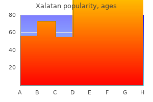
Xalatan
Xalatan dosages: 2.5 ml
Xalatan packs: 1 bottles, 2 bottles, 3 bottles, 4 bottles, 5 bottles, 6 bottles, 7 bottles, 8 bottles, 9 bottles, 10 bottles
2.5 ml xalatan discount overnight delivery
This is a condition in which the olfactory bulbs fail to type medications to avoid during pregnancy 2.5 ml xalatan discount otc, as do areas of the mind related to regulation of sex hormones medicine 1920s xalatan 2.5 ml buy. A variety of receptors sensitive to one or a couple of stimulus types provide sensory perform of the pores and skin and underlying tissues. Information about somatic sensation enters both specific and nonspecific ascending pathways. The somatic sensations embrace touch, pressure, the senses of posture and movement, temperature, and ache. Rapidly adapting mechanoreceptors of the pores and skin give rise to sensations such as vibration, touch, and motion, whereas slowly adapting ones give rise to the feeling of stress. Skin receptors with small receptive fields are concerned in fantastic spatial discrimination, whereas receptors with larger receptive fields signal less spatially precise contact or strain sensations. A major receptor sort liable for the senses of posture and kinesthesia is the muscle-spindle stretch receptor. Cold receptors are sensitive to reducing temperature; heat receptors sign details about growing temperature. Tissue injury and immune cells launch chemical agents that stimulate specific receptors that give rise to the sensation of ache. An eyeball too long or too brief relative to the focusing energy of the lens and cornea causes nearsighted (myopic) or farsighted (hyperopic) vision, respectively. The photopigments of the rods and cones are made up of a protein component (opsin) and a chromophore (retinal). The rods and every of the three cone types have different opsins, which make every of the 4 receptor types delicate to completely different ranges of sunshine wavelengths. When light strikes retinal, it changes form, triggering a cascade of events leading to hyperpolarization of photoreceptors and decreased neurotransmitter release from them. When exposed to darkness, the rods and cones are depolarized and subsequently release extra neurotransmitter than in gentle. The optic nerve fibers from the medial half of each retina cross to the opposite aspect of the mind in the optic chiasm. The fibers from the optic nerves terminate within the lateral geniculate nuclei of the thalamus, which sends fibers to the visual cortex. Photoreceptors also send information to areas of the mind coping with organic rhythms. Coding within the visible system happens alongside parallel pathways by which totally different elements of visual info, corresponding to color, form, movement, and depth, are kept separate from each other. The three cone photopigments differ within the strength of their response to light over differing ranges of wavelengths. Certain ganglion cells are excited by enter from one kind of cone cell and inhibited by enter from a special cone sort. Our sensation of color is determined by the output of the various opponent colour cells and the processing of this output by brain areas concerned in shade vision. Color blindness is as a end result of of abnormalities of the cone pigments resulting from genetic mutations. Six skeletal muscle tissue management eye movement to scan the visible subject for objects of curiosity, hold the fixation point centered on the fovea centralis despite movements of the item or the pinnacle, and forestall adaptation of the photoreceptors. The semicircular canals detect angular acceleration during rotation of the head, which causes bending of the stereocilia on their hair cells. Otoliths within the gelatinous substance of the utricle and saccule (a) transfer in response to modifications in linear acceleration and the place of the pinnacle relative to gravity and (b) stimulate the stereocilia on the hair cells. The receptors for taste lie in taste buds throughout the mouth, principally on the tongue. Different types of taste receptors have completely different sensory transduction mechanisms. Olfactory receptors, that are part of the afferent olfactory neurons, lie within the higher nasal cavity. Odorant molecules, once dissolved within the mucus that bathes the olfactory receptors, bind to particular receptors (protein-binding sites). Each olfactory receptor cell has one or at most a number of of the one thousand completely different receptor sorts. Olfactory pathways go directly to the olfactory cortex and limbic system, rather than to the thalamus. Sound waves enter the external auditory canal and press towards the tympanic membrane, inflicting it to vibrate. The vibrating membrane causes movement of the three small center ear bones; the stapes vibrates against the oval window membrane. Movements of the oval window membrane set up strain waves in the fluid-filled scala vestibuli, which trigger vibrations in the cochlear duct wall, establishing stress waves within the fluid there. These strain waves trigger vibrations within the basilar membrane, which is located on one side of the cochlear duct. As this membrane vibrates, the hair cells of the organ of Corti move in relation to the tectorial membrane. Separate parts of the basilar membrane vibrate maximally in response to specific sound frequencies; excessive frequency is detected close to the oval window and low frequency toward the far end of the cochlear duct. What are the sensory implications of the different crossover factors of the anterolateral and dorsal column ascending pathways in sufferers with accidents that harm half of the spinal twine at a given level List no less than two methods the retina has tailored to decrease the potential downside caused by the photoreceptors being the last layer of the retina that light reaches. Detail the separate mechanisms activated in photoreceptor cells in the presence and in the absence of sunshine. List the sequence of events that occurs between the entry of a sound wave into the external auditory canal and the firing of motion potentials in the cochlear nerve. Describe the useful relationship between the scala vestibuli, scala tympani, and the cochlear duct. What is the relationship between head motion and cupula motion in a semicircular canal What causes the discharge of neurotransmitter from the utricle and saccule receptor cells A vestibular apparatus lies within the temporal bone on each side of the head and consists of three semicircular canals, a utricle, and a saccule. Over the past 3 months, he had experienced a few events when he suddenly grew to become dizzy. Lasting anywhere from a few seconds to a couple of hours, the episodes were typically accompanied by complications, nausea, and vomiting. Because these could be indicators of great underlying sickness, nevertheless, the physician elected to do a more thorough examination. He held a bloody towel pressed tightly to the right facet of his head, and his pores and skin was pale and sweaty. As the emergency room doctor cleaned and stitched the wound, the man and his spouse defined what had occurred.
Generic xalatan 2.5 ml
Just as with ion channels medicine etymology 2.5 ml xalatan generic with visa, the plasma membranes of various cells comprise differing kinds and numbers of transporters; consequently medicine 3604 pill xalatan 2.5 ml cheap visa, they exhibit variations in the forms of substances transported and in their rates of transport. A single ion channel may open and shut many times every second, suggesting that the channel protein fluctuates between these conformations. Over an prolonged time frame, at any given electrochemical gradient, the total number of ions that cross through a channel is dependent upon how typically the channel opens and the way long it stays open. Three elements can alter the channel protein conformations, producing adjustments in how lengthy or how often a channel opens. First, the binding of specific molecules to channel proteins might directly or not directly produce either an allosteric or covalent change within the form of the channel protein. Such channels are therefore termed ligand-gated ion channels, and the ligands that affect them are sometimes chemical messengers, such as those released from the ends of neurons onto goal cells. The precise conformational change is more more probably to be simply adequate to permit or stop an ion to fit through. A change in the conformation of the transporter exposes the transporter binding web site first to one floor of the membrane then to the opposite, thereby transferring the sure solute from one side of the membrane to the opposite. This mannequin reveals net mediated transport from the extracellular fluid to the within of the cell. The size of the conformational change is exaggerated for illustrative functions in this and subsequent figures. Among the most important facilitated-diffusion methods in the physique are those who mediate the transport of glucose throughout plasma membranes. It may be anticipated that as a result of facilitated diffusion the glucose focus inside cells would turn into equal to the extracellular focus. This Four components determine the magnitude of solute flux via a mediated-transport system: (1) the solute focus, (2) the affinity of the transporters for the solute, (3) the number of transporters in the membrane, and (4) the rate at which the conformational change in the transport protein occurs. The flux via a mediated-transport system could be altered by changing any of these 4 components. For any transported solute, a finite number of specific transporters reside in a given membrane at any explicit moment. As with any binding web site, as the focus of the solute to be transported is elevated, the number of occupied binding websites will increase until the transporters turn into saturated-that is, until all the binding websites are occupied. When the transporter binding websites are saturated, the maximal flux across the membrane has been reached and no additional increase in solute flux will happen with will increase in solute concentration. In truth, two kinds of mediated transport exist-facilitated diffusion and active transport. Net facilitated diffusion continues till the concentrations of the solute on the 2 sides of the membrane turn out to be equal. At this level, equal numbers of molecules are binding to the transporter on the outer surface of the lipid bilayer of a plasma membrane (green line) will increase repeatedly in proportion to the extracellular focus, whereas the flux of molecules through a mediated-transport system (purple line) reaches a maximal worth. In this manner, facilitated diffusion contributes significantly to metabolic homeostasis. As with facilitated diffusion, lively transport requires a substance to bind to the transporter in the membrane. As with facilitated-diffusion transporters, active-transport transporters exhibit specificity and saturation-that is, the flux via the transporter is maximal when all transporter binding websites are occupied. The web motion of solute from decrease to higher focus and the maintenance of a better steady-state focus on one side of a membrane may be achieved solely with continuous enter of vitality into the active-transport process. This transporter, which is current in all cells, strikes Na1 from intracellular to extracellular fluid, and K1 in the reverse direction. In both instances, the actions of the ions are in opposition to their respective concentration gradients. This returns the transporter to its original conformation, leading to reduced affinity of the K1 binding sites and elevated affinity of the Na1 binding websites. Together, the activities of these and different active-transport systems account for a significant share of the entire energy usage of the human physique. In the plasma membrane, the direction of energetic Ca21 transport is from cytosol to extracellular fluid. All enzymes in the physique require a narrow range of pH for optimal activity; consequently, this active-transport process is important for cell metabolism and survival. Thus, transporters that mediate secondary active transport have two binding sites, one for an ion-typically but not all the time Na1 -and another for a second molecule. In this example, the electrochemical gradient for Na1 is directed into the cell due to the upper focus of Na1 within the extracellular fluid and the surplus adverse costs contained in the cell. The other solute to be transported, nonetheless, must move towards its focus gradient, uphill into the cell. In this example, the binding of a sodium ion to the transporter produces an allosteric increase in the affinity of the solute binding site on the extracellular floor of the membrane. Binding of Na1 and solute causes a conformational change within the transporter that exposes the binding sites to the intracellular fluid. Na1 diffuses down its electrochemical gradient into the cell, which returns the solute binding web site to a low-affinity state. This, in turn, would lead to a failure of the secondary activetransport techniques that depend on the Na1 concentration gradient for their source of energy. As famous earlier, the net motion of Na1 by a secondary active-transport protein is at all times from high extracellular concentration into the cell, where the focus of Na1 is decrease. Therefore, in secondary active transport, the motion of Na1 is all the time downhill, whereas the online movement of the actively transported solute on the same transport protein is uphill, moving from decrease to larger concentration. High-affinity binding sites for Na1 exist on the extracellular surface of the transporter. Binding of Na1 will increase the affinity of the binding site for the transported solute. The transporter then undergoes a conformational change, which exposes each binding websites to the intracellular facet of the membrane. When the transporter changes conformation, its affinity for Na1 decreases, and Na1 strikes into the intracellular fluid by simple diffusion down its electrochemical gradient. At the identical time, the affinity of the solute binding site decreases, which releases the solute into the intracellular fluid. Once the transporter releases both molecules, the protein assumes its original conformation. The Na1 is then actively transported back out of the cell by primary lively transport, so that the electrochemical gradient for Na1 is maintained. The most essential distinction, due to this fact, between main and secondary energetic transport is that secondary energetic transport uses the saved energy of an electrochemical gradient to transfer both an ion and a second solute across a plasma membrane. The creation and maintenance of the electrochemical gradient, nevertheless, depend upon the motion of main active transporters. The creation of a Na1 concentration gradient across the plasma membrane by the first active transport of Na1 is a way of not directly "storing" power that may then be used to drive secondary active-transport pumps linked to Na1. Sodium ions at all times transfer down their concentration gradient right into a cell, and the transported solute always moves up its gradient. Both Na1 and the transported solute X move in the identical course during cotransport, however in reverse instructions throughout countertransport. In summary, the distribution of drugs between the intracellular and extracellular fluid is usually unequal (Table four.
Diseases
- Macular corneal dystrophy
- Beemer Ertbruggen syndrome
- Lipogranulomatosis
- Mental retardation short stature hand contractures genital anomalies
- Cerebro facio thoracic dysplasia
- Netherton syndrome ichthyosis
- Nephrosis deafness urinary tract digital malformation
Xalatan 2.5 ml cheap
Consequently medicine 1800s order xalatan 2.5 ml overnight delivery, insulin secretion increases greater than it might if plasma glucose had been the one controller medicine 74 xalatan 2.5 ml cheap with mastercard, thereby minimizing the absorptive peak in plasma glucose concentration. This mechanism minimizes the chance of enormous increases in plasma glucose after a meal, which among different things might exceed the capability of the kidneys to fully reabsorb the entire glucose that appears within the filtrate within the renal nephrons. Incretins are gastrointestinal hormones that act as feedforward signals to the pancreas. The clinical features of the completely different forms of diabetes mellitus shall be lined later on this chapter. Finally, enter of the autonomic neurons to the islets of Langerhans additionally influences insulin secretion. Activation of the parasympathetic neurons, which occurs during the ingestion of a meal, stimulates the secretion of insulin and constitutes a second sort of feedforward regulation. In distinction, activation of the sympathetic neurons to the islets or an increase in the plasma focus of epinephrine (the hormone secreted by the adrenal medulla) inhibits insulin secretion. In summary, insulin has the primary function in controlling the metabolic changes required for feasting or fasting. They all oppose the action of insulin in a technique or one other and are generally recognized as glucose-counterregulatory controls. As described subsequent, crucial of these are glucagon, epinephrine, sympathetic nerves, cortisol, and progress hormone. Begin Plasma glucose Pancreatic islet alpha cells Glucagon secretion Plasma glucagon Liver Glycogenolysis Gluconeogenesis Ketone synthesis Plasma glucose Plasma ketones glucagon secretion. Thus, glucagon (1) stimulates glycogenolysis, (2) stimulates gluconeogenesis, and (3) stimulates the synthesis of ketones. The total outcomes are to increase the plasma concentrations of glucose and ketones, which are essential for the postabsorptive state, and to prevent hypoglycemia. The effects, if any, of glucagon on adipocyte function in people are nonetheless unresolved. The main stimulus for glucagon secretion is a lower in the circulating concentration of glucose (which in flip causes a decrease in plasma insulin). The adaptive value of such a reflex is obvious; a decreased plasma glucose focus induces an increase within the secretion of glucagon into the blood, which, by its results on metabolism, serves to restore regular blood glucose concentration by glycogenolysis and gluconeogenesis. Conversely, an elevated plasma glucose focus inhibits the secretion of glucagon, thereby serving to to return the plasma glucose concentration toward regular. As a result, through the postabsorptive state, there is an increase in the glucagon/insulin ratio in the plasma, and this accounts nearly entirely for the transition from the absorptive to the postabsorptive state. The dual and reverse actions of glucagon and insulin on glucose homeostasis clearly illustrate the final precept of physiology that the majority physiological capabilities are controlled by a quantity of regulatory methods, often working in opposition. The secretion of glucagon, like that of insulin, is managed not solely by the plasma concentration of glucose but also by amino acids and by neural and hormonal inputs to the islets. For example, vital increases in certain amino acids-as may occur 574 Chapter 16 Given the effects of glucagon on plasma glucose concentrations, what impact do you think fight-or-flight (stress) reactions would have on the circulating stage of glucagon Glucagon secreted in such conditions helps forestall hypoglycemia that may happen following the rise in insulin in a protein-rich meal. As another example, the sympathetic nerves to the islets stimulate glucagon secretion-just the other of their impact on insulin secretion. This is a method during which extra vitality in the type of glucose is supplied in instances of stress or emergency. Epinephrine and Sympathetic Nerves to Liver and Adipose Tissue As famous earlier, epinephrine and the sympathetic nerves to the pancreatic islets inhibit insulin secretion and stimulate glucagon secretion. Its main direct effects embody stimulation of (1) glycogenolysis in both the liver and skeletal muscle, (2) gluconeogenesis within the liver, and (3) lipolysis in adipocytes. Activation of the sympathetic nerves to the liver and adipose tissue elicits the same responses from these organs as does circulating epinephrine. Both are then released into the blood, the place they serve instantly as an power Begin Plasma glucose Reflex through glucose receptors in the central nervous system Adrenal medulla Epinephrine secretion Activity of sympathetic nerves to liver and adipose tissue Plasma epinephrine Skeletal muscle Glycogenolysis Liver Glycogenolysis Gluconeogenesis Adipose tissue Lipolysis Plasma glucose, fatty acids, glycerol stimulation of both gluconeogenesis and lipolysis; nevertheless, neither of those crucial metabolic transformations occurs to the usual diploma in a person poor in cortisol. Therefore, in response to fasting, people with a cortisol deficiency can develop hypoglycemia important enough to intervene with cellular operate. Moreover, cortisol can have more than a permissive operate when its plasma focus does increase, because it does throughout stress. At high concentrations, cortisol elicits many metabolic events ordinarily associated with fasting (Table 16. In truth, cortisol truly decreases the sensitivity of muscle and adipose cells to insulin, which helps to keep plasma glucose focus during fasting, thereby offering a daily source of energy for the mind. Clearly, here is one other hormone that, along with glucagon and epinephrine, can exert actions opposite these of insulin. Glycogenolysis in skeletal muscle contributes to restoring plasma glucose by releasing lactate, which is transformed to glucose in the liver and launched into the blood. Thus, enhanced sympathetic nervous system activity exerts results on organic metabolism-specifically, elevated plasma concentrations of glucose, glycerol, and fatty acids-that are reverse those of insulin. As might be predicted from these effects, low blood glucose leads to increases in both epinephrine secretion and sympathetic nerve activity to the liver and adipose tissue. This is the same stimulus that leads to increased glucagon secretion, although the receptors and pathways are totally different. When the plasma glucose focus decreases, glucose-sensitive cells within the central nervous system (and, probably, the liver) initiate the reflexes that lead to increased activity in the sympathetic pathways to the adrenal medulla, liver, and adipose tissue. The adaptive value of the response is identical as that for the glucagon response to hypoglycemia; blood glucose returns towards regular, and fatty acids are provided for cell utilization. Growth Hormone the primary physiological effects of development hormone are to stimulate each growth and protein synthesis. Compared to these results, these it exerts on carbohydrate and lipid metabolism are less significant. Nonetheless, as is true for cortisol, either deficiency or excess of progress hormone does produce important abnormalities in lipid and carbohydrate metabolism. Growth hormone (1) increases the responsiveness of adipocytes to lipolytic stimuli, (2) stimulates gluconeogenesis by the liver, and (3) reduces the ability of insulin to stimulate glucose uptake by muscle and adipose tissue. Basal concentrations are permissive for stimulation of gluconeogenesis and lipolysis in the postabsorptive state. Net end result: Increased plasma concentrations of amino acids, glucose, and free fatty acids Regulation of Organic Metabolism and Energy Balance 575 Cortisol Cortisol, the most important glucocorticoid produced by the adrenal cortex, has a vital permissive operate in the adjustments to fasting. Epinephrine stimulates glycogenolysis in each liver and skeletal muscle, whereas glucagon does so only in liver. Hypoglycemia Hypoglycemia is broadly outlined as an abnormally low plasma glucose concentration. The plasma glucose concentration can decrease to very low values, usually through the postabsorptive state, in persons with several forms of disorders.
Order xalatan 2.5 ml otc
The ensuing improve in cytosolic Ca21 concentration prompts proteins required for the vesicle membrane to fuse with the plasma membrane and release the vesicle contents into the extracellular fluid medications available in mexico generic xalatan 2.5 ml with amex. Material stored in secretory vesicles is on the market for speedy secretion in response to a stimulus symptoms menopause xalatan 2.5 ml buy discount line, without delays that might happen if the fabric had to be synthesized after the stimulus arrived. Exocytosis is the mechanism by which most neurons communicate with one another through the discharge of neurotransmitters stored in secretory vesicles that merge with the plasma membrane. These two membranes often include completely different ion channels and different transporters for mediated transport. As a result of these variations, substances can undergo a net motion from a low focus on one facet of an epithelium to the next concentration on the opposite facet. Examples include the absorption of material from the gastrointestinal tract into the blood, the motion of gear between the kidney tubules and the blood during urine formation, and the secretion of ions and water by glands such as sweat glands. The organic substance then moves out of the cell at the blood facet down a concentration gradient by the use of facilitated diffusion. Shown beneath the cell is the focus profile of the transported solute throughout the epithelium. The motion of Na1 into the cell across the plasma membrane on the other side is all the time downhill. In this case, the entry of an organic molecule X throughout the apical plasma membrane occurs by way of a secondary lively transporter linked to the downhill motion of Na1 into the cell. In the method, X moves from a lower concentration in the luminal fluid to a higher concentration in the cell. The substance exits across the basolateral membrane by facilitated diffusion, which moves the fabric from its higher focus in the cell to a decrease concentration in the extracellular fluid on the blood side. The concentration of the substance could also be considerably greater on the blood facet than in the lumen because the blood-side concentration can strategy equilibrium with the excessive intracellular focus created by the apical membrane entry step. The energetic transport of Na1, as beforehand described, results in a decrease in the Na1 focus on one facet of an epithelial layer (the luminal facet in our example) and an increase on the opposite. Therefore, internet movement of solute across an epithelium is accompanied by a circulate of water in the same path. It can additionally be the major method by which water is absorbed from the intestines into the blood (Chapter 15). The energetic transport of Na1 throughout the cells and into the surrounding interstitial areas produces an elevated osmolarity on this region and a decreased osmolarity in the lumen. This results in the osmotic move of water across the epithelium in the identical course as the web solute movement. The water diffuses by way of aquaporins within the membrane (transcellular pathway) and across the tight junctions between the epithelial cells (paracellular pathway). The binding sites on transporters exhibit chemical specificity, affinity, and saturation. The magnitude of the flux by way of a mediated-transport system depends on the degree of transporter saturation, the number of transporters within the membrane, and the rate at which the conformational change in the transporter happens. Facilitated diffusion is a mediated-transport process that moves molecules from larger to decrease concentrations throughout a membrane via a transporter until the 2 concentrations turn out to be equal. Active transport is a mediated-transport process that strikes molecules towards an electrochemical gradient throughout a membrane via a transporter and an input of energy. Secondary active transport uses the binding of ions (often Na1) to the transporter to drive the secondary-transport process. In secondary lively transport, the downhill circulate of an ion is linked to the uphill movement of a second solute both in the same course because the ion (cotransport) or in the opposite direction of the ion (countertransport). Water crosses membranes by (a) diffusing by way of the lipid bilayer, and (b) diffusing through protein channels within the membrane. Osmosis is the diffusion of water throughout a membrane from a area of upper water focus to a area of decrease water concentration. The osmolarity-total solute concentration in a solution-determines the water concentration: the upper the osmolarity of a solution, the lower the water focus. Application of enough pressure to an answer will stop the osmotic move of water into the answer from a compartment of pure water. Net water movement happens from a region of decrease osmotic strain to one of larger osmotic pressure. Because water comes to diffusion equilibrium throughout cell membranes, the intracellular fluid has an osmolarity equal to that of the extracellular fluid. Na1 and Cl2 are the most important successfully nonpenetrating solutes in the extracellular fluid; K1 and varied organic solutes are the major effectively nonpenetrating solutes in the intracellular fluid. What options of epithelial cells shown in this figure lend support to that precept Simple diffusion is the movement of molecules from one location to another by random thermal motion. The net flux between two compartments always proceeds from higher to decrease concentrations. Diffusion equilibrium is reached when the concentrations of the diffusing substance within the two compartments become equal. The magnitude of the web flux J across a membrane is directly proportional to the concentration difference throughout the membrane Co 2 Ci, the floor space of the membrane A, and the membrane permeability coefficient P. Nonpolar molecules diffuse via the hydrophobic portions of membranes rather more rapidly than do polar or ionized molecules because nonpolar molecules can dissolve in the fatty acyl tails within the lipid bilayer. Ions diffuse throughout membranes by passing via ion channels fashioned by integral membrane proteins. The diffusion of ions throughout a membrane depends on each the focus gradient and the membrane potential. The flux of ions across a membrane may be altered by opening or closing ion channels. During endocytosis, regions of the plasma membrane invaginate and pinch off to form vesicles that enclose a small volume of extracellular material. The three classes of endocytosis are (i) fluid endocytosis, (ii) phagocytosis, and (iii) receptor-mediated endocytosis. Most endocytotic vesicles fuse with endosomes, which in flip switch the vesicle contents to lysosomes for digestion by lysosomal enzymes. Potocytosis is a special kind of receptor-mediated endocytosis by which vesicles called caveolae ship their contents directly to the cytosol. Exocytosis, which occurs when intracellular vesicles fuse with the plasma membrane, provides a means of adding parts to the Movement of Molecules Across Cell Membranes 113 Mediated-Transport Systems I. The mediated transport of molecules or ions across a membrane entails binding the transported solute to a transporter protein within the membrane. Changes within the conformation of the transporter move the binding site to the alternative side of the membrane, where the solute dissociates from the protein.
Xalatan 2.5 ml proven
An improve in firing of the arterial baroreceptors because of symptoms estrogen dominance cheap 2.5 ml xalatan with visa an increase in pressure causes medicine zetia 2.5 ml xalatan cheap with mastercard, by the use of the medullary cardiovascular heart, an increase in parasympathetic outflow to the center and a decrease in sympathetic outflow to the heart, arterioles, and veins. The baroreceptor reflexes are short-term regulators of arterial strain but adapt to a maintained change in strain. Blood stress could be influenced by many factors other than baroreceptors, including arterial blood fuel concentrations, pain, feelings, and sexual exercise. Write the equation relating imply arterial pressure to cardiac output and whole peripheral resistance. What variable accounts for the reality that imply pulmonary arterial stress is lower than mean systemic arterial stress Draw a circulate diagram illustrating the components that decide mean arterial pressure. Identify the receptors, afferent pathways, integrating center, efferent pathways, and effectors in the arterial baroreceptor reflex. When the arterial baroreceptors decrease or increase their rate of firing, what modifications in autonomic outflow and cardiovascular perform occur Describe the position of blood quantity within the long-term regulation of arterial pressure. Mean arterial strain, the primary regulated variable within the cardiovascular system, equals the product of cardiac output and complete peripheral resistance. The primary baroreceptors are the arterial baroreceptors, together with the two carotid sinuses and the aortic arch. Other baroreceptors are positioned within the systemic veins, pulmonary vessels, and partitions of the guts. Thus, the preliminary events-blood loss and decreased blood volume-are partially compensated for by the movement of interstitial fluid into the vascular system. This mechanism, referred to as autotransfusion, can restore the blood volume to nearly normal ranges within 12 to 24 hours after a reasonable hemorrhage (Table 12. At this time, the whole restoration of blood quantity is due to expansion of the plasma quantity; subsequently, the hematocrit actually decreases. The early compensatory mechanisms for hemorrhage (the baroreceptor reflexes and interstitial fluid absorption) are highly environment friendly, so that losses of as a lot as 30% of total blood quantity could be sustained with solely slight reductions of mean arterial pressure or cardiac output. We must emphasize that absorption of interstitial fluid solely redistributes the extracellular fluid. Ultimate restoration of blood quantity involves increasing fluid ingestion and minimizing water loss through the kidneys. These slower-acting compensations include an increase in thirst and a reduction in the excretion of salt and water in the urine. They are mediated by hormones, including renin, angiotensin, and aldosterone, and are described in Chapter 14. Replacement of the lost erythrocytes, which requires the hormone erythropoietin to stimulate erythropoiesis (maturation of immature red blood cells), was described in Section A of this chapter. The most severe consequence of hypotension is lowered blood move to the mind and cardiac muscle. The values of things modified as a direct result of the hemorrhage-stroke quantity, cardiac output, and imply arterial pressure-are restored by the baroreceptor reflex towards, however not all the method in which to , normal. In distinction, values not altered directly by the hemorrhage but only by the reflex response to hemorrhage-heart rate and total peripheral resistance- improve above their prehemorrhage values. The increased peripheral resistance results from increases in sympathetic outflow to the arterioles in many vascular beds (but not these of the heart and brain). Thus, skin blood flow might decrease significantly because of arteriolar vasoconstriction; this is why the skin can become pale and cold following a major hemorrhage. Note that the lower in arterial pressure immediately following hemorrhage is secondary to the lower in stroke volume and, due to this fact, cardiac output. The circulatory system, particularly the guts, suffers harm if shock is prolonged. As the guts deteriorates, cardiac output additional declines and shock becomes progressively worse. Ultimately, shock might become irreversible even though blood transfusions and other appropriate remedy might quickly restore blood strain. Why that is so requires an understanding of the action of gravity upon the lengthy, continuous columns of blood in the vessels between the guts and the feet. The pressures described in previous sections of this chapter are for a person in the horizontal position, during which all blood vessels are at nearly the identical level as the guts. In contrast, when a person is standing, the intravascular stress in all places becomes equal to the pressure generated by cardiac contraction plus an extra stress equal to the burden of a column of blood from the center to the purpose of measurement. In an average grownup, for instance, the burden of a column of blood extending from the center to the feet would quantity to 80 mmHg. In a foot capillary, therefore, the pressure could doubtlessly enhance from 25 (the common capillary strain resulting from cardiac contraction) to a hundred and five mmHg, the additional 80 mmHg being due to the load of the column of blood. This increase in strain as a outcome of gravity influences the efficient circulating blood quantity in a number of methods. First, the increased hydrostatic strain that occurs in the legs (as nicely because the buttocks and pelvic area) when a person is standing pushes outward on the highly distensible vein walls, inflicting marked distension. Simultaneously, the rise in capillary stress caused by the gravitational pressure produces increased filtration of fluid out of the capillaries into the interstitial space. The combined effects of venous pooling and increased capillary filtration cut back the effective circulating blood quantity very similarly to the results caused by a mild hemorrhage. Venous pooling explains why an individual could sometimes feel faint when standing up all of a sudden. The decreased venous return causes a transient decrease in end-diastolic volume and subsequently decreased stretch of the ventricles. This reduces stroke volume, which in flip reduces cardiac output and blood stress. The effects of gravity may be offset by contraction of the skeletal muscular tissues in the legs. Hemorrhage is a hanging instance of hypotension because of a decrease in blood volume. It might happen by way of the pores and skin, as in extreme sweating or burns, or through the gastrointestinal tract, as in diarrhea or vomiting, or by way of the kidneys, as with unusually massive urinary losses. Regardless of the route, the lack of fluid decreases blood volume and can result in signs and compensatory cardiovascular adjustments much like these seen in hemorrhage. One main trigger is a decrease in cardiac contractility (for instance, during a heart attack). Another trigger is strong emotion, which in uncommon cases can cause hypotension and fainting. The higher mind facilities concerned with feelings inhibit sympathetic exercise to the circulatory system and improve parasympathetic activity to the guts, leading to a markedly decreased arterial stress and mind blood move. It ought to be famous that the fainting that typically occurs in a person donating blood is normally because of hypotension brought on by emotion, not due to the blood loss, as a end result of a gradual donation of zero.
Xalatan 2.5 ml generic without prescription
After remaining pretty low and secure for the first week symptoms dust mites order 2.5 ml xalatan with amex, the plasma focus of estrogen increases quickly in the course of the second week because the dominant ovarian follicle grows and secretes more estrogen symptoms jock itch buy xalatan 2.5 ml line. This is adopted by a second enhance because of secretion by the corpus luteum and, finally, a rapid decrease during the last days of the cycle. Very small amounts of progesterone are launched by the ovaries through the follicular part till just earlier than ovulation. Very soon after ovulation, the developing corpus luteum begins to release large quantities of progesterone; from this level, the progesterone sample is just like that for estrogen. Its pattern is much like that of estrogen: It increases through the late follicular part, remains high during the luteal section, and then decreases because the corpus luteum degenerates. The following dialogue will explain how these hormonal adjustments are interrelated to produce a self-cycling sample. The suggestions results of the ovarian hormones to be described in the text are summarized for reference in Table 17. The events marked by the circled numbers are described later within the text and are listed right here to present a abstract. There are at all times numerous preantral and early antral follicles within the ovary between puberty and menopause. Some of the estrogen produced diffuses into the blood and maintains a comparatively secure plasma concentration 3. The androgens diffuse into the granulosa cells and are transformed to estrogen by aromatase. Therefore, the secretion of estrogen by the granulosa cells requires the interaction of both kinds of follicle cells and both pituitary gland gonadotropins. The dashed line with an arrow throughout the ovaries indicates that estrogen will increase granulosa cell perform (local constructive feedback). The dominant follicle now starts to secrete sufficient estrogen that the plasma concentration of this steroid begins to increase 5. Estrogen, at these still comparatively low concentrations, is exerting a adverse suggestions inhibition on the secretion of gonadotropins (Table 17. The main oocyte completes its first meiotic division and undergoes cytoplasmic modifications that prepare the ovum for implantation should fertilization happen. The granulosa cells start releasing progesterone and lowering the release of estrogen, which accounts for the midcycle lower in plasma estrogen focus and the small rise in plasma progesterone concentration simply earlier than ovulation. Enzymes and prostaglandins, synthesized by the granulosa cells, break down the follicular�ovarian membranes. These weakened membranes rupture, allowing the oocyte and its surrounding granulosa cells to be carried out onto the surface of the ovary. The remaining granulosa cells of the ruptured follicle (along with the theca cells of that follicle) are reworked into the corpus luteum, which begins to release progesterone and estrogen. During its quick life within the nonpregnant lady, the corpus luteum secretes large quantities of progesterone and estrogen 12, in addition to inhibin. In the presence of estrogen, the excessive plasma focus of progesterone causes a decrease in the secretion of the gonadotropins by the pituitary gland. Consequently, through the luteal section of the cycle, plasma concentrations of the gonadotropins are very low thirteen. The corpus luteum has a finite life in the absence of an increase in gonadotropin secretion. With degeneration of the corpus luteum, plasma progesterone and estrogen concentrations decrease 15. This completes the description of the management of ovarian perform throughout a typical menstrual cycle. When the corpus luteum degenerates, the decrease in hormone secretion allows the gonadotropin concentrations to increase sufficient to promote the expansion of another group of follicles. This illustrates that ovarian occasions, by way of hormonal suggestions, management the hypothalamus and anterior pituitary gland. Day 1 is the primary day of menstrual move, and the whole duration of menstruation is known as the menstrual section (generally about three to 5 days in a typical 28-day cycle). During this time, the epithelial lining of the uterus-the endometrium-degenerates, resulting in the menstrual circulate. The menstrual move then ceases, and the endometrium begins to thicken as it regenerates underneath the influence of estrogen. This interval of development, the proliferative section, lasts for the ten days or so between cessation of menstruation and the incidence of ovulation. Soon after ovulation, under the influence of progesterone and estrogen from the corpus luteum, the endometrium begins to secrete glycogen in the glandular epithelium, followed by glycoproteins and mucopolysaccharides. The part of the menstrual cycle between ovulation and the onset of the subsequent menstruation known as the secretory part. During the proliferative phase, an rising plasma estrogen concentration stimulates progress of both the endometrium and the underlying uterine smooth muscle (called the myometrium). In addition, it induces the synthesis of receptors for progesterone in endometrial cells. Then, following ovulation and formation of the corpus luteum (during the secretory phase), progesterone acts upon this estrogen-primed endometrium to convert it to an actively secreting tissue. The endometrial glands become coiled and crammed with glycogen, the blood vessels turn out to be more numerous, and enzymes accumulate within the glands and connective tissue. These modifications are essential to make the endometrium a hospitable surroundings for implantation and nourishment of the developing embryo. Progesterone additionally inhibits myometrial contractions, in large part by opposing the stimulatory actions of estrogen and regionally generated prostaglandins. This is very important to ensure that a fertilized egg can safely implant as quickly as it arrives within the uterus. Uterine quiescence is maintained by progesterone throughout being pregnant and is important to prevent premature delivery. Estrogen and progesterone even have essential results on the secretion of mucus by the cervix. All of those characteristics are most pronounced at the time of ovulation and allow sperm deposited in the vagina to transfer simply through Reproduction 621 the decrease in plasma progesterone and estrogen concentrations that outcomes from degeneration of the corOvarian occasion pus luteum deprives the highly developed endometrium of its hormonal help and causes menstruation. The first occasion is constriction of the uterine blood vessels, which Ovum leads to a diminished provide of oxygen and vitamins to Progesterone Estrogen Estrogen the endometrial cells. Disintegration starts in the complete lining, aside from a skinny, underlying layer that may regenerate the endometrium in the next cycle. Both the vasoconstriction and uterine contractions Endometrial are mediated by prostaglandins produced by the endomethickness trium in response to the lower in plasma estrogen and progesterone concentrations. The major explanation for menDay 1 5 10 15 20 25 28 5 strual cramps, dysmenorrhea, is overproduction of those prostaglandins, leading to excessive uterine contractions. Menstrual Proliferative Secretory Menstrual Uterine section the prostaglandins additionally affect clean muscle elsewhere within the physique, which accounts for a number of the systemic Ovarian Follicular Follicular Luteal symptoms that typically accompany the cramps, such section as nausea, vomiting, and headache.
American Mistletoe. Xalatan.
- Dosing considerations for American Mistletoe.
- What is American Mistletoe?
- Increasing muscle contractions, causing abortion, or any other uses.
- Are there safety concerns?
- How does American Mistletoe work?
Source: http://www.rxlist.com/script/main/art.asp?articlekey=96319

Buy cheap xalatan 2.5 ml online
Although medicine ethics cheap 2.5 ml xalatan free shipping, theoretically medicine for high blood pressure 2.5 ml xalatan cheap amex, hypertension might result from an increase both in cardiac output or in complete peripheral resistance, it seems that generally of well-established primary hypertension, increased complete peripheral resistance attributable to decreased arteriolar radius is essentially the most vital factor. A number of environmental threat elements contribute to the development of main hypertension. Recent research present that lifestyle modifications that reduce those components end in lowered blood stress, both in hypertensive and healthy individuals. Obesity and the regularly associated insulin resistance (discussed in Chapter 16) are threat elements, and weight reduction significantly reduces blood pressure in most people. Chronic, excessive salt consumption can also be related to hypertension, and up to date analysis has revealed mechanisms by which even slight elevations in plasma Na1 ranges result in continual overstimulation of the sympathetic nervous system, constriction of arterioles, and narrowing of the lumen of arteries. In addition to obesity and extreme salt intake, other environmental factors hypothesized to contribute to major hypertension embody smoking; extra alcohol consumption; diets low in fruits, vegetables, and entire grains; diets low in vitamin D and calcium; lack of train; chronic stress; excess caffeine consumption; maternal smoking; low birth weight; and not being breast-fed as an toddler. These sufferers respond well to a low-sodium diet or to medication called diuretics, which trigger elevated Na1 and water loss in the urine (see Chapter 14). A number of endocrine problems end in hypertension, corresponding to syndromes involving hypersecretion of cortisol, aldosterone, or thyroid hormone (see Chapters eleven and 14). Medications corresponding to oral contraceptives and nonsteroidal antiinflammatory drugs also can contribute to hypertension. Finally, a link has been established between hypertension and the irregular nighttime respiratory pattern, sleep apnea (see Chapter 13). The major classes of medication used to treat hypertension are summarized in Table 12. You will observe in subsequent sections of this chapter that these similar medicine are also used to treat coronary heart failure and in each the prevention and therapy of coronary heart attacks. One reason for this overlap is that these three ailments are causally interrelated. For instance, as famous in this section, hypertension is a major threat factor for the development of heart illness. In addition, although, the drugs usually have a number of cardiovascular effects, which can contribute in several methods to the therapy of the totally different ailments. They are most likely to lower cardiac output with little or no change in complete peripheral resistance. Beta-adrenergic receptor blockers (beta-blockers): these medication exert their antihypertensive effects primarily by decreasing cardiac output. Calcium channel blockers: these medication reduce the entry of Ca21 into vascular smooth muscle cells, causing them to contract much less strongly and reducing complete peripheral resistance. Drugs that antagonize a quantity of elements of the sympathetic nervous system: the main effect of these drugs is to scale back sympathetic mediated stimulation of arteriolar smooth muscle and thereby reduce total peripheral resistance. Examples embrace drugs that inhibit the mind centers that mediate the sympathetic outflow to arterioles, and drugs that block a-adrenergic receptors on the arterioles. It has turn out to be commonplace apply to separate folks with heart failure into two classes: (1) those with diastolic dysfunction (problems with ventricular filling) and (2) those with systolic dysfunction (problems with ventricular ejection). Its irregular stiffness ends in a reduced ability to fill adequately at normal diastolic filling pressures. In pure diastolic dysfunction, ventricular compliance is decreased but ventricular contractility is regular. Several conditions may lead to decreased ventricular compliance, but by far the most common is the existence of systemic hypertension. As noted within the previous part, hypertrophy results when the left ventricle pumps in opposition to a chronically elevated arterial pressure (afterload). The structural and biochemical modifications associated with this hypertrophy make the ventricle stiff and less able to expand. In contrast to diastolic dysfunction, systolic dysfunction outcomes from myocardial harm, like that resulting from a heart attack (discussed next). This kind of dysfunction is characterised by a lower in cardiac contractility-a lower stroke volume at any given end-diastolic volume. Fluid retention causes an increase in end-diastolic quantity and restores stroke quantity toward normal by the Frank�Starling mechanism. Note that this compensation occurs despite the very fact that contractility-the fundamental defect-has not been altered by the fluid retention. The decreased cardiac output of coronary heart failure, regardless of whether or not it is due to diastolic or systolic dysfunction, triggers the arterial baroreceptor reflexes. In this example, these reflexes are elicited greater than traditional as a end result of, for unknown reasons, the afferent baroreceptors are much less sensitive. In other words, the baroreceptors discharge much less quickly than normal at any given imply or pulsatile arterial strain and the brain interprets this decreased discharge as a larger-than-usual lower in stress. The reflex will increase in coronary heart rate and total peripheral resistance are initially useful in restoring cardiac output and arterial stress, just as if the changes in these parameters had been triggered by hemorrhage. Maintained chronically all through the interval of cardiac failure, the baroreceptor reflexes also result in fluid retention and an expansion-often massive-of the extracellular volume. This is as a end result of, as Chapter 14 describes, the neuroendocrine efferent parts of the reflexes cause the kidneys to scale back their excretion of sodium and water. Because the plasma volume is part of the extracellular fluid quantity, plasma volume additionally will increase. Therefore, fluid retention can be, no much less than initially, an adaptive response to decreased cardiac output. For one factor, when a ventricle with systolic dysfunction (as opposed to a normal ventricle) becomes very distended with blood, its force of contraction actually decreases and the state of affairs worsens. Second, the fluid retention, with its accompanying enhance in venous stress, causes edema-accumulation of interstitial fluid. Therefore, a lot of the fluid retained by the kidneys ends up as additional interstitial fluid rather than additional plasma. Most important in this regard, failure of the left ventricle- whether or not because of diastolic or systolic dysfunction-leads to pulmonary edema, the buildup of fluid within the interstitial spaces of the lung or in the air spaces themselves. The reason for such accumulation is that the left ventricle fails to pump blood to the identical extent as the proper ventricle, so the amount of blood in all of the pulmonary vessels increases. The ensuing engorgement of pulmonary capillaries will increase the capillary strain above its usually very low value, causing filtration to happen at a rate sooner than the lymphatics can take away the fluid. They get rid of the extreme fluid accumulation contributing to edema and/or worsening myocardial perform. Cardiac inotropic medicine: Drugs that improve beta-adrenergic receptor pathways and drugs such as digitalis, which increases ventricular contractility by growing cytosolic Ca21 concentration in the myocardial cell, can improve cardiac output in the brief term. Vasodilator medication: Drugs that lower total peripheral resistance and therefore the arterial blood strain (afterload) in opposition to which the failing coronary heart pumps. Beta-adrenergic receptor blockers: Drugs that block the main adrenergic receptors in the myocardium. You might predict that such an motion, by blocking sympathetically induced increases in cardiac contractility, can be counterproductive (note above that beta-agonists are sometimes used, which is more intuitive). One hypothesis is that extra sympathetic stimulation of the heart reflexively produced by the decreased cardiac output of heart failure might trigger an extreme elevation of cytosolic Ca21 focus, which would result in cell apoptosis and necrosis; beta-adrenergic receptor blockers would stop this. By chronically maintaining the arterial blood stress the failing heart must pump against, this elevated resistance makes the failing heart work more durable.
2.5 ml xalatan buy mastercard
First medications 2 times a day 2.5 ml xalatan generic free shipping, protein ingestion increases the concentration of peptides in the lumen of the abdomen medications identification buy xalatan 2.5 ml free shipping. The second cause is extra complicated and reflects the effects of proteins on luminal acidity. During the cephalic part, earlier than food enters the stomach, the H1 concentration within the lumen increases because there are few buffers present to bind any secreted H1. The protein in food is a superb buffer, nonetheless, in order it enters the abdomen, the H1 concentration decreases as H1 binds to proteins and begins to denature them. The more protein in a meal, the larger the buffering of acid and the more acid secreted. High acidity in the duodenum triggers reflexes that inhibit gastric acid secretion. This inhibition is useful as a result of the digestive exercise of enzymes and bile salts within the small gut is strongly inhibited by acidic options. This reflex limits gastric acid manufacturing when the H1 focus within the duodenum increases as a end result of the entry of chyme from the abdomen. Acid, distension, hypertonic options, solutions containing amino acids, and fatty acids in the small gut reflexively inhibit gastric acid secretion. The extent to which acid secretion is inhibited through the intestinal section varies, depending upon the quantities of those substances within the intestine; the net end result is the same, however-balancing the secretory activity of the stomach with the digestive and absorptive capacities of the small intestine. Exposure to low pH in the lumen of the abdomen activates a very rapid, autocatalytic course of by which pepsin is produced from pepsinogen. These enzymes are synthesized and stored intracellularly in inactive forms, collectively referred to as zymogens. The primary pathway for exciting pepsinogen secretion is input to the chief cells from the enteric nervous system. During the cephalic, gastric, and intestinal phases, a lot of the components that stimulate or inhibit acid secretion exert the identical impact on pepsinogen secretion. However, pepsin accelerates protein digestion and normally accounts for about 20% of whole protein digestion. It can also be necessary within the digestion of collagen contained within the connective-tissue matrix of meat. This is beneficial as a outcome of it helps shred meat into smaller, more simply processed pieces with larger floor area for digestion. The pepsin thus shaped additionally catalyzes its own production by appearing on further molecules of pepsinogen. As within the esophagus, the stomach produces peristaltic waves in response to the coming meals. Each wave begins in the physique of the abdomen and produces solely a ripple as it proceeds toward the antrum; this contraction is too weak to produce a lot mixing of the luminal contents with acid and pepsin. As the wave approaches the larger mass of wall muscle surrounding Gastric Motility An empty abdomen has a quantity of solely about 50 mL, and the diameter of its lumen is just barely bigger than that of the small gut. As a consequence of the sphincter closing, solely a small amount of chyme is expelled into the duodenum with each wave. This backward movement of chyme, known as retropulsion, generates sturdy shear forces that helps to disperse the meals particles and improve mixing of the chyme. Recall that the decrease esophageal sphincter Esophagus Duodenum Lower esophageal sphincter Pyloric sphincter Stomach Peristaltic wave prevents this retrograde motion of abdomen contents from coming into the esophagus. Their rhythm (three per minute) is generated by pacemaker cells in the longitudinal easy muscle layer. These smooth muscle cells undergo spontaneous depolarization� repolarization cycles (slow waves) generally identified as the essential electrical rhythm of the abdomen. In the absence of neural or hormonal enter, nevertheless, these depolarizations are too small to cause vital contractions. Excitatory neurotransmitters and hormones act upon the sleek muscle to additional depolarize the membrane, thereby bringing it closer to threshold. The variety of spikes fired with each wave determines the energy of the muscle contraction. Therefore, whereas the frequency of contraction is determined by the intrinsic primary electrical rhythm and stays essentially constant, the force of contraction-and, consequently, the quantity of gastric emptying per contraction- is determined reflexively by neural and hormonal enter to the antral easy muscle. The initiation of those reflexes relies upon upon the contents of each the abdomen and small gut. For example, gastrin in sufficiently high concentrations will increase the pressure of antral smooth muscle contractions. Distension of the stomach also increases the drive of antral contractions by way of lengthy and brief reflexes triggered by mechanoreceptors in the stomach wall. Black arrows indicate motion of luminal material; purple arrows indicate motion of the peristaltic wave in the stomach wall. Membrane depolarization brings the gradual wave nearer to threshold, increasing the motion potential frequency and thus the drive of clean muscle contraction. The price of gastric emptying has vital scientific implications notably when contemplating what meals kind is eaten with oral drugs. A meal rich in fats content tends to sluggish oral drug absorption because of a delay of the drug getting into the small gut via the pyloric sphincter. It does so by slowing the speed of entry of chyme and thereby the delivery of huge molecules that may rapidly be broken down into many small molecules by enzymes in the small intestine. An increase in parasympathetic activity will increase gastric motility, whereas a rise in sympathetic exercise decreases motility. The endocrine portion secretes insulin, glucagon, and different hormones into the blood. Furthermore, the dearth of regular water movement into the lumen results in a thickening of pancreatic secretions; this could result in clogging of the pancreatic ducts and pancreatic injury. In reality, the cystic and fibrotic (scarring) look of the diseased pancreas was the origin of the name of this disease. The enzymes the pancreas secretes digest fat, polysaccharides, proteins, and nucleic acids to fatty acids and monoglycerides, sugars, amino acids, and nucleotides, respectively. The proteolytic enzymes are secreted in inactive types (zymogens), as described for pepsinogen in the abdomen, after which activated in the duodenum by other enzymes. Like pepsinogen, the secretion of zymogens protects pancreatic cells from autodigestion. A key step on this activation is mediated by enterokinase, which is embedded within the apical plasma membranes of the intestinal epithelial cells. Enterokinase is a proteolytic enzyme that splits off a peptide from pancreatic trypsinogen, forming the active enzyme trypsin. This activating function is along with the perform of trypsin in digesting ingested protein.
Xalatan 2.5 ml discount free shipping
Keeping in thoughts the neural and hormonal mechanisms out there for regulating gastrointestinal activity medicine 44175 xalatan 2.5 ml discount otc, we are ready to now look at the specific contractile and secretory processes that happen in each section of the digestive system treatment viral conjunctivitis buy xalatan 2.5 ml on-line. The lack of salivary gland function, which frequently happens on this syndrome, could be handled by taking frequent sips of water and with oral fluoride remedy to stop tooth decay. In addition, these patients-mostly women-can have an impaired sense of style, issue chewing, and even ulcers (holes) in the mucosa of the mouth. Swallowing Swallowing is a complex reflex initiated when Mouth, Pharynx, and Esophagus Chewing Chewing is managed by the somatic nerves to the skeletal muscle tissue of the mouth and jaw. In addition to the voluntary control of those muscle tissue, rhythmic chewing motions are reflexively activated by the strain of meals against the gums, onerous palate at the roof of the mouth, and tongue. Activation of these mechanoreceptors leads to reflexive inhibition of the muscle tissue holding the jaw closed. The ensuing leisure of the jaw reduces the pressure on the assorted mechanoreceptors, leading to a model new cycle of contraction and relaxation. Attempting to swallow a large particle of meals can result in choking if the particle lodges over the trachea, blocking the entry of air into the lungs. The secretion of saliva is managed by each sympathetic and parasympathetic neurons. Unlike their antagonistic exercise in most organs, both techniques stimulate salivary secretion, with the parasympathetic neurons producing the larger response. In the absence of ingested materials, a low fee of salivary secretion retains the mouth moist. This reflex is initiated by chemoreceptors (acidic foods are particularly robust stimuli) and strain receptors within the walls of the mouth and on the tongue. Increased saliva secretion is achieved by a large increase in blood move to the salivary glands, which is mediated primarily by a rise in parasympathetic neural exercise. These receptors send afferent impulses to the swallowing center in the medulla oblongata of the brainstem. This center then elicits swallowing by way of efferent fibers to the muscle tissue in the pharynx and esophagus as properly as to the respiratory muscles. Impulses from the swallowing heart inhibit respiration, elevate the larynx, and shut the glottis (the area across the vocal cords and the area between them), preserving food from shifting into the trachea. This prevents aspiration of food, a potentially harmful scenario during which food travels down the trachea and can trigger choking, or when regurgitated stomach contents are allowed into the lungs inflicting harm. Skeletal muscle surrounds the upper third of the esophagus, and easy muscle surrounds the decrease two-thirds. In distinction, the luminal stress in the pharynx at the opening to the esophagus is equal to atmospheric pressure, and the stress on the opposite finish of the esophagus in the stomach is slightly greater than atmospheric pressure. Therefore, strain variations may are probably to pressure each air (from above) and gastric contents (from below) into the esophagus. The esophageal part of swallowing begins with rest of the higher esophageal sphincter. Immediately after the food has passed, the sphincter closes, the glottis opens, and respiratory resumes. Once in the esophagus, the food moves towards the stomach by a progressive wave of muscle contractions that proceed alongside the esophagus, compressing the lumen and forcing the food ahead. Such waves of contraction in the muscle layers surrounding a tube are often identified as peristaltic waves. Upper esophageal sphincter Lower esophageal sphincter Trachea Esophagus Diaphragm Stomach sphincters. After the food passes, the sphincter closes, resealing the junction between the esophagus and the stomach. The act of swallowing is a neural and muscular reflex coordinated by a bunch of brainstem nuclei collectively called the swallowing center. Both skeletal and smooth muscle tissue are involved, so the swallowing center should direct efferent exercise in each somatic nerves (to skeletal muscle) and autonomic nerves (to easy muscle). Simultaneously, afferent fibers from receptors within the esophageal wall send info to the swallowing middle; this could alter the efferent exercise. In different phrases, if the strain in the belly cavity increases, for example, throughout cycles of respiration or contraction of the abdominal muscles, the pressures on both the gastric contents and the terminal segment of the esophagus are elevated collectively. During being pregnant, the expansion of the fetus not solely will increase the strain on the abdominal contents but also can push the terminal section of the esophagus via the diaphragm into the thoracic cavity. The sphincter is due to this fact no longer assisted by Chapter 15 adjustments in stomach strain. Consequently, during the last half of pregnancy, elevated abdominal pressure tends to drive a number of the gastric contents up into the esophagus (gastroesophageal reflux, or simply acid reflux). The hydrochloric acid from the stomach irritates the esophageal walls, producing pain generally known as heartburn (because the pain seems to be situated within the space of the heart). Heartburn often subsides in the final weeks of pregnancy previous to delivery, as the uterus descends lower into the pelvis, reducing the pressure on the stomach. Gastroesophageal reflux and the pain of heartburn additionally happen in the absence of being pregnant. Some individuals have much less efficient lower esophageal sphincters, leading to repeated episodes of gastric contents refluxing into the esophagus. In excessive cases, ulceration, scarring, obstruction, or perforations (holes) of the decrease esophagus may occur. Gastroesophageal reflux can also happen after a big meal, which may sufficiently increase the strain in the stomach to force acid into the esophagus. It can also trigger coughing and irritation of the larynx within the absence of any esophageal symptoms, and it has even been implicated in the onset of asthmatic signs in vulnerable people. The decrease esophageal sphincter undergoes temporary intervals of leisure not solely throughout a swallow but additionally in the absence of a swallow. During these intervals of rest, small quantities of the acid contents from the abdomen usually reflux into the esophagus. The acid within the esophagus triggers a secondary peristaltic wave and also stimulates elevated salivary secretion, which helps to neutralize the acid and clear it from the esophagus. At the junction between the antrum and the small intestine is a hoop of contractile clean muscle called the pyloric sphincter. Lining the walls of the glands are parietal cells, which secrete acid and intrinsic issue, and chief cells, which secrete pepsinogen. This once more illustrates the final principle of physiology that structure (increased floor area) is a determinant of perform (efficient secretion). Thus, every of the three main exocrine secretions of the stomach-mucus, acid, and pepsinogen-is secreted by a special cell type. The gastric glands within the antrum additionally contain enteroendocrine cells referred to as G cells, which secrete gastrin. The uppermost a half of the body of the stomach is recognized as the fundus and is functionally part of the body. The fundus is the uppermost portion of the body of the abdomen and is functionally thought of part of the physique. Not proven are D and G cells, which release somatostatin and gastrin, respectively, and are positioned primarily in and across the glands within the antral region.
Xalatan 2.5 ml generic on line
Our capability to discriminate colour additionally depends on the intensity of light hanging the retina symptoms vitamin b12 deficiency 2.5 ml xalatan sale. In brightly lit conditions medicine reviews buy xalatan 2.5 ml with amex, the differential response of the cones allows for good shade vision. Under bright lighting situations, the three forms of cones reply over totally different frequency ranges. Hold very still and stare at the triangle contained in the yellow circle for 30 seconds. Thus, objects that appear vividly colored in bright daylight are perceived in shades of gray as night falls and lighting turns into so dim that solely rods can respond. Color Blindness At high gentle intensities, as in daylight vision, most people-92% of the male population and over 99% of the female population- have normal color vision. However, there are several kinds of defects in color imaginative and prescient that result from mutations in the cone pigments. The most typical form of color blindness, red�green color blindness, is present predominantly in men, affecting 1 out of 12. Men with red�green color blindness lack either the red or the green cone pigments completely or have them in an irregular form. Color blindness outcomes from a recessive mutation in a quantity of genes encoding the cone pigments. Genes encoding the purple and green cone pigments are situated very near one another on the X chromosome, whereas the gene encoding the blue chromophore is positioned on chromosome 7. In males, the presence of only a single X chromosome signifies that a single recessive allele from the mom will result in colour blindness, even though the mother herself might have normal color imaginative and prescient Inferior indirect as a result of having one normal X chromo(transparent view) some. It also implies that 50% of the male offspring of that mother might be expected to be shade blind. With normal shade imaginative and prescient, the quantity fifty seven is seen; no quantity is clear to those with a red�green defect. In addition, gentle rays are scattered less on the means in which to the outer segment of those cones than in other retinal areas, as a result of the interneuron layers and the blood vessels are displaced to the perimeters. To focus an important point within the visible picture (the fixation point) on the fovea and hold it there, the eyeball have to be Superior indirect Lateral rectus Medial rectus Superior rectus Superior levator faraway from either side Eye Movement the macula lutea region of the retina, inside which the fovea centralis is situated, is specialized in a quantity of methods to provide the best visual acuity. It is comprised of densely packed cones with minimal convergence by way of the bipolar and ganglion cell layers. The fast actions, known as saccades, are small, jerking movements that quickly bring the eye from one fixation point to one other to enable a search of the visible area. In addition, saccades transfer the visual image over the receptors, thereby preventing adaptation that might end result from persistent photobleaching of photoreceptors in a given area of the retina. Slow eye movements are concerned each in tracking visual objects as they transfer via the visual area and during compensation for movements of the pinnacle. Control techniques for the other slow actions of the eyes require the continual feedback of visible information about the transferring object. Anything capable of disturbing molecules-for instance, vibrating objects-can function a sound supply. When struck, the tuning fork vibrates, creating disturbances of air molecules that make up the sound wave. The sound wave consists of zones of compression, in which the molecules are shut together and the strain is elevated, alternating with zones of rarefaction, during which the molecules are farther apart and the pressure is lower. As the air molecules bump against each other, the zones of compression and rarefaction ripple outward and the sound wave is transmitted over distance. The human ear can detect volume variations over an enormous vary, from the sound of somebody breathing in the room to a jet taking off on a nearby runway. Because of this incredible range, sound loudness is measured in decibels (dB), that are a logarithmic function of sound pressure. The threshold for human listening to is assigned a value of 0 dB, and an increase of 30 dB, for instance, would characterize a 1000-fold improve in sound stress. The sounds heard most keenly by human ears are those from sources vibrating at frequencies between 1000 and 4000 Hz, however the whole vary of frequencies audible to human beings extends from 20 to 20,000 Hz. Sequences of pure tones of varying frequencies are typically Common Diseases of the Eye Of the many illnesses of the attention, three account for a large proportion of all serious issues related to human imaginative and prescient, significantly as we age. The first is called cataract, an opacity (clouding) of the lens as a end result of the buildup of proteins in the lens tissue. As the opacity of the lens progresses, vital blurring, loss of night vision, and difficulty focusing on nearby objects happen. Cataracts are related to smoking, trauma, certain medicines, heredity, and diseases such as diabetes. Because long-term publicity to ultraviolet radiation may have an effect, many consultants suggest wearing sun shades to delay the onset. With assistance from an implanted artificial lens or compensating corrective lenses, efficient vision could be restored. A second major cause of eye harm is glaucoma, in which retinal cells are broken on account of increased stress within the eye. These two fluids are colorless and allow the transmission of sunshine from the front of the eye to the retina. The aqueous humor is constantly fashioned by special vascular tissue that overlies the ciliary muscle and drains away through a canal in entrance of the iris on the edge of the cornea. Glaucoma is a major cause of irreversible blindness, however it could be treated either with medicines that cut back the manufacturing of aqueous humor or with laser surgery that reshapes the drainage buildings within the eye, thereby bettering removal of aqueous humor. Its causes are in many cases unknown, but glaucoma has been linked with diabetes, sure medications, bodily trauma to the eye, and genetics. In a third main illness, the macula lutea area of the retina turns into impaired in a situation often known as macular degeneration, producing a defect characterized by loss of imaginative and prescient in the center of the visible field. The shapes of the outer ear (the pinna, or auricle) and the exterior auditory canal assist to amplify and direct the sound. The sound waves reverberate from the perimeters and finish of the external auditory canal, filling it with the continual vibrations of strain waves. The tympanic membrane (eardrum) is stretched across the tip of the exterior auditory canal, and as air molecules push in opposition to the membrane, they cause it to vibrate at the similar frequency as the sound wave. Under larger stress during a zone of compression, the tympanic membrane bows inward. The distance the membrane moves, though at all times very small, is a perform of the pressure with which the air molecules hit it and is related to the sound stress and due to this fact its loudness. During the subsequent zone of rarefaction, the membrane bows outward; when the sound ceases, it returns towards a midpoint. The exquisitely delicate tympanic membrane responds to all the varying pressures of the sound waves, vibrating slowly in response to low-frequency sounds and rapidly in response to high-frequency sounds. The Middle and Inner Ear the tympanic membrane separates the external auditory canal from the center ear, an air-filled cavity within the temporal bone of the skull. The pressures within the external auditory canal and center ear cavity are 216 Chapter 7 normally equal to atmospheric strain. The middle ear cavity is exposed to atmospheric stress through the eustachian tube, which connects the middle ear to the pharynx. The slitlike ending of this tube within the pharynx is generally closed, but muscle movements open the tube throughout yawning, swallowing, or sneezing.